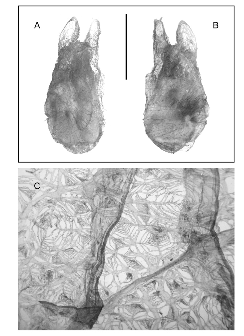
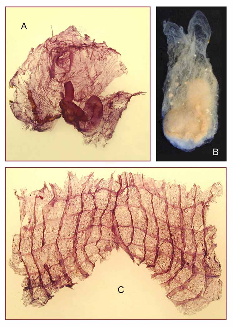
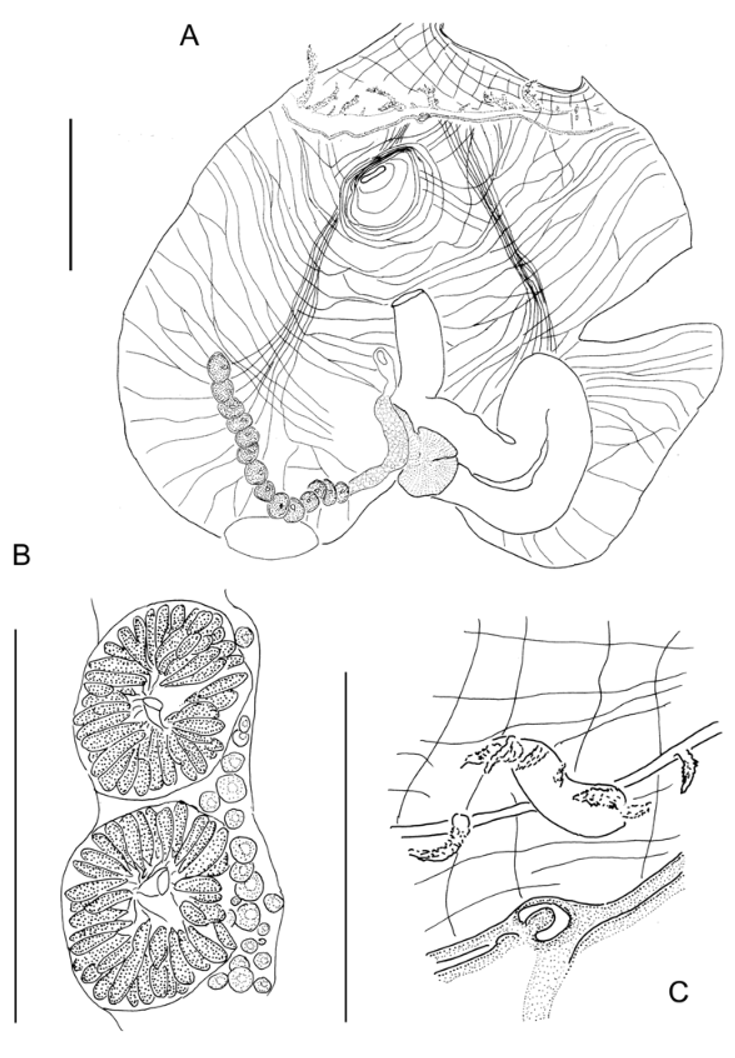
Description. All specimens are of about the same size 4 to 5 cm in length, colorless in formalin, particularly soft and consequently shapeless, embedded in a gelatinous thin tunic. They are not incrusted with sand but some particles adhere to the sticky tunic and fill the digestive tract. Amphipods inhabit some specimens. The body is easily seen through the transparent tunic. The siphons are in apical position, widely open (Fig. 1 A, B). The oral one is directed towards the ventral side, its rim partially damaged in all specimens. Four lobes are seen at the edge of the cloacal siphon in one animal. Both siphons have a long funnel of invaginated tunic. Even when the tunic is partially torn, the body remains in good condition. Its general shape is elongate, wider in the posterior part (Fig. 1 A, B). It does not seem contracted. The body wall is particularly thin and transparent with a musculature made of spaced long fibres (Figs 1 A, B, 2 B, 3 A). On the siphons, circular muscles are not branched and do not form a true sphincter; they are crossed by a few longitudinal short fibres. Around the body, circular muscles are irregularly spaced and sparsely branched (Fig. 1 A, B). On each side there is a narrow longitudinal musculature composed of a single ribbon of 8 to 10 fibres, starting from the peripharyngeal band (Figs 2 A, 3 A), they extend posteriorly remaining parallel along most of their length, fanning out at the posterior end of the body. This ribbon lies in the middle of each body side. About 12 widely separated oral tentacles (Figs 2 A, 3 A), stout with short branches, are located above a low membrane. They are unequal in length but there is not a definite pattern in the arrangement of the sizes (in the specimens where they can be observed). The peripharyngeal band has 2 crests and is not dorsally curved. The dorsal tubercle is flat, C-shaped, opened towards the right (Fig. 3 C). The dorsal lamina, smooth edged, increases in height posteriorly (Fig. 2 C). The branchial tissue is thin with an imperforated band along each side of the endostyle. There are 7 longitudinal vessels in high blades on each side (Fig. 2 C), prolonged a short distance anterior to the stigmata. Five wide transverse vessels delimit square meshes (Fig. 2 C). The stigmata are more or less numerous in a mesh, irregular in size and disposition (Fig. 1 C). They are made of a double spiral protruding in a cone for the largest. Parastigmatic vessels form an irregular web in a mesh (Fig. 1 C). The digestive loop (Fig. 2 A, 3 A) occupies half of the left side. In all specimens it is full of material. The oesophagus is short, not distinct from the stomach. There are small hepatic papillae on the external side of the stomach. No folds have been detected on the stomach wall. The intestine is wide, it extends in a long primary closed loop, and forms a secondary wide loop. The gaping anus opens with a smooth rim. The base of the rectum is attached to the stomach. There is a single tubular gonad in a ventral position on the right side (Figs 2 A, B; 3 A). The long ovary forms a half-circle along the posterior ventral line and joins the base of the rectum to which it is attached. The testis comprises a long series of lobules arranged in successive rosettes along the internal side of the ovary (Fig. 3 A, B). A short sperm duct opens internally from the centre of each rosette of testes (Fig. 3 B). The kidney is an oval vesicle at the very posterior end of the body, against the ovary (Fig. 3 A).
Monniot, Françoise, Monniot, Claude (2008): A new species of Bostrichobranchus (Ascidiacea, Molgulidae) from the eastern tropical Atlantic. Zootaxa 1742: 42-46, DOI: 10.5281/zenodo.181566