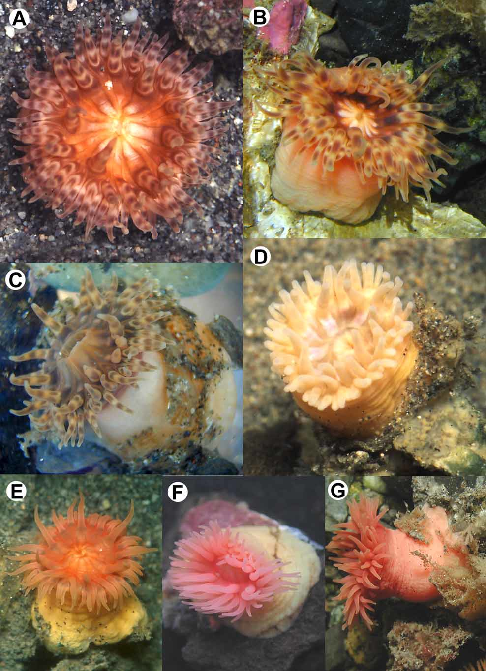
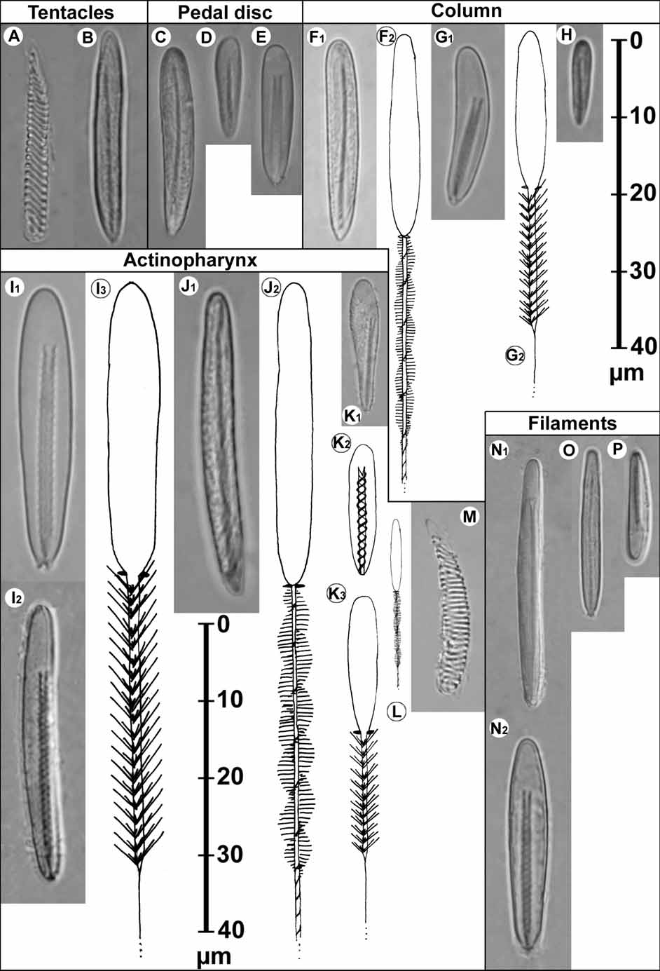
Colour (Figs. 3 A – G). Oral disc red, orange, yellowish, pale rose, pink, light brown or ochre; uniformly coloured or with (often 12) reddish-brown radial lines visible on endocoels, or with 12 - lobed, petal-like white and yellow pattern (Fig. 2 B). Mesenterial insertions visible through oral disc. Actinopharynx yellow to light brown. Tentacles slightly transparent, rose to red or pink, yellowish, light brown or ochre; uniformly coloured (Figs. 2 D – G) or with 3 – 6 brown transverse bands at inner and outer or only inner side (Figs. 2 A – C). Column rose-coloured, reddish, yellowish, orange to ochre, or light brown; uniformly coloured or distalmost 1 / 4 – 1 / 5 more intensely coloured (often brown; Fig. 2 D) with a short transition to paler proximal portion of column; colour generally continuously fading towards pedal disc. Pedal disc coloured as column, not transparent. Preserved specimens whitish to brown. Oral disc and tentacles. Between 66 and 110 conical tentacles, hexamerously arranged in 5 – 6 cycles, last cycle generally not complete, length about half diameter of oral disc, inner longer than outer, situated on outer third to half of oral disc, innermost 12 (two cycles) in many animals slightly more central (Fig. 2 A), more intensely coloured and directed upward. Oral disc circular, mesenterial insertions in many specimens visible as darker lines (Figs. 2 A, C, D, E). Mouth opening central, slightly oval, slightly elevated in many specimens. Column. In situ higher than broad, after sampling broader than high, proximally broader than distally; smooth, often with loose ring of mucus and dirt in proximal part (Figs. 2 C, D, G). Small fosse. Column can completely cover tentacles when retracted. Pedal disc. More or less circular, generally wider than column and oral disc, limbus slightly lobed. Internal anatomy. In most parts of column, 24 mesenteries hexamerously arranged in four cycles, first cycle (six pairs) including directives fertile macrocnemes with strong circumscript retractors, second and third cycle (six plus 12 pairs) sterile microcnemes without retractors, fourth cycle incomplete, pairs of extremely small microcnemes only just below margin, unequal proximal extension of mesenteries of a pair. Mesenteries of second cycle wider than those of third cycle. More tentacles than mesenteries in mid-column and at base. Actinopharynx deeply furrowed, with two distinct siphonoglyphs, about half length of column; two pairs of FIGURE 3. Histological sections of Paraisanthus fabiani: A, macrocneme with retractor and parietobasilar muscle; B, transverse section of the upper column; C, transverse section of the lower column; D, longitudinal section of the upper column with sphincter; E, cross section through tentacle; F, longitudinal section of the pedal disc with basilar muscles. 1 st to 3 rd cycle of mesenteries I, II, III, basilar muscles bm, directives d, ectoderm ec, endoderm en, ectodermal longitudinal muscles of tentacle et, mesogloea m, macrocnemes ma, filaments mf, microcnemes mi, parietobasilar muscles pb, actinopharynx ph, retractor muscles r, sperms s, siphonoglyph si, sphincter sp. TABLE 2. Size and distribution of cnidae of Paraisanthus fabiani n. sp. (ZSM 20070247 / 1, letters refer to Fig. 4), in each tissue in order of abundance: s: sporadic, f: few, c: common and v: very common. “ m l “ and “ m w ” are the means, “ d l ” and “ d w ” are the standard deviations (all in µm), “ t ” is the apparent total number of turns of spine-rows on the shaft, “ # ” is the number of capsules measured, “ p ” is the proportion of specimens having this cnida type. Exceptional sizes in parentheses. Spirocysts (A, M), basitrichs (B, C, D, F, H, J, L, O), microbasic p-mastigophores (E, N, P), microbasic pmastigophores B 1 (G, I, K). directives. Oral and marginal stomata; marginal stomata circular, in the centre of stronger mesenteries (stomata not always visible in smaller specimens). Sexes separate, no signs of asexual reproduction. Five of the sectioned specimens with reproductive tissue, collected in February and March, four male (ZSM 20070246, ZSM 20070247 / 1, ZSM 20070249, USNM 1101612), one female (ZSM 20051690). No zooxanthellae. Sphincter mesogloeal, strong, nearly entire width of mesogloea (Fig. 3 D), restricted to uppermost part (~ 1 / 8 – 1 / 10) of column. Macrocneme retractors strong, strongly restricted to circumscribed (Figs. 3 A – C). Parietobasilar muscles distinct on perfect mesenteries (Figs. 3 A – C); basilar muscles distinct (Fig. 3 F). Longitudinal muscles of tentacles (Fig. 3 E) and radial muscles of oral disc ectodermal. Endodermal circular muscles of column well marked, weaker at sphincter level (Fig. 3 D). Cnidom. Spirocysts, basitrichs, microbasic p-mastigophores B (Fig. 4). Cnidae of eight specimens were examined.
Häussermann, Verena, Försterra, Günter (2008): A new species of sea anemone from the Chilean fjord region, Paraisanthus fabiani (Actiniaria: Isanthidae), with a discussion of the family Isanthidae Carlgren, 1938. Zootaxa 1897: 27-42, DOI: 10.5281/zenodo.184465