AnimaliaNot EvaluatedacceptedspeciesAccepted
Rumarcanella gusuku
GBIF:119384688
ABOUT
Descriptions(6)
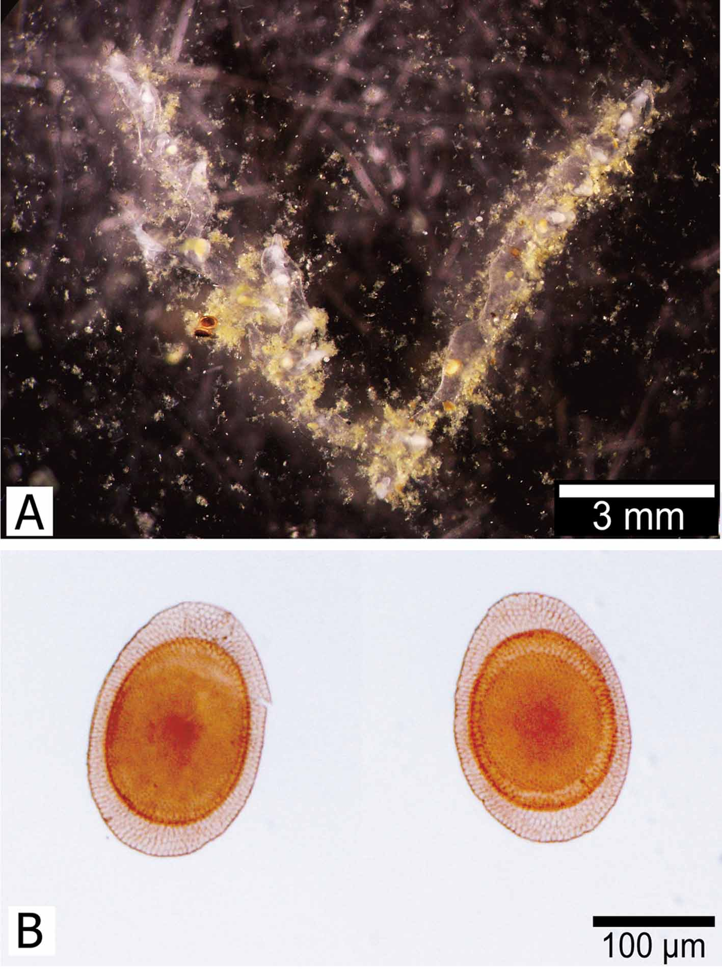
Description. Laboratory-cultured colony composed of narrow tubes, branching, weakly chitinized, transparent (Fig. 4 A). Floatoblasts oval, small, 315 – 346 (328 ± 11) μm long (n = 14) by 215 – 248 (228 ± 10) μm wide; length / width ratio about 1.4 (Figs 4 B, C). Ventral fenestra oval; 182 – 223 (203 ± 12) μm long by 158 – 185 (175 ± 7) μm wide. Dorsal fenestra almost circular, 176 – 202 (194 ± 9) μm long by 143 – 162 (155 ± 6) μm wide. In SEM images, surface of fenestrae entirely covered with rounded tubercles, each topped with a small hypertubercle (Fig. 5). Floatoblast valves asymmetrical in lateral and transverse views; ventral valve strongly convex, dorsal valve less convex, sometimes almost flat. Floatoblast suture with small irregular protuberances. Annuli sculptured with irregular pavement; ventral annulus narrow (~ 20 μm) along sides, wider (~ 50 μm) at both ends. Sessoblast unknown.
Hirose, Masato, Mawatari, Shunsuke F. (2011): Freshwater Bryozoa of Okinawa, Japan, with descriptions of Rumarcanella gen. nov. (Phylactolaemata: Plumatellidae) and two new species. Zootaxa 2732: 1-19, DOI: 10.5281/zenodo.276554
Plumatella sp. 2. Hirose et al. 2008: 65.
Hirose, Masato, Mawatari, Shunsuke F. (2011): Freshwater Bryozoa of Okinawa, Japan, with descriptions of Rumarcanella gen. nov. (Phylactolaemata: Plumatellidae) and two new species. Zootaxa 2732: 1-19, DOI: 10.5281/zenodo.276554
Remarks. Cultured colonies of R. gusuku resemble those of R. minuta and R. vorstmani in having a thin, transparent colony wall; unfortunately, we did not find any colonies in the wild, and these might differ in morphology from cultured colonies. Although the floatoblast of R. gusuku is very similar to that of R. minuta; the annulus is uniform in width in R. minuta, but narrower on both sides in R. gusuku; symmetrical in lateral view in R. minuta, but asymmetrical in R. gusuku, with a strongly convex ventral valve.
Hirose, Masato, Mawatari, Shunsuke F. (2011): Freshwater Bryozoa of Okinawa, Japan, with descriptions of Rumarcanella gen. nov. (Phylactolaemata: Plumatellidae) and two new species. Zootaxa 2732: 1-19, DOI: 10.5281/zenodo.276554
Distribution. Presently known only from southern and central Okinawa.
Hirose, Masato, Mawatari, Shunsuke F. (2011): Freshwater Bryozoa of Okinawa, Japan, with descriptions of Rumarcanella gen. nov. (Phylactolaemata: Plumatellidae) and two new species. Zootaxa 2732: 1-19, DOI: 10.5281/zenodo.276554
Etymology. The species name is from the Ryukyuan language gusuku, meaning “ castle, ” in reference to the type locality.
Hirose, Masato, Mawatari, Shunsuke F. (2011): Freshwater Bryozoa of Okinawa, Japan, with descriptions of Rumarcanella gen. nov. (Phylactolaemata: Plumatellidae) and two new species. Zootaxa 2732: 1-19, DOI: 10.5281/zenodo.276554
Material examined. Holotype. Cultured colony originating from a floatoblast, collected 21 January 2008 by Tohru Iseto from Ryutan-ike pond next to the castle, city of Naha; specimen deposited in National Science Museum, Tokyo (NSMT-Te 678). Paratype. Cultured colony originating from a floatoblast, collection data as for holotype; specimen deposited in National Science Museum, Tokyo (NSMT-Te 679). Other material. Floatoblasts from Ryutan-ike pond next to the castle, city of Naha.
Hirose, Masato, Mawatari, Shunsuke F. (2011): Freshwater Bryozoa of Okinawa, Japan, with descriptions of Rumarcanella gen. nov. (Phylactolaemata: Plumatellidae) and two new species. Zootaxa 2732: 1-19, DOI: 10.5281/zenodo.276554
Export occurrence data
Darwin Core Archive (ZIP)
CLASSIFICATION
Taxonomic Classification Tree

MULTIMEDIA
Media Files(2)
FIGURE 4. Rumarcanella gusuku n. sp. A, colony. B, floatoblast, ventral (left) and dorsal (right) valves.
Imageimage/png© Hirose, Masato;Mawatari, Shunsuke F.Hirose, Masato;Mawatari, Shunsuke F.
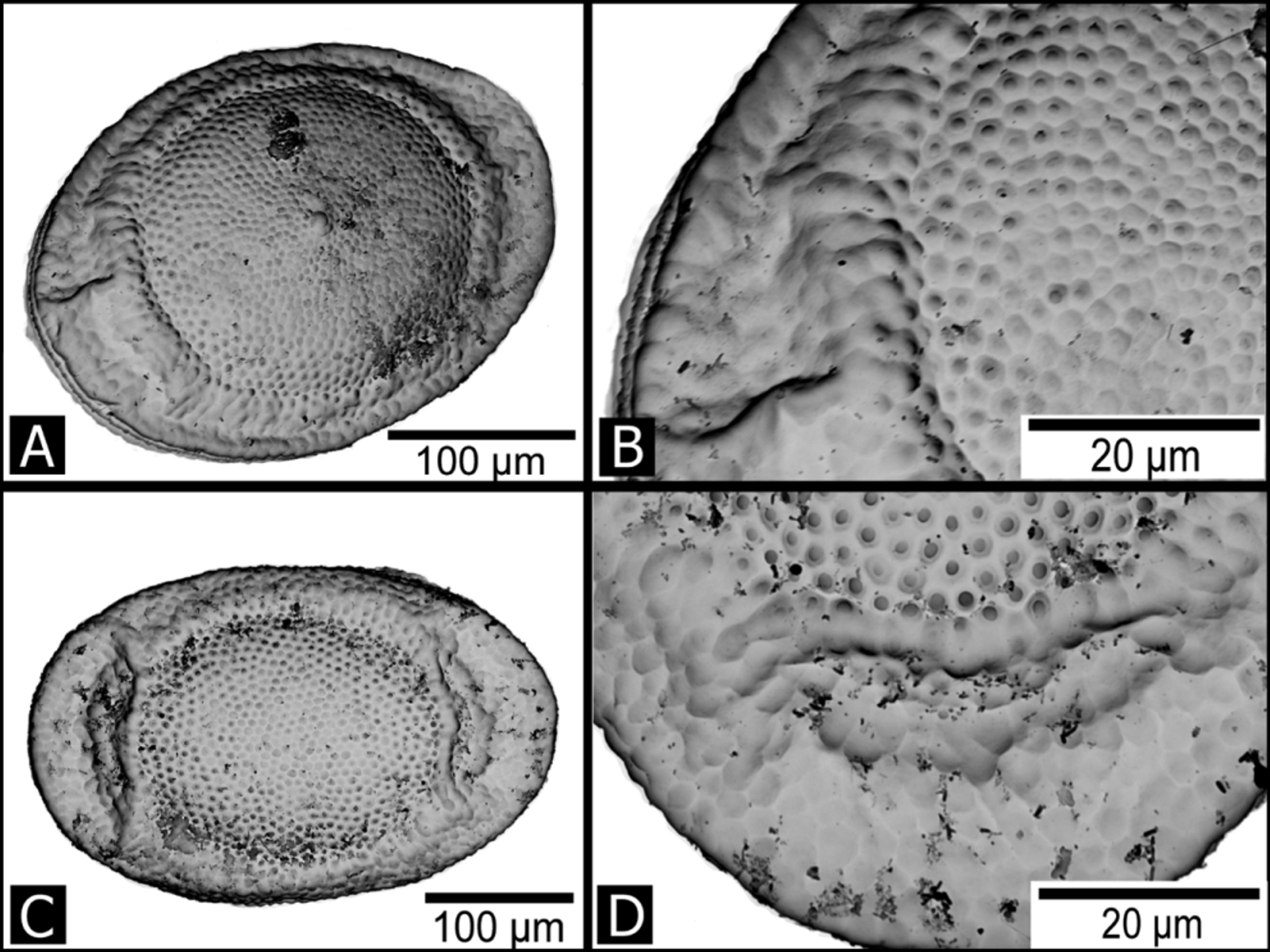
FIGURE 5. Rumarcanella gusuku n. sp. SEM micrographs of floatoblast. A, ventral valve. B, enlargement of A, showing hypertubercles on the fenestra. C, dorsal valve. D, enlargement of C, showing hypertubercles on the fenestra.
Imageimage/png© Hirose, Masato;Mawatari, Shunsuke F.Hirose, Masato;Mawatari, Shunsuke F.
IMAGES