AnimaliaNot EvaluatedacceptedspeciesAccepted
Rumarcanella vorstmani
(Toriumi, 1952) Toriumi, 1952
GBIF:119384690
0year
0
Synonyms
ABOUT
Descriptions(4)
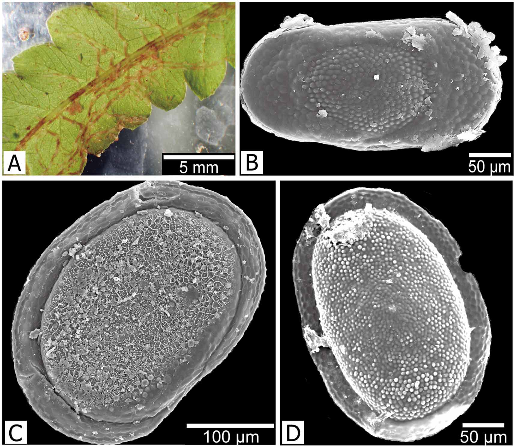
Description. Colony with narrow branches, transparent, almost entirely recumbent (Fig. 8 A). Tentacle number 20 – 27 (Wood et al. 2006). Floatoblast (Fig. 8 B) oblong-elliptical, small, 295 – 322 (310 ± 12) μm long by 155 – 182 (168 ± 14) μm wide (n = 4), with length / width ratio of about 1.8; symmetrical in lateral view; both ventral and dorsal fenestrae subcircular, both with tubercles bearing single hypertubercles. Sessoblast (Figs 8 C, D) small, 288 – 358 (324 ± 28) μm long by 215 – 248 (229 ± 14) μm wide (n = 5); fenestra with tubercles, annulus with weak reticulation.
Hirose, Masato, Mawatari, Shunsuke F. (2011): Freshwater Bryozoa of Okinawa, Japan, with descriptions of Rumarcanella gen. nov. (Phylactolaemata: Plumatellidae) and two new species. Zootaxa 2732: 1-19, DOI: 10.5281/zenodo.276554
Remarks. Rumarcanella vorstmani was originally described as Plumatella javanica, but Toriumi (1952 a) recognized the former as a distinct species on the basis of tentacle number, floatoblast size, and the surface microsculpture of the sessoblast. The transparent, weakly chitinized colony wall of R. vorstmani has made generic placement difficult. Lacourt (1968) included this species in Hyalinella owing to the soft, transparent ectocyst, but because the species produces sessoblasts, Wiebach (1973) transferred it to Plumatella. In a similar case, R. minuta had long been reported to produce only floatoblasts and was placed in Hyalinella. Toriumi (1972) reported the occurrence of sessoblasts, and Wood et al. (2006) transferred the species to Plumatella. Toriumi reported tubercles on the sessoblast of R. vorstmani. When Wood & Wood (2000) reexamined specimens that Mukai (1984) had identified as P. vorstmani, they found the surface of the capsule to be reticulate rather than tuberculate. However, Wood et al. (2006) reported R. vorstmani sessoblasts with a tuberculate fenestra from many sites in Thailand. In our collection, the reticulation (Fig. 8 C) on the sessoblast fenestra is easily lost from the surface, and the strong tubercles underneath become apparent (Fig. 8 D). Thus, the reticulation may be associated with the membrane that covers the sessoblast surface.
Hirose, Masato, Mawatari, Shunsuke F. (2011): Freshwater Bryozoa of Okinawa, Japan, with descriptions of Rumarcanella gen. nov. (Phylactolaemata: Plumatellidae) and two new species. Zootaxa 2732: 1-19, DOI: 10.5281/zenodo.276554
Distribution. Rumarcanella vorstmani has been reported from mainland Asia (Bushnell 1973; Wood et al. 2006), India (Lacourt 1968), Java and Indonesia (Vorstman 1928 a, b), and from Okinawa to Miyagi Prefecture in Japan (Toriumi 1952 a; Mukai 1984).
Hirose, Masato, Mawatari, Shunsuke F. (2011): Freshwater Bryozoa of Okinawa, Japan, with descriptions of Rumarcanella gen. nov. (Phylactolaemata: Plumatellidae) and two new species. Zootaxa 2732: 1-19, DOI: 10.5281/zenodo.276554
Material examined. Floatoblasts from Fukuji Dam, village of Higashi; several mature colonies with floatoblasts and sessoblasts, Kanna Dam, village of Ginoza.
Hirose, Masato, Mawatari, Shunsuke F. (2011): Freshwater Bryozoa of Okinawa, Japan, with descriptions of Rumarcanella gen. nov. (Phylactolaemata: Plumatellidae) and two new species. Zootaxa 2732: 1-19, DOI: 10.5281/zenodo.276554
Export occurrence data
Darwin Core Archive (ZIP)
CLASSIFICATION
Taxonomic Classification Tree

MULTIMEDIA
Media Files(1)
FIGURE 8. Rumarcanella vorstmani (Toriumi, 1952). A, colony on a leaf of an aquatic plant. B, floatoblast, ventral view (SEM). C, sessoblast (SEM) with villous reticulation over tubercles on the fenestra. D, Sessoblast with tubercles but no reticulation on the fenestra.
Imageimage/png© Hirose, Masato;Mawatari, Shunsuke F.Hirose, Masato;Mawatari, Shunsuke F.
IMAGES