
Pyuridae
GBIF:119396072
ABOUT
Descriptions(1)
Pyuridae
Bathypera hastaefera Vinogradova, 1962 (Figures 22 BD, 23B, 24B)
Vinogradova, 1962: 206, fig. 3. Kott, 1969: 140 fig.194. Monniot & Monniot 1983: 85, part of specimens included in Bathyfera splendens .
Stations (events when several trawling operations per station): 8-21-30 (66)-42-57.
The specimens are normally spherical. When fixed the tunic has numerous parallel furrows due to the contraction of the body wall which closely adheres to the tunic (Fig. 22 D). The spicules are uniformly distributed at the colony surface, implanted on a round base and protruding in an asymmetric group of few spines of inequal length (Fig.22B). The spicules reach 600µm in length. The musculature has the same disposition as in B. splendens but the crossed longitudinal and transverse fibres are stronger and more extended on the body wall (Fig. 24B). All other characters are the same in both species taking in consideration the individual variations: branchial sac (Fig. 23 B), gut gonads and endocarps. Monniot and Monniot (1983) with few specimens estimated the differences insufficient to separate the species; the study of numerous samples collected during the CEAMARC cruise now confirms the validity of B. hastaefera .
B. hastaefera was only recorded from the eastern part of the Antarctic continent down to 2000 m. It was present in some of the CEAMARC stations in sympatry with. B. splendens .
Export occurrence data
Darwin Core Archive (ZIP)
CLASSIFICATION
Taxonomic Classification Tree

MULTIMEDIA
Media Files(3)
FIGURE 22. A, B, spicules: A, Bathypera splendens; B, Bathypera hastaefera. C, Bathypera splendens; D, Bathypera hastaefera. Scale bars: A, B = 100 µm; C, D = 3 cm.

FIGURE 23. Branchial tissues: A, Bathypera splendens. B, Bathypera hastaefera.
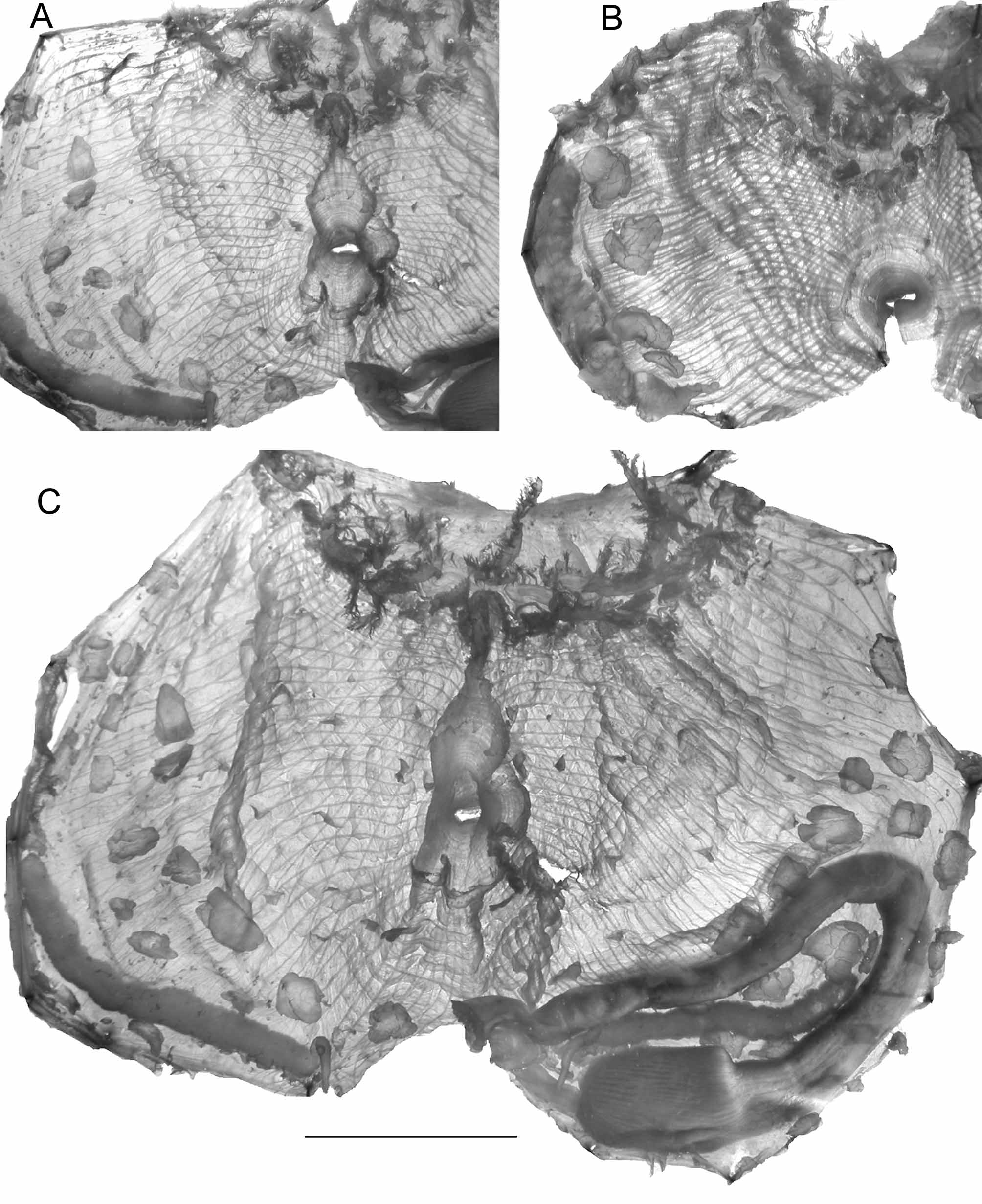
FIGURE 24. A, B, musculature of the left body side: A, Bathypera splendens; B, Bathypera hastaefera. C, Bathypera splendens, specimen ventrally opened, scale bar C = 1 cm.
IMAGES