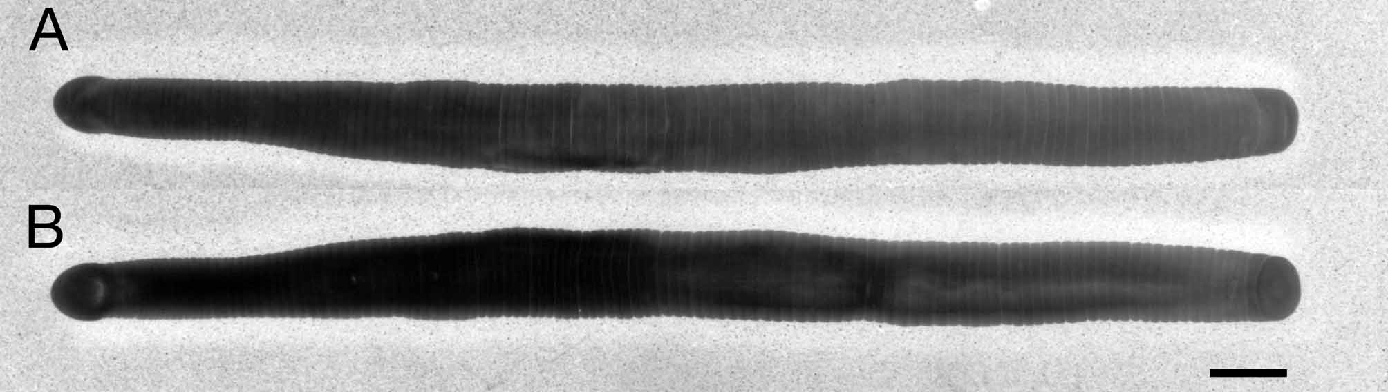
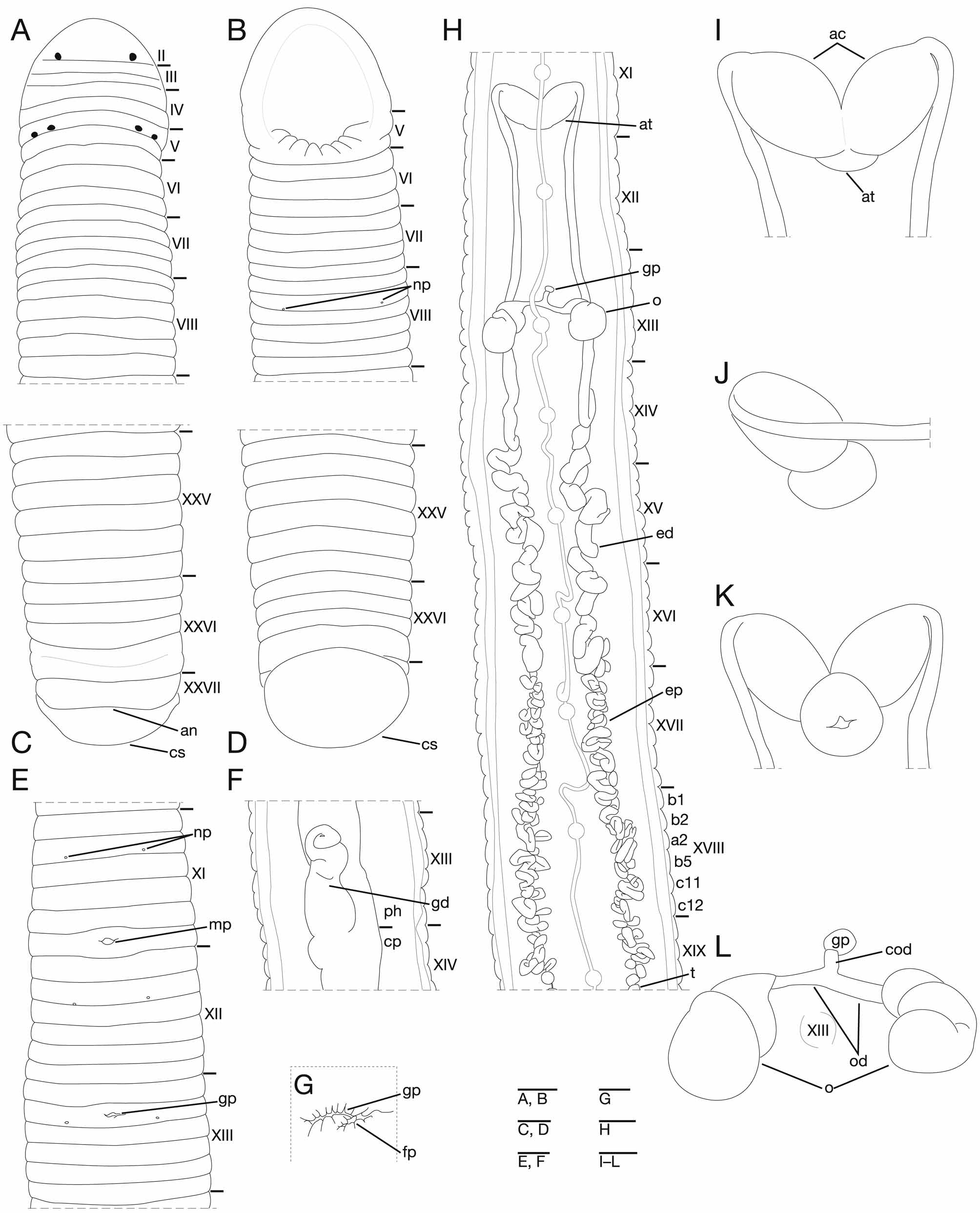
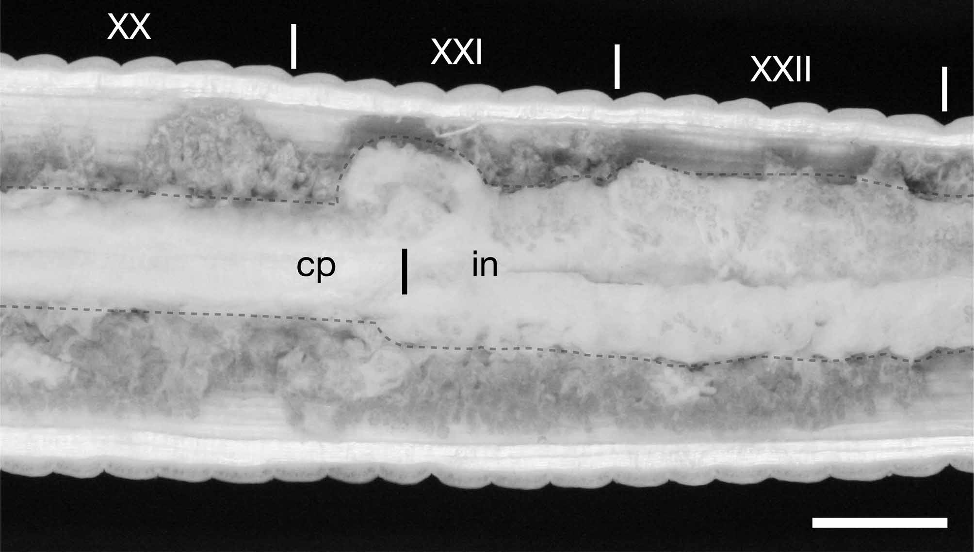
Type specimens. The ten specimens, which were used in the original description of O. ijimai (Oka 1895), are automatically considered the syntypes in accordance with the Article 73.2 of the International Code of Zoological Nomenclature (International Commisson on Zoological Nomenclature 1999). I surveyed leech collection at NSMT and UMUT (see Materials and Methods above), however, I could not find out any type series of O. ijimai. According to the Art. 75.1 of the Code, a neotype should be designated when 1) no name-bearing type specimen is believed to be extant and 2) a name-bearing type is necessary to define the nominal taxon objectively (International Commisson on Zoological Nomenclature 1999). The nomenclatural status of Orobdella ijiami does not apply the condition 2) above, because its type locality is only one place, Nikko, Japan and I did not collect any other sexannulate Orobdella species from the type locality. As shown at the Example of the Art. 75.2 of the Code, a neotype designation for such species like O. ijimai would be invalid (International Commisson on Zoological Nomenclature 1999). Therefore, I have not designated a neotype for O. ijimai in this study. Materials examined. A total of six specimens newly collected from around the Inarigawa Sand-tap Dams at the type locality, Nikko, Tochigi Pref., Japan by Takafumi Nakano. Four specimens collected on May 26, 2010: KUZ Z 108 (alt. 960 m, 36 ° 47´01 ˝ N, 139 ° 34´54 ˝ E), dissected; KUZ Z 109 (alt. 946 m, 36 ° 46´59 ˝ N, 139 ° 34´55 ˝ E); KUZ Z 110, dissected, and KUZ Z 111 (alt. 940 m, 36 ° 46´59 ˝ N, 139 ° 34´56 ˝ E). Two specimens collected on May 28, 2010: KUZ Z 112 (alt. 990 m, 36 ° 47´08 ˝ N, 139 ° 34´54 ˝ E); and KUZ Z 113, dissected (alt. 1014 m, 36 ° 47´13 ˝ N, 139 ° 34´51 ˝ E). Emended description. Body firm, muscular, elongated, maximum BL 93.3 mm (KUZ Z 111), maximum BW 6.3 mm (KUZ Z 110) (Fig. 2). Caudal sucker ventral, oval, its diameter slightly smaller than maximum body width (Fig. 3 D). In life, color variable, generally dorsal surface yellowish green, ventral surface whitish yellow, dorsal surface darker than ventral surface. Color faded in preservative, without any dorsal dark lines. Somite I completely merged with prostomium (Fig. 3 A). Somite II uniannulate, slightly separated from I (Fig. 3 A). Somite III biannulate, (a 1 + a 2)> a 3 (Fig. 3 A) or uniannulate (KUZ Z 110). Somite IV biannulate, (a 1 + a 2)> a 3 (Fig. 3 A). Somite V biannulate, a 3 forming posterior margin of oral sucker (Fig. 3 A, B). Somites VI complete triannulate (Fig. 3 A, B). Somite VII complete quadrannulate. Somites VIII – XXV complete sexannulate, b 1 = b 2 = a 2 = b 5 = c 11 = c 12 (Fig. 3 A – E, H), X b 5 the first annulus of the clitellum, XIII a 2 the last annulus of the clitellum (KUZ Z 110). Somite XXVI quadrannulate, a 1 = a 2 = b 5> b 6, b 6 the last complete annulus on the venter (Fig. 3 C, D). Somite XXVII uniannulate or biannulate (Fig. 3 C). Anus behind XXVII (Fig. 3 C). Post-anal annulus absent (Fig. 3 C). Anterior ganglionic mass in VI a 1 – a 3. Ganglion VII in a 2. Ganglia VIII – XXV in a 2 of each somite, except ganglion XIV in b 5 (Fig. 3 H). Ganglion XXVI in a 1. Posterior ganglionic mass in XXVI b 5 and b 6. Eyes three pairs, first pair dorsally on posterior margin of II, second and third pairs dorsolaterally on posterior margin of V (a 1 + a 2) (Fig. 3 A). Nephridiopores 17 pairs, ventrally at posterior margin of b 2 of each somite, at VIII – XXIV (Fig. 3 B, E). Papillae numerous, minute, hardly visible, one row on every annulus. Pharynx agnathous, euthylaematous, reaching to XIII / XIV. Crop tubular, acecate, reaching to XX c 11 / c 12 – XXI a 2 (Fig. 4). Gastropore conspicuous, ventral, at middle of XIII b 2 (Fig. 3 E, G). Gastroporal duct muscular, tortuous at junction with gastropore, bulbiform, joining with crop in XIV b 1 – b 2 (Fig. 3 F). Intestine tubular, acecate, reaching to XXIV a 2 / b 5 – c 11, one pair of undeveloped chambers at junction with crop or chamber absent (Fig. 4). Rectum tubular, thin-walled. Male gonopore at the middle or posterior of XI c 12 (Fig. 3 E). Female gonopore at XIII b 2, inconspicuous, diagonally behind gastropore (Fig. 3 G). Gonopores separated by 1 / 2 + 7 + 1 / 2 annuli (Fig. 3 E). Testisacs multiple, two or three testisacs on each side in each annulus, in XVIII c 11 – XIX a 2 to XXV b 1 – b 5 (Fig. 3 H). Epididymides in XVI b 5 / c 11 – c 11 / c 12 to XVIII c 11 – XIX a 2 (Fig. 3 H). Ejaculatory bulbs absent. Ejaculatory ducts in XI b 5 to XVI b 5 / c 11 – c 11 / c 12, loosely coiled, widen gradually from each junction with epididymis, narrow at each junction with atrial cornu, then turning sharply inward toward atrial cornu without pre-atrial loop (Fig. 3 H – K). Atrial cornua in XI b 5 to c 12, muscular, nearly oviform (Fig. 3 H – K). Atrium short, muscular, globular in XI c 12 (Fig. 3 I – K). Ovisacs one pair, thin-walled, globular, in XIII a 2 to c 11 (Fig. 3 H, L). Oviduct thin-walled, left oviduct crossing ventrally beneath nerve cord, both oviducts converging into common oviduct in XIII a 2 (Fig. 3 H, L). Common oviduct thin-walled, short, directly descending to female gonopore (Fig. 3 L).
Nakano, Takafumi (2011): Redescription of Orobdella ijimai (Hirudinida: Arhynchobdellida: Gastrostomobdellidae), and two new species of Orobdella from the Ryukyu Archipelago, Japan. Zootaxa 2998: 1-15, DOI: 10.5281/zenodo.207870