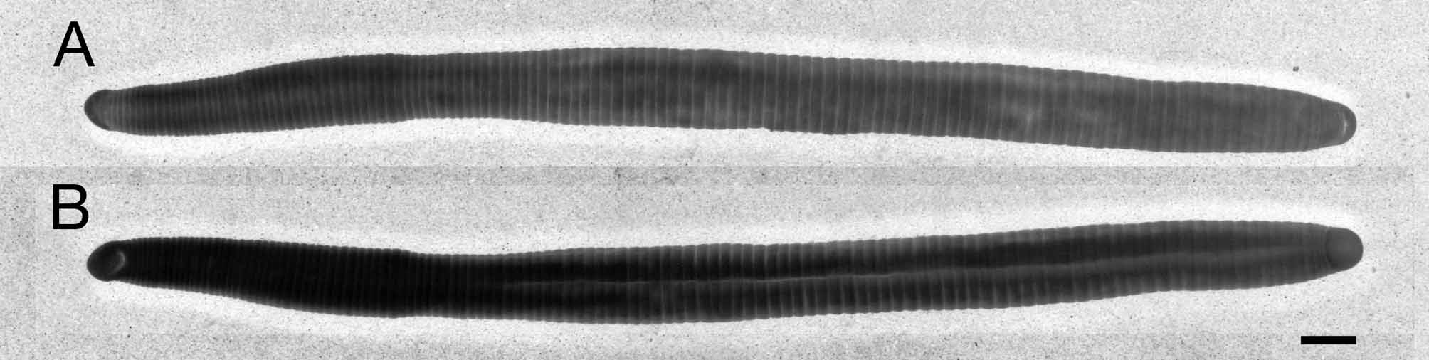
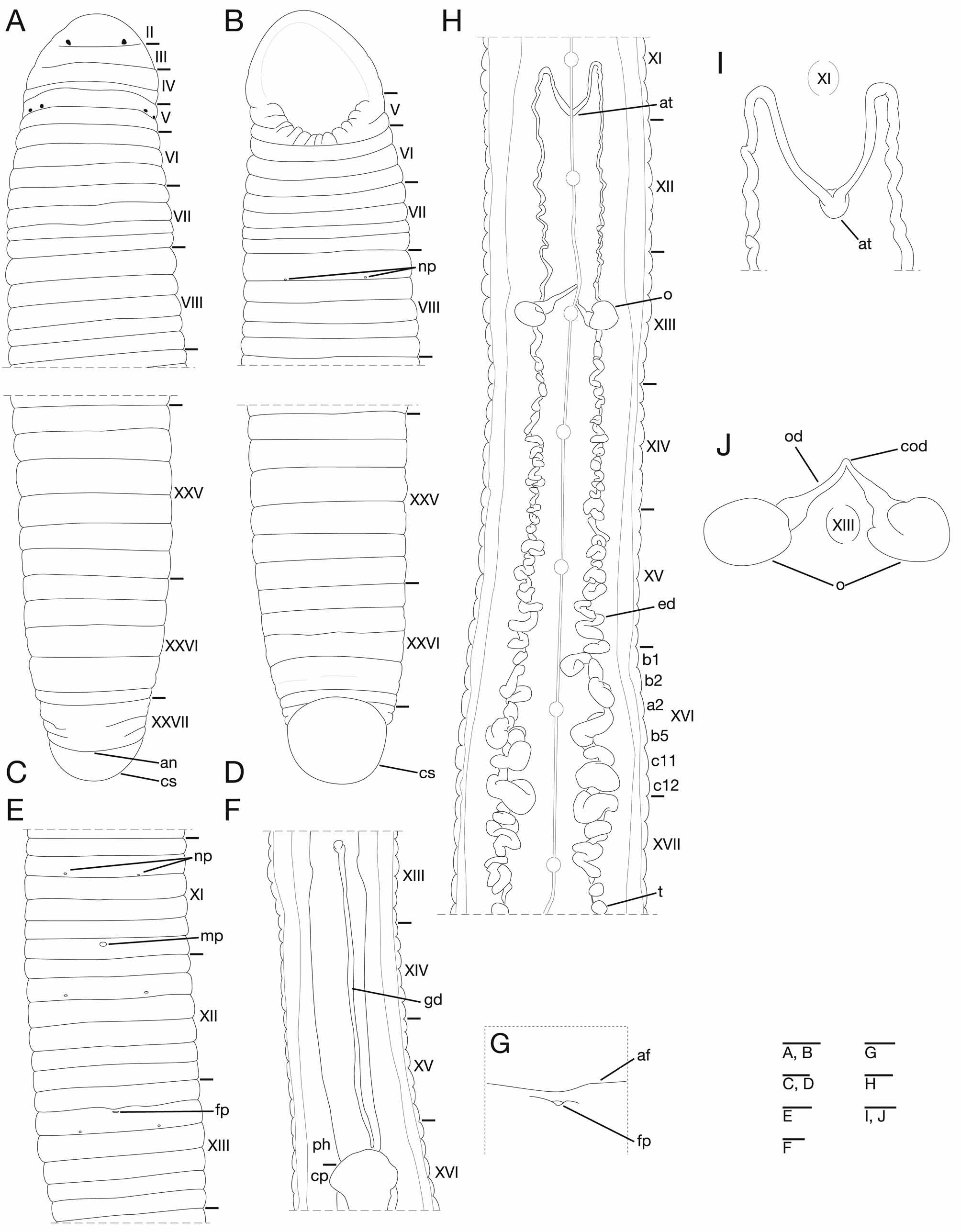
Description of holotype. Body firm, muscular, BL 106.7 mm, BW 6.6 mm (Fig. 5). Caudal sucker ventral, oval, its diameter slightly smaller than maximum body width (Fig. 6 D). In life, dorsal surface yellowish green, ventral surface whitish yellow, dorsal surface darker than ventral surface. Color faded in preservative, without any dark lines. Somite I completely merged with prostomium (Fig. 6 A). Somite II uniannulate, not separated from I (Fig. 5 A). Somite III uniannulate (Fig. 6 A). Somite IV biannulate (Fig. 6 A). Somite V biannulate, a 3 forming posterior margin of oral sucker (Fig. 6 A, B). Somites VI triannulate (Fig. 6 A, B). Somite VII complete quadrannulate, a 1 = a 2> b 5 = b 6 (Fig. 6 A, B). Somite VIII complete quinquannulate, a 1> a 2 = b 5> c 11 = c 12 (Fig. 6 A, B). Somite IX complete sexannulate, b 1 c 11 = c 12. Somites X – XXV complete sexannulate, b 1 = b 2 = a 2 = b 5 = c 11 = c 12 (Fig. 6 C – E, H), X a 2 the first annulus of the clitellum, XIII b 5 the last annulus of the clitellum. Somite XXVI quinquannulate, b 1 = b 2 = a 2 b 6, b 6 the last complete annulus on the venter (Fig. 6 C, D). Somite XXVII incomplete biannulate (Fig. 6 C). Anus behind XXVII (Fig. 6 C). Post-anal annulus absent (Fig. 6 C). Anterior ganglionic mass in VI a 2 and a 3. Ganglion VII in a 2. Ganglia VIII – XXI in a 2 of each somite (Fig. 6 H). Ganglia XXII – XXV in b 2 and a 2 of each somite. Ganglion XXVI in b 1 and b 2. Posterior ganglionic mass in XXVI a 2 – b 6. Eyes three pairs, first pair dorsally in II, second and third pairs dorsolaterally on posterior margin of V (a 1 + a 2) (Fig. 6 A). Nephridiopores 17 pairs, ventrally at posterior margin of a 1 at VIII and b 2 of each somite, at IX – XXIV (Fig. 6 B, E). Papillae numerous, minute, hardly visible, one row on every annulus. Pharynx agnathous, euthylaematous, reaching to XVI b 2 / a 2. Crop tubular, acecate, in XVI b 2 / a 2 to XXII a 2. Gastropore absent (Fig. 6 E, G). Gastroporal duct narrow, tubular but slightly bulbous at female gonopore, in XIII b 1 / b 2 to XVI b 2, not joining with crop (Fig. 6 F). Intestine tubular, acecate, in XXII a 2 to XXIV / XXV. Rectum tubular, thin-walled. Male gonopore at anterior of XI c 12, close to annular furrow of c 11 / c 12 (Fig. 6 E). Female gonopore at anterior of XIII b 2, close to annular furrow of b 1 / b 2 (Fig. 6 E, G). Gonopores separated by 8 annuli (Fig. 6 E). Testisacs multiple, two or three testisacs on each side in each annulus, in XVII b 5 to XXV b 5 (Fig. 6 H). Epididymides absent. Ejaculatory bulbs absent (Fig. 6 H). ejaculatory ducts in XI a 2 to XVII a 2, coiled, widen from each junction with testisacs, narrow gradually toward each junction with atrium, with pre-atrial loop extending to ganglion XI (Fig. 6 H, I). Atrial cornua absent. Atrium rudimentary in XI c 11 and c 12 (Fig. 6 H, I). Ovisacs one pair, thin-walled, globular, in XIII a 2 (Fig. 6 H, J). Oviduct thin-walled, right oviduct crossing ventrally beneath nerve cord, both oviducts converging into common oviduct in XIII b 2 (Fig. 6 H, J). Common oviduct thin-walled, very short, directly descending to female gonopore (Fig. 6 H, J). Variation. Somite VII triannulate (KUZ Z 124) or ventrally triannulate (KUZ Z 118, Z 122). Somite XXVII incomplete biannulate or triannulate. Clitellum from X b 5 to XIII a 2 – XIV b 1. Pharynx reaching to XVI b 1 – c 11. Crop reaching to XXII b 1 – XXIII b 1. Gastroporal duct tends to attach ventral sinus (Fig. 7). Intestine reaching to XXIV c 11 – XXV b 1 / b 2. Male gonopore in XI c 11 / c 12 or at anterior XI c 12. Female gonopore in XIII b 1 / b 2 or at anterior of XIII b 2. Testisacs in XVIII b 1 – XVIII b 5 to XXV a 2 – c 11.
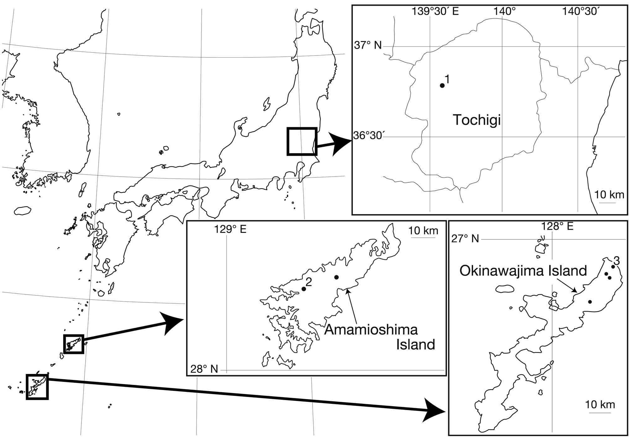
Nakano, Takafumi (2011): Redescription of Orobdella ijimai (Hirudinida: Arhynchobdellida: Gastrostomobdellidae), and two new species of Orobdella from the Ryukyu Archipelago, Japan. Zootaxa 2998: 1-15, DOI: 10.5281/zenodo.207870