AnimaliaNot EvaluatedacceptedspeciesAccepted
Microhyla mymensinghensis

Microhyla mymensinghensis

GBIF:119412855

0

Synonyms

ABOUT

Descriptions(6)

Description of holotype. Body small (SVL: 21.3) and stocky. Vomerine teeth absent, tongue elliptical. Head width greater than length (HW: 7.2; HL: 5.9), snout truncate, projecting slightly beyond the lower jaw. Canthus rostralis rounded, lore sloping and weakly concave. Nostril nearer to tip of snout than to eye (S-N: 1.3; N-E: 2.3). Tympanum hidden. Inter-orbital space wider than eyelid and inter-nostril space (E-E: 2.9; ELW: 2.1 and N-N: 2.1). Fingers slender, free, and tips not swollen. Finger length F 1
Hasan, Mahmudul, Islam, Mohammed Mafizul, Kuramoto, Mitsuru, Kurabayashi, Atsushi, Sumida, Masayuki (2014): Description of two new species of Microhyla (Anura: Microhylidae) from Bangladesh. Zootaxa 3755 (5): 401-418, DOI: 10.11646/zootaxa.3755.5.1
Variation. The Sylhet population has a longer head than the Mymensingh population (HL / SVL: 0.39 vs. 0.29, HL / HW: 1.17 vs. 0.91), the differences being highly significant. Dorsal pattern is variable among individuals from the Mymensingh population (Figs. 6 A, 6 B), but Netrokona, Sylhet and Sunamganj specimens follow the dorsal pattern noted in Fig. 6 A. Usually, the dorsal color of Mymensingh population is reddish to yellowish, while a few individuals have a browner dorsal color (Fig. 6 B). Of the 15 specimens examined from Mymensingh, 9 individuals (60 %) have a whitish ventral throat while 6 individual (40 %) have a slightly ash ventral throat, along with a few speckles from their chin to base of forelimbs and rarely between the bases of forelimbs, not correlated with the male vocal sac. Of the examined 14 specimens from Sylhet, 12 individuals (86 %) have two parallel small dark lines at the terminus of their X-shaped distinct pattern, located near the crescent-shaped markings on the anus, but the remaining two individuals (IABHU 3945 and 3950) lacked these lines. These lines are irregular in the Mymensingh population. Chromosomes. Microhyla mymensinghensis has 2 n = 24 chromosomes (Fig. 7). The frogs of genus Microhyla from Southeast Asia have been reported to possess 2 n = 24 or 2 n = 22 chromosomes (King 1990; Kuramoto 1990; Kuramoto & Yong 1992). M. okinavensis (Ryukyu Archipelago, Japan), M. fissipes (China) and M. berdmorei (Thailand) have 2 n = 24 chromosomes. Joshy & Kuramoto (2011) suggested that the 2 n = 24 and 2 n = 22 karyotypes derive from the 2 n = 26 karyotype of the Indian M. ornata and M. rubra. The karyotype of M. mymensinghensis supports a close relationship with M. fissipes. Natural history. Microhyla mymensinghensis is sympatric with M. berdmorei, in the northeastern part of Bangladesh. We observed M. mymensinghensis either in grass under large trees (locally called “ Lendi Korui ”) or in open fields with some vegetation and slightly wet and loose soil. After dissection, we found many small insect parts, as well as some sand in the gut. Their breeding season is likely June-July, as we caught several females in June from Bangladesh Agricultural University Campus (BAUC) just after light rains, which contained about 40 – 50 mature ova in the ovaries. Each ovum is very small, approximately 650 µm in diameter.
Hasan, Mahmudul, Islam, Mohammed Mafizul, Kuramoto, Mitsuru, Kurabayashi, Atsushi, Sumida, Masayuki (2014): Description of two new species of Microhyla (Anura: Microhylidae) from Bangladesh. Zootaxa 3755 (5): 401-418, DOI: 10.11646/zootaxa.3755.5.1
Diagnosis. The new species M. mymensinghensis is allocated to genus Microhyla due to its smooth or warty skin, absence of vomerine teeth, hidden tympanum, and sister relationships with other microhylid frogs (Hasan et al. 2012; Hasan et al. unpublished data). Comparison of the 16 S-rrn and / or cytb sequences of the new species with its congeners in DDBJ / EMBL / GenBank databases from South and Southeast Asia including Bangladesh, India and Myanmar, denotes that any known microhylid frogs do not correspond to this new species and merits its distinctness. Considering the molecular analyses and morphological characters i. e. a crescent-shaped marking on the anus, an X-shaped marking on the dorsum and the tibio-tarsal articulation of the new species deduct it from all of the known Microhyla species from South and Southeast Asia, particularly from Bangladesh and its neighbor countries; but make confusion with only M. mukhlesuri, M. fissipes, M. heymonsi and M. ornata. However, the new species differs from M. ornata by the presence of an outer metatarsal tubercle (vs. outer metatarsal tubercle is absence in M. ornata) and from M. heymonsi by smaller size (SVL = 14.2 – 21.3 mm) (vs. 22 – 26 mm in M. heymonsi) (Chanda 2002) and presence of X-shaped marking on the dorsal (this kind of marking absence in M. heymonsi). The new species can be distinguished from its most near congener M. fissipes by the extension of tibiotarsal articulation until eye to the tip of snout (vs. reached only until eye in M. fissipes). Most importantly, the new species differs from M. mukhlesuri by smaller body size, SVL = 16.5 ± 1.8 mm (vs. SVL = 17.8 ± 1.4 mm in M. mukhlesuri), higher forearm width, FAW = 0.9 ± 0.1 mm (vs. FAW = 0.7 ± 0.1 mm in M. mukhlesuri) (see Table 1), and presence of crescent shape marking on the anus (vs. this marking looks “ U ” shape in the M. mukhlesuri). Further, like with M. mukhlesuri, none of the old nomina of Microhyla corresponds to this new species. In conclusion, this new species is small frog with SVL of 14.2 – 17.6 mm in males and 15.2 – 21.3 mm in females. Head length subequal head width, finger formula 1 <4 <2 <3, toe formula 1 <5 <2 <3 <4, fingers free and slender, tips of fingers and toes not widened, rudimentary web between toes and subarticular tubercles distinct (Figs. 4 G, 4 H). TIL / SVL ratio was 0.57 ± 0.04, whereas this value was 0.54 ± 0.03 in M. mukhlesuri, and 0.50 ± 0.02 in M. fissipes from Taiwan. Tibiotarsal articulation extends from between the eyes to the tip of the snout, while it reaches near the eye in M. fissipes and in front of the shoulders in M. ornata. Phylogenetically, it shows sister relationship with M. mukhlesuri plus M. fissipes with high genetic divergences (Hasan et al. 2012; Hasan et al. unpublished data; this study).
Hasan, Mahmudul, Islam, Mohammed Mafizul, Kuramoto, Mitsuru, Kurabayashi, Atsushi, Sumida, Masayuki (2014): Description of two new species of Microhyla (Anura: Microhylidae) from Bangladesh. Zootaxa 3755 (5): 401-418, DOI: 10.11646/zootaxa.3755.5.1
Distribution. The known occurrence of M. mymensinghensis includes the Mymenisngh, Netrokona, Sylhet and Sunamganj districts in the in the central and northeastern regions of Bangladesh (Fig. 1).
Hasan, Mahmudul, Islam, Mohammed Mafizul, Kuramoto, Mitsuru, Kurabayashi, Atsushi, Sumida, Masayuki (2014): Description of two new species of Microhyla (Anura: Microhylidae) from Bangladesh. Zootaxa 3755 (5): 401-418, DOI: 10.11646/zootaxa.3755.5.1
Etymology. The specific name refers to Mymensingh, the type locality of this species.
Hasan, Mahmudul, Islam, Mohammed Mafizul, Kuramoto, Mitsuru, Kurabayashi, Atsushi, Sumida, Masayuki (2014): Description of two new species of Microhyla (Anura: Microhylidae) from Bangladesh. Zootaxa 3755 (5): 401-418, DOI: 10.11646/zootaxa.3755.5.1
Holotype. IABHU 4116, adult female (SVL: 21.3 mm, if not otherwise specified, the following body parts are measured in mm) collected from Bangladesh Agricultural University Campus (24 º 44 ′ 50 ″ N, 90 º 24 ′ 24 ″ E,> 18 m asl.), Mymensingh, Bangladesh on June 25, 2012 by M. Hasan (Figs. 4 E, 4 F). Paratypes. IABHU 4004, adult male (SVL: 14.8); IABHU 4117, adult female (SVL: 20.2); and IABHU 4120, adult female (SVL: 20.5) collected from Bangladesh Agricultural University Campus, Mymensingh, Bangladesh on 9 June 2011 and 25 June 2012 by M. Hasan. IABHU 3947, adult male (SVL: 17.6); IABHU 3948, adult male (SVL: 17.3); and IABHU 3899, adult female (SVL: 16.7) collected from Golapganj, Sylhet on 6 June 2011 by M. M. R. Khan.
Hasan, Mahmudul, Islam, Mohammed Mafizul, Kuramoto, Mitsuru, Kurabayashi, Atsushi, Sumida, Masayuki (2014): Description of two new species of Microhyla (Anura: Microhylidae) from Bangladesh. Zootaxa 3755 (5): 401-418, DOI: 10.11646/zootaxa.3755.5.1

Export occurrence data

Darwin Core Archive (ZIP)

CLASSIFICATION

Taxonomic Classification Tree

MULTIMEDIA

Media Files(5)

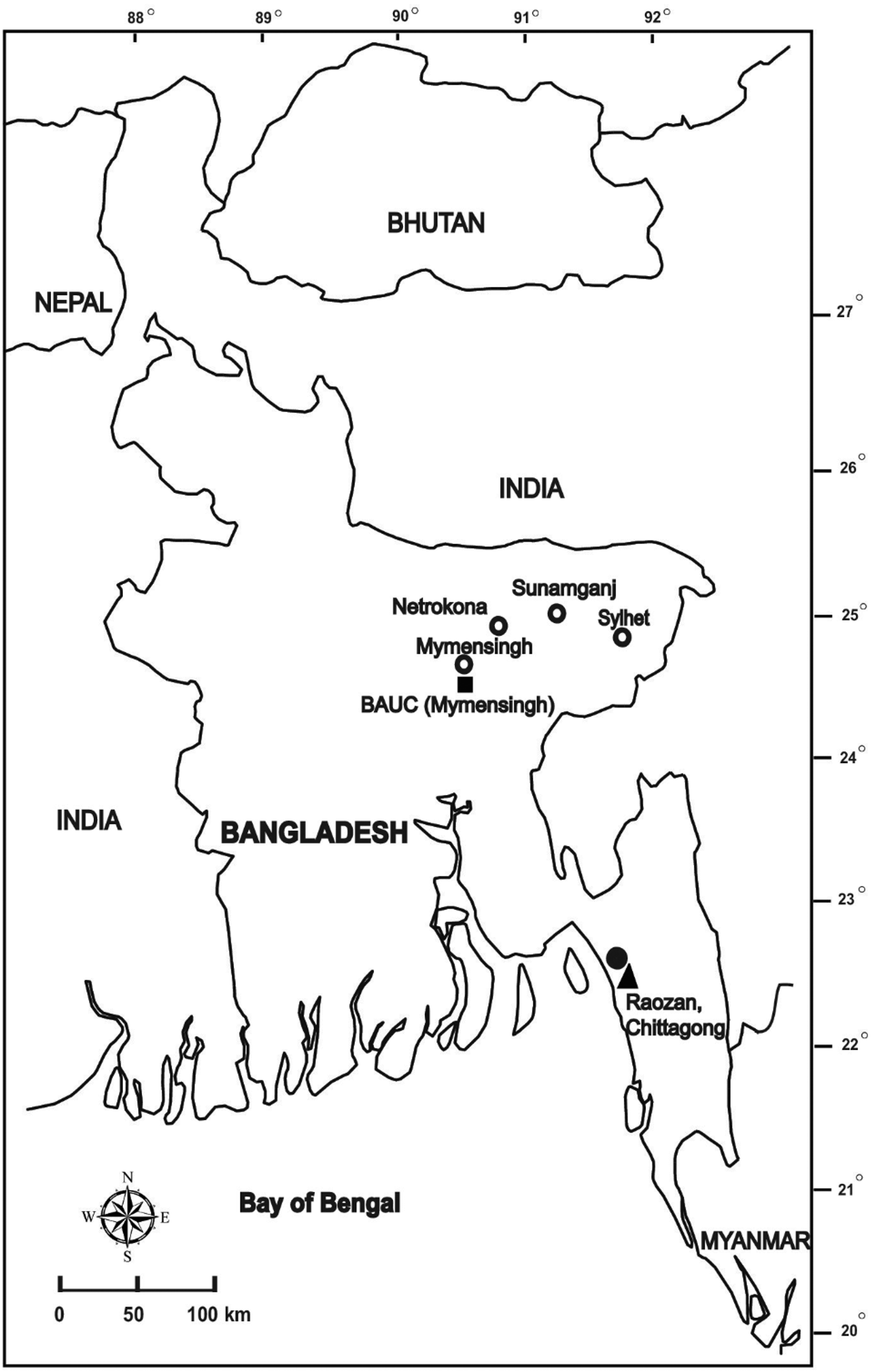
FIGURE 1. Map showing the collection sites and known occurrences of M. sp. C and M. sp. M in Bangladesh, indicated by closed and open circles, respectively. The type locality of holotype IABHU 3956 for M. sp. C and holotype IABHU 4116 for M. sp. M is indicated by closed triangles and squares, respectively.

Imageimage/png© Hasan, Mahmudul;Islam, Mohammed Mafizul;Kuramoto, Mitsuru;Kurabayashi, Atsushi;Sumida, MasayukiHasan, Mahmudul;Islam, Mohammed Mafizul;Kuramoto, Mitsuru;Kurabayashi, Atsushi;Sumida, Masayuki

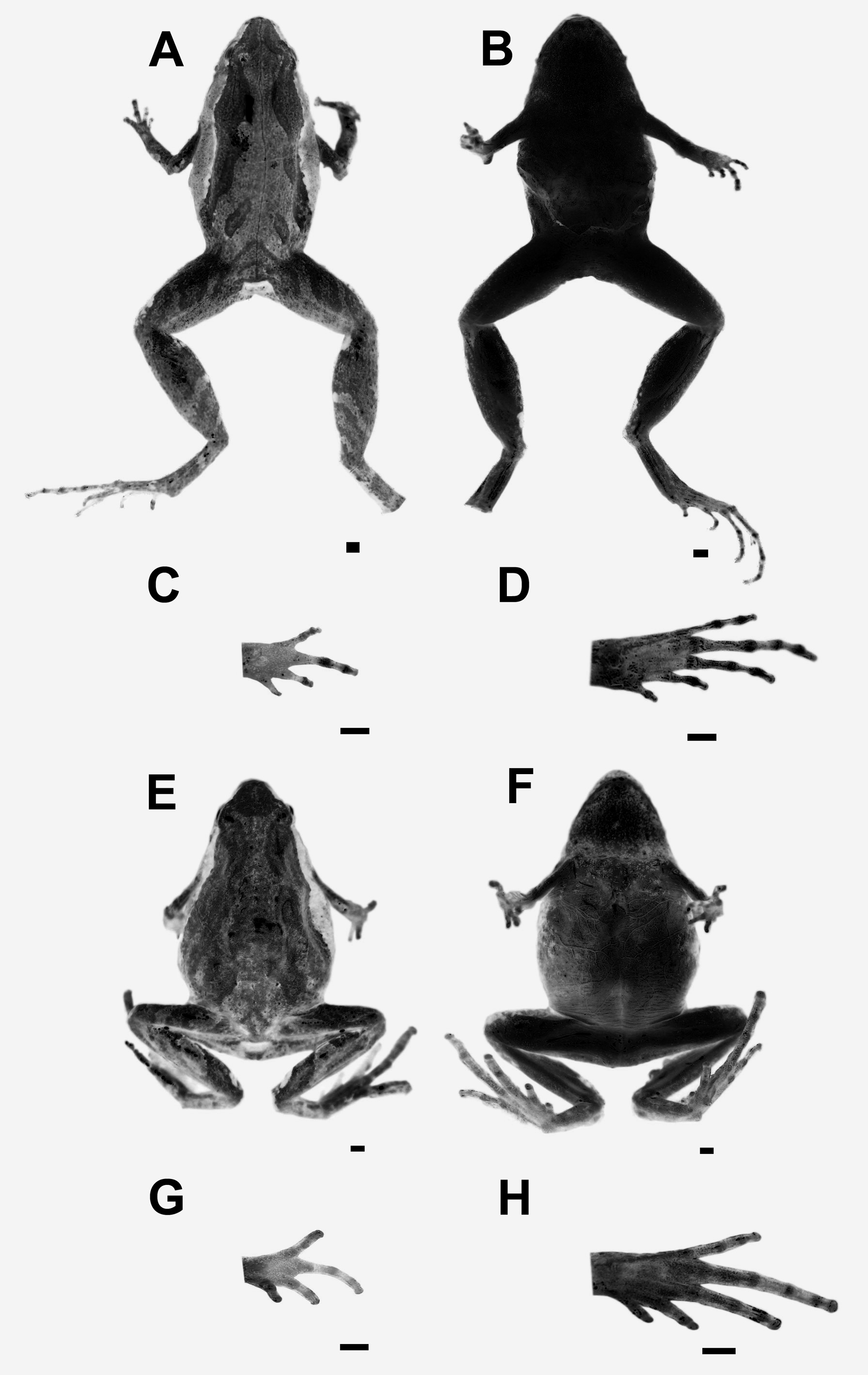
FIGURE 4. (A) Dorsal view and (B) Ventral view of the holotype of Microhyla mukhlesuri sp. nov (IABHU 3956). Ventral view of right (C) hand and (D) foot of paratype (3960) of M. mukhlesuri sp. nov .. (E) Dorsal view and (F) Ventral view of the holotype of M. mymensinghensis sp. nov (IABHU 4116). Ventral view of right (G) hand and (H) foot of holotype (4116) of M. mymensinghensis sp. nov .. All pictures of specimens were taken after preservation in alcohol. Scale bar = 1 mm.

Imageimage/png© Hasan, Mahmudul;Islam, Mohammed Mafizul;Kuramoto, Mitsuru;Kurabayashi, Atsushi;Sumida, MasayukiHasan, Mahmudul;Islam, Mohammed Mafizul;Kuramoto, Mitsuru;Kurabayashi, Atsushi;Sumida, Masayuki

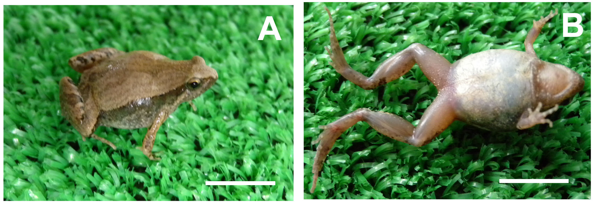
FIGURE 5. Holotype (IABHU 4116) of M. mymensinghensis sp. nov. in life. (A) Dorsal view. (B) Ventral view. Scale bar = 10 mm.

Imageimage/png© Hasan, Mahmudul;Islam, Mohammed Mafizul;Kuramoto, Mitsuru;Kurabayashi, Atsushi;Sumida, MasayukiHasan, Mahmudul;Islam, Mohammed Mafizul;Kuramoto, Mitsuru;Kurabayashi, Atsushi;Sumida, Masayuki

FIGURE 6. Microhyla mymensinghensis sp. nov. from Bangladesh Agricultural University Campus (BAUC), Mymensingh, Bangladesh. A and B showing color variations between individuals from BAUC. Scale bar = 10 mm.

Imageimage/png© Hasan, Mahmudul;Islam, Mohammed Mafizul;Kuramoto, Mitsuru;Kurabayashi, Atsushi;Sumida, MasayukiHasan, Mahmudul;Islam, Mohammed Mafizul;Kuramoto, Mitsuru;Kurabayashi, Atsushi;Sumida, Masayuki

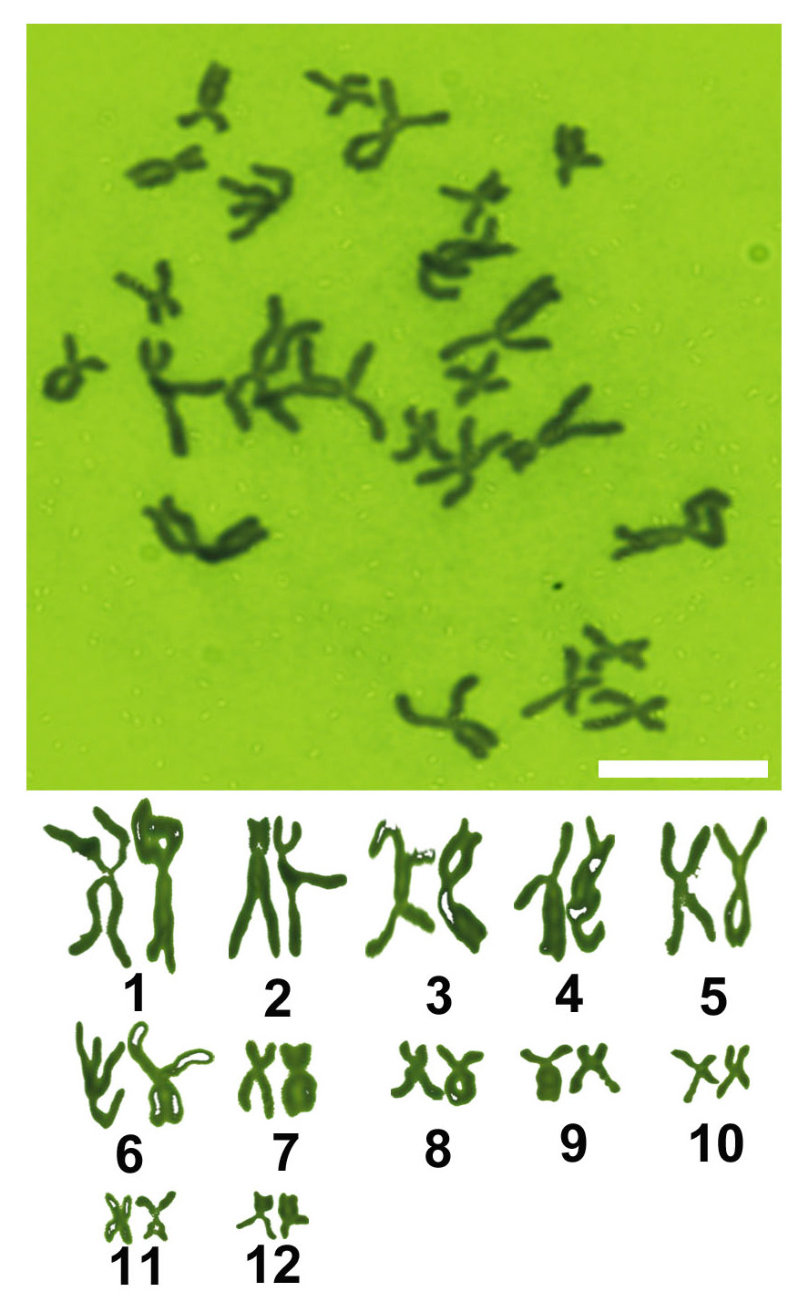
FIGURE 7. Metaphase spread and karyotype from bone marrow cells of M. mymensinghensis. Scale bar = 10 µm.

Imageimage/png© Hasan, Mahmudul;Islam, Mohammed Mafizul;Kuramoto, Mitsuru;Kurabayashi, Atsushi;Sumida, MasayukiHasan, Mahmudul;Islam, Mohammed Mafizul;Kuramoto, Mitsuru;Kurabayashi, Atsushi;Sumida, Masayuki

IMAGES

Gallery(5)

See Gallery

Occurrences with images

Source Information

Description of two new species of Microhyla (Anura: Microhylidae) from Bangladesh

checklist

This dataset contains the digitized treatments in Plazi based on the original journal article Hasan, Mahmudul, Islam, Mohammed Mafizul, Kuramoto, Mitsuru, Kurabayashi, Atsushi, Sumida, Masayuki (2014): Description of two new species of Microhyla (Anura: Microhylidae) from Bangladesh. Zootaxa 3755 (5): 401-418, DOI: 10.11646/zootaxa.3755.5.1

Abstract

Two new frog species belonging to genus Microhyla from the southeast, central and northeast regions of Bangladesh are described. Based on a molecular phylogeny derived from mitochondrial DNA sequences, one of the new species forms a clade with M. fissipes, while the second new species is sister to this clade. The DNA sequences of the mitochondrial cytochrome b gene from these new species are substantially diverged from M. fissipes (8.9 and 10.2% [3.6 and 4.2% for 16S ribosomal RNA gene] uncorrected pairwise divergence, respectively), and the estimated phylogenetic splits from their closest relative is in the Pliocene (3.4 Mya) and middle Miocene (10.5 Mya). The first new species (Microhyla mukhlesuri sp. nov.) can be diagnosed from its nearest congener (M. fissipes) by the following characteristics: SVL: 16.5–21.0 mm, finger length 1 <4 <2 <3, tips of finger and toes not swollen, subarticular tubercles distinct, an inverse U-shaped mark on the anus, and a distinct X-shaped marking on the dorsum. Although the second new species (M. mymensinghensis sp. nov.) shares some morphological characteristics with the first new species, it can be readily diagnosed from its close congeners by its longer hindlimbs (HLL / SVL), tibia (TIL/SVL) and forearm width (FAW /SVL), in addition to a combination of the following characteristics: SVL: 14.2–21.3 mm, snout truncate, a crescent-shaped marking on the anus, and an X-shaped marking on the dorsum. The tibiotarsal articulation extends to the eye in M. fissipes but ranges from the eye to the tip of the snout in the two new species.

Key words: Microhyla mukhlesuri sp. nov., Microhyla mymensinghensis sp. nov., Microhylidae, Mitochondrial DNA, Divergence time, Morphology, Bangladesh

Hasan M, Islam M M, Kuramoto M, Kurabayashi A, Sumida M, plazi (2014). Description of two new species of Microhyla (Anura: Microhylidae) from Bangladesh. Plazi.org taxonomic treatments database. Checklist dataset https://doi.org/10.11646/zootaxa.3755.5.1 accessed via GBIF.org on 2026-06-14.

CC0Published 12/31/2014View dataset
GBIF Usage Key
119412855
Dataset Key
004e46b2-437c-42fb-af76-245a4d8e411c
Origin
source
Backbone Key
7981031
Taxon ID
F3648799FFF5FFA842D91F10FBDC105F.taxon
Last Crawled
6/11/2026
Last Interpreted
6/11/2026