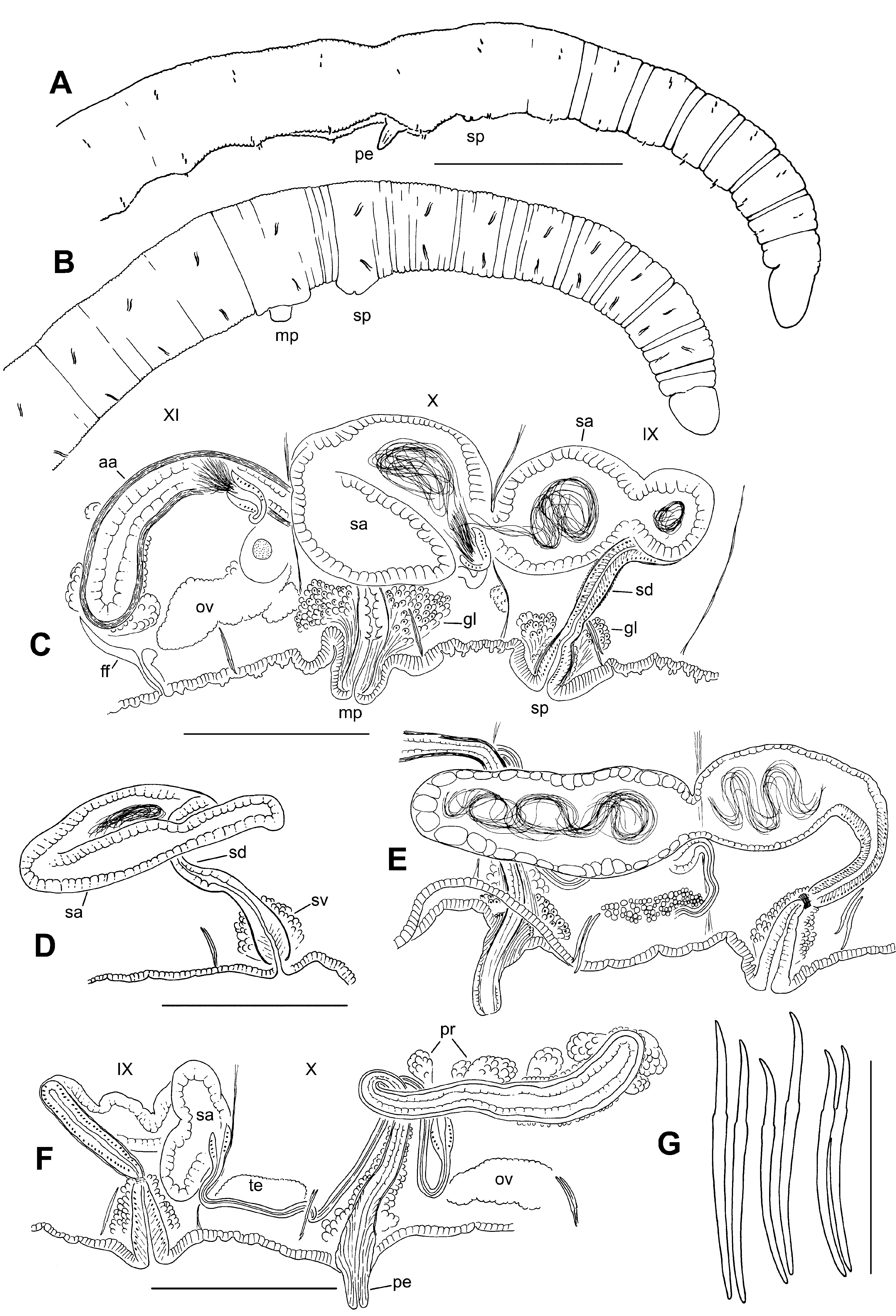
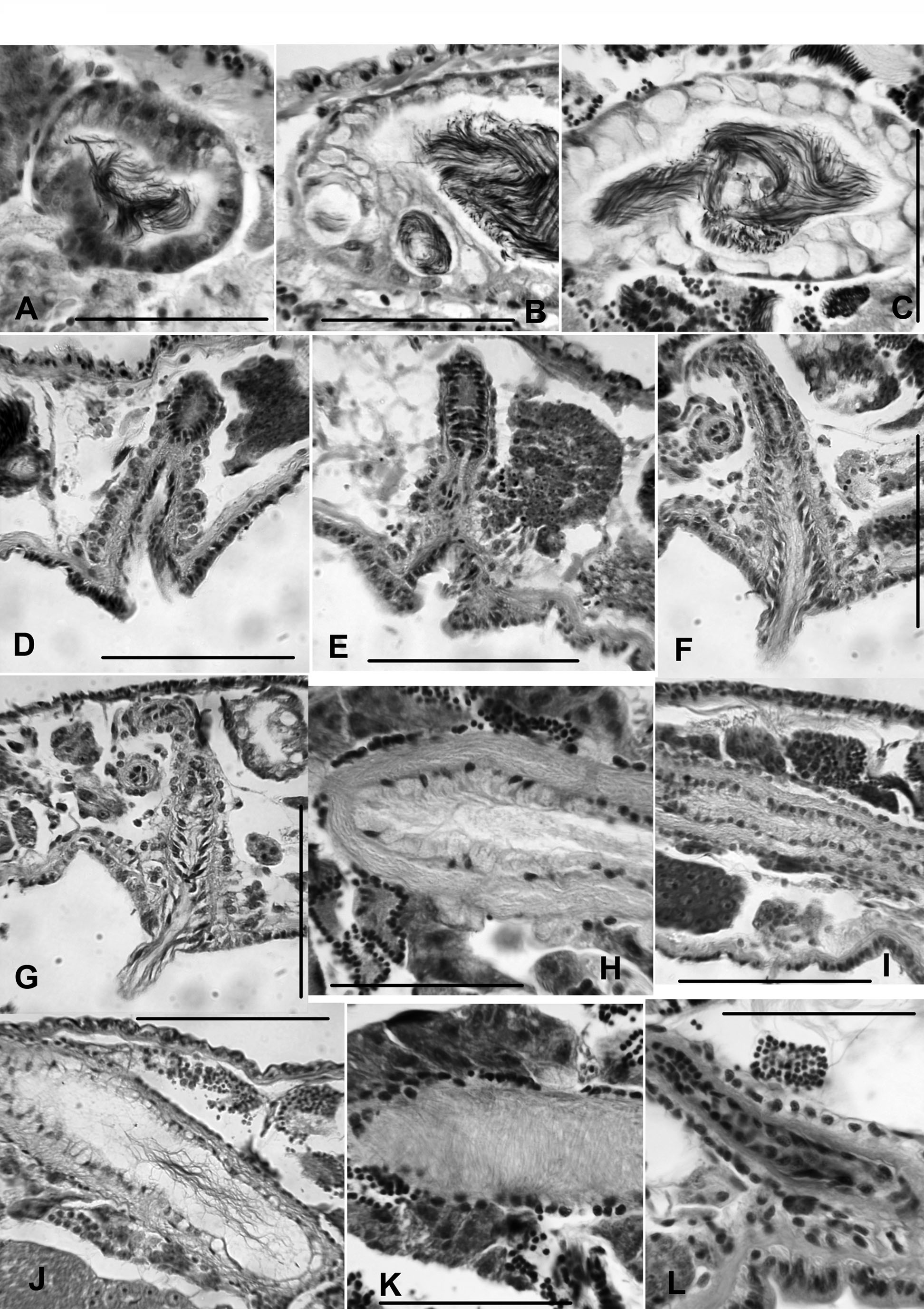
Description. Small, very thin worms, length (preserved) 14 – 19 mm, 81 – 107 segments; width 0.19 – 0.25 mm in X, maximum width to 0.21 – 0.28 mm. Secondary segmentation a narrow anterior ring in preclitellar segments, from about III – IX (Fig. 5 A – B). Clitellum indistinct, only slightly thicker or more glandular than surrounding epidermis, (IX) X – XII. Chaetae sigmoid, simple-pointed, with nodulus 31 – 39 % of chaeta length from tip (Fig. 5 G). Chaeta length 44 – 70 μm in mid-body; dorsal and ventral chaetae similar in size; slightly shorter in posterior segments, but proportions similar in anterior and posterior segments. Within each bundle, the outer (more lateral) chaeta may be slightly shorter than the inner. Prostomium rounded to conical, about 0.12 – 0.15 mm long, width about equal to length; prolobous. Brain in the peristomium, deeply lobed. Pharynx in II to mid-IV; dorsal wall concave with columnar, ciliated cells, about 12 μm thick; ventral wall very thin (2 – 4 μm), with cuboidal, non-ciliated cells. Pharyngeal glands well developed dorsolaterally in V – VII or VIII. Longitudinal muscle layer 4 – 7 μm thick in preclitellar segments; circular muscle layer 2 μm. Septa 1 / 2 and 2 / 3 inconspicuous. Epidermis 5 – 10 μm thick anterior to clitellum; to about 10 – 12 μm in clitellum; 5 – 7 μm in post-clitellar segments; to 24 μm in prostomium. Lateral, commissural blood vessels in preclitellar segments; these vessels thin and convoluted, typically joining ventral vessel 1 segment behind junction with dorsal vessel. No lateral blood vessels in middle or posterior segments. Dorsal blood vessel separate in preclitellar segments; adjacent or appressed to top of gut posterior to about X. Chloragogen cells begin in VI or VII. First nephridia usually paired on 6 / 7, the next on 12 / 13; nephridia in a few posterior segments, paired or on one side. Each nephridium has a small anteseptal funnel and a granular postseptal thickening; the posterior duct forms a loop, which extends ventrally, entering one or more posterior segments, and terminating in a short ectal duct in the originating segment. Ectal ducts usually terminate in a nearly spherical vesicle (diameter about 20 μm) at the inconspicuous nephropore. Spermatheca single, median in IX; pore may be distinct, just behind the ventral chaetae; pore surrounded by a pad of slightly thickened epidermis, 70 – 120 μm wide, externally appearing as a low mound (Fig. 5 B). Ectal end of spermathecal duct widens into a narrowly-conical vestibule, 80 – 120 μm high by 30 – 45 μm wide near spermathecal pore; the vestibule lined with columnar epithelium, a 4 – 6 μm thick muscle layer, and surrounded by an irregular (10 – 22 μm) layer of granular cells (Fig. 5 C – F, 6 D). Narrowed ental end of vestibule joined by a cylindrical spermathecal duct, 110 – 130 μm long by 22 – 24 μm wide; duct has columnar epithelium, a very thin muscle layer (about 1 μm) and a narrow lumen; duct narrowed and surrounded by a ring of circular muscle fibers, forming a short sphincter at junction with vestibule (Fig. 6 D – E). Spermathecal ampulla of fully mature worms (with developed eggs) usually extends into X, sometimes to XI; elongate-sacciform, 360 – 710 μm long, 60 – 200 μm wide, containing unordered, loosely-packed sperm (Fig. 5 C, E). Ampullar epithelium cuboidal in about the ectal 1 / 5; the remainder thick (12 – 28 μm), with irregular, vacuolated cells (Fig. 6 A – C); muscle layer very thin (1 μm). In mated specimens, the sperm is in a loose, unordered bundle (Fig. 6 C); vacuoles of spermathecal wall may contain sperm (Fig. 6 B). Spermathecal ampulla of recently-mated worms (without mature eggs) may be folded within IX; with an ovate ectal chamber and a narrower ental portion (Fig. 5 D); in unmated worms the entire ampulla is narrowly tubular and typically folded (Fig. 5 F). Testes paired in IX and X, medium to large size, often extending beyond mid-segment. Ovaries paired in XI, usually large; in some worms extending through XI into XII. Sperm sacs paired, extending back as far as XVII, usually not extending anteriorly from IX; egg sacs may extend 1 or 2 segments beyond sperm sacs. Female funnels 50 – 95 μm tall, with the posterior side much taller than the anterior; female pores intersegmental, on 11 / 12. Male funnels paired on 9 / 10 and 10 / 11; simple v-shaped; anterior and posterior pairs about the same size (35 – 60 μm high); both directed anteriad, or the posterior may extend back into XI within the sperm sac. Both anterior and posterior male funnels functional, with associated sperm. Both anterior and posterior vasa deferentia very thin (5 – 8 μm diameter). Posterior vasa deferentia enter X directly, without penetrating 10 / 11 and forming a loop in XI. All vasa deferentia approach the atrial duct near the beginning of the ampulla, but it is not clear where they enter. Male pore single, median in X, behind the ventral chaetae (Fig. 5 A – B); body wall usually concave ventrally in X – XI around and behind pore (Fig. 5 A, E); concavity not associated with distinct internal musculature (Fig. 6 G); pore area sometimes protruding as a short, truncate porophore (Fig. 5 C). Penis cylindrical to truncate-conical, 50 – 100 μm long by 22 – 35 μm wide when extended (Figs. 5 E, 6 F – G), apparently formed by extruded lining cells from the end of the atrial duct, but with small vacuoles visible near the tip. Expanded ectal part of duct (penial structure) about 70 – 100 μm high, with a 5 μm thick muscle layer, and lined with elongate, outwardly-directed cells having basal nuclei (Fig. 6 F). Penial structure surrounded by an irregular layer of cells, possibly accessory glands, 17 – 25 μm thick. Atrium usually extends into XI or XII. Ectal duct elongate-cylindrical, ascending vertically in X, often forming a loop before junction with the ampulla (Fig. 5 F). Duct length 130 – 290 μm, width 16 – 25 μm, gradually widening entally at ampulla. At the ectal end, duct gradually widens, forming the penial structure. Atrial duct surrounded by a thin (2 – 5 μm) muscle layer (Fig. 6 L). Atrial ampulla elongate, cylindrical or club-shaped; length 170 – 370 μm, maximum width (middle or near ental end) 46 – 85 μm. Ampullar muscle layer 7 – 13 μm thick, mostly composed of irregularly transverse fibers, but with a very sparse outer layer of variably oriented diagonal fibers (Fig. 6 H – K). Ampullar epithelium variable: granular, with indistinct cell boundaries in some specimens, 7 – 14 μm thick, with basal nuclei (Fig. 6 H – I); in other specimens thinner and vacuolate (Fig. 6 J); lumen variable, 5 – 12 μm wide. Prostate glands multicellular and petiolate, to 30 – 50 μm tall, bundles generally sparse (Fig. 5 F, 6 I, K); single prostatelike cells may also cover the atria.
Fend, Steven V., Lenat, David R. (2012): New Eclipidrilus species (Annelida, Clitellata, Lumbriculidae) from southeastern North America. Zootaxa 3194: 51-67, DOI: 10.5281/zenodo.210008