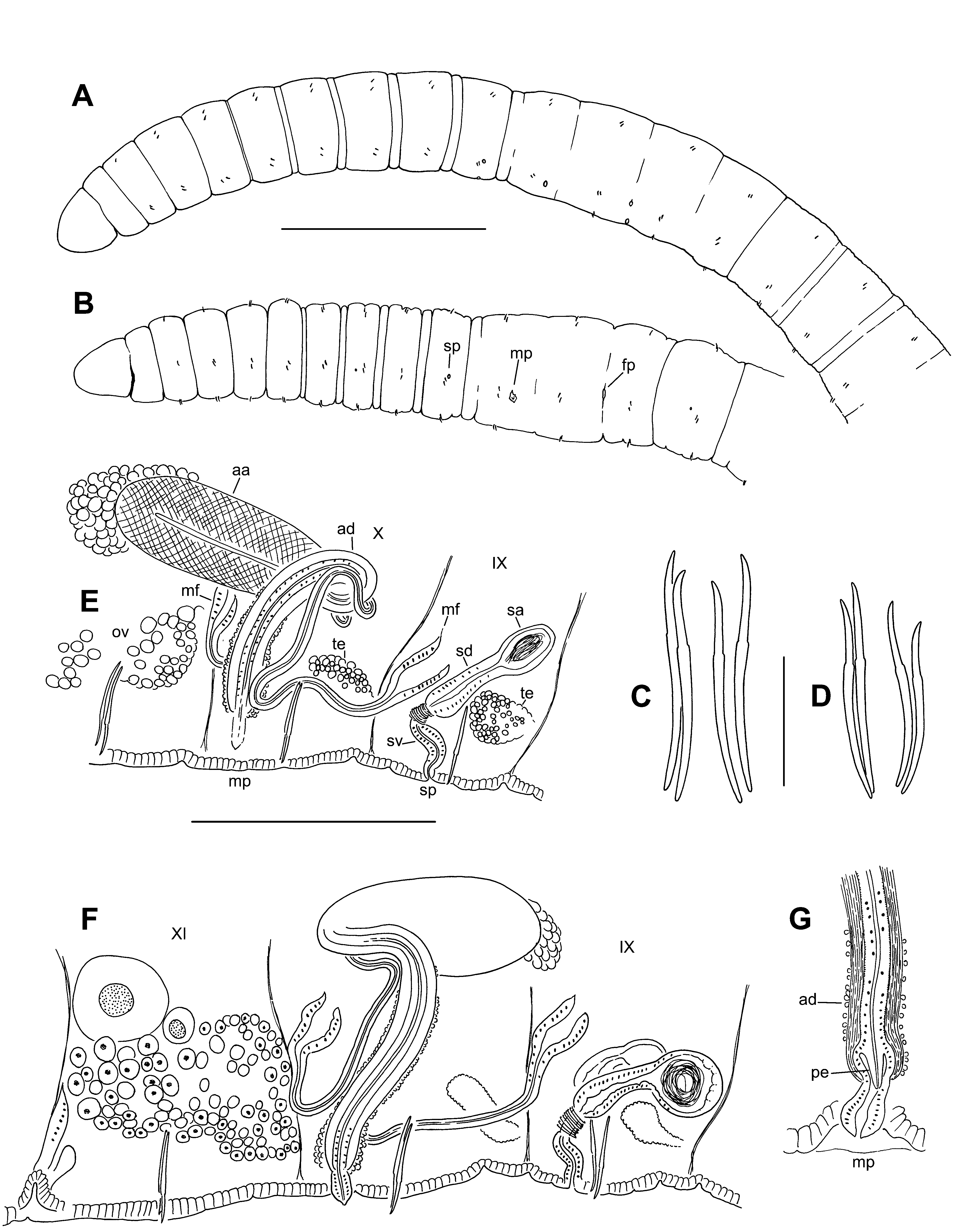
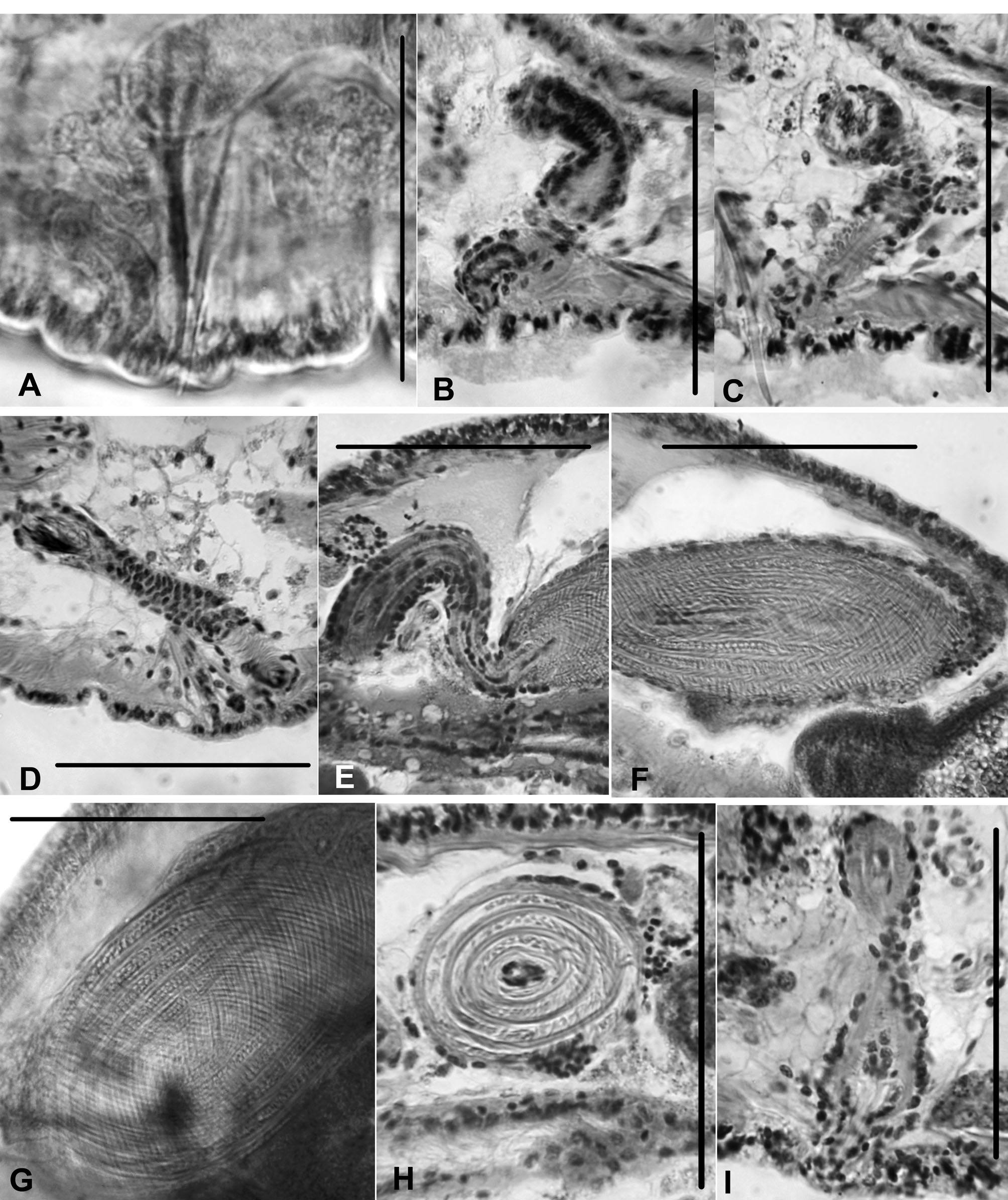
Description. Small, thin worms, length (preserved) 7.7 – 9.5 mm, 48 – 64 segments; width 0.20 – 0.30 mm in X, maximum width 0.24 – 0.3 mm. Segmentation usually obscured in clitellum, weakly expressed in some post-clitellar segments; secondary segmentation sometimes weak, a narrow anterior ring from about VI – X (Fig. 3 A – B). Clitellum X – XII. Chaetae sigmoid, simple-pointed, with nodulus 35 – 40 % of chaeta length from tip (Fig. 3 C – D). Chaeta length 58 – 84 μm in mid-body; 55 – 80 μm in posterior segments; proportions similar in anterior and posterior segments. Dorsal and ventral chaetae approximately equal in length; within each bundle, the outer (more lateral) chaeta may be slightly shorter than the inner. Prostomium rounded-conical, length slightly less than width; prolobous. Brain in the peristomium, strongly lobed. Pharynx in II – IV; dorsal wall with columnar, ciliated cells; ventral wall very thin, with cuboidal, non-ciliated cells. Pharyngeal glands in (IV) V – VII (VIII), relatively small, irregular lobes, dorsal and / or lateral to gut. Longitudinal muscle layer 12 – 14 μm thick in preclitellar segments; circular muscle layer 3 μm. Septa 1 / 2 and 2 / 3 inconspicuous. Epidermis 4 – 12 μm thick anterior to clitellum; to about 12 – 15 μm in clitellum; 4 – 5 μm in post-clitellar segments; to 12 – 27 μm in prostomium. Lateral, commissural blood vessels in at least some preclitellar segments; these vessels thin and convoluted, typically joining ventral vessel 1 segment behind junction with dorsal vessel. No lateral blood vessels in middle or posterior segments. Dorsal blood vessel separate in anterior segments; on top of gut posterior to about XI. Chloragogen cells begin in about VII. First nephridia usually paired on 6 / 7, the next on 12 / 13; nephridia in few posterior segments, usually on one side only. Each nephridium has a small anteseptal funnel and a granular, narrow postseptal thickening 30 – 40 μm long; the posterior duct forms a loop that extends ventrally, entering one or more posterior segments, and terminating in a short ectal duct in the originating segment. Nephropores anterior to ventral chaetae, inconspicuous; ectal ducts without distinct vesicles. Spermathecae paired in IX; pores small, on ventral chaetal lines, just behind the ventral chaetae; accessory glands weak or absent (Fig. 3 E – F). Ectal vestibule of duct is narrow and usually curved, about 30 – 50 μm long by up to 14 μm wide; ental 1 / 2 – 1 / 3 narrower, surrounded by a ring of circular (transverse) muscle at the junction with the main duct (Figs. 3 E – F, 4 A – C). Main part of spermathecal duct 38 – 60 μm long, 18 – 22 μm wide in ectal part, narrowing to 12 – 17 μm entally, with densely- packed, columnar epithelium and a narrow lumen; spermathecal duct well differentiated from the ampulla (Fig. 3 E – F). Spermathecal ampullae entirely in IX; ovate and compact, to 37 – 64 μm long by 25 – 40 μm wide. Ampullar epithelium thin (3 – 4 μm) (Fig. 4 D). In mated specimens the sperm is in an unordered bundle. Testes paired in IX and X, small to medium size, extending at most to mid-segment. Ovaries paired in XI; usually extending through XI, sometimes into XII. Sperm sacs not developed (sperm in testicular segments); egg sacs usually paired; may extend to XIV. Female funnels up to 50 μm tall; female pore intersegmental, on 11 / 12. Male funnels single on 9 / 10 and 10 / 11; anterior and posterior similar in size (height 35 – 48 μm), directed anteriad within IX and X. Both anterior and posterior male funnels simple, conical; functional, with associated sperm. Both anterior and posterior vasa deferentia narrow (7 – 10 μm diameter); they approach the atrial duct near the male pore, then loosely follow the duct, joining the atrium near the base (ectal end) of the ampulla. Posterior vas deferens enters X directly, without penetrating 10 / 11 (Fig. 3 E – F) Male pore inconspicuous; single, slightly lateral to ventral midline in X, near 10 / 11 (Fig. 3 A – B). Atrial duct widens slightly at the ectal end (Fig. 4 E), subtending a small, narrowly conical penis within narrow sac (length 20 – 33 μm, width 10 – 16 μm); the entire structure terminates in a small, conical papilla, which may be contained within a shallow depression (Fig. 3 G, 4 I). A few small (to 20 μm) accessory glands may be present at male pore; absent or inconspicuous in most specimens. The main atrial duct extends dorsally around one side of gut, then usually loops posteriorly and widens abruptly to form the ampulla (Fig. 3 E – F, 4 E). Duct length 180 – 295 μm, width to 25 – 29 μm near male pore, narrowing entally to 17 – 25 μm. Duct musculature more or less longitudinal, 5 – 7 μm thick in ectal part; lumen narrow ectally, widening to 6 μm entally; epithelium very thin. Atrial ampulla narrowly ovate (Fig. 3 E – F, 4 F); length 152 – 215 μm, width at middle 52 – 98 μm. Ampulla with a very thick, cross-hatched muscle layer, to 29 – 31 μm, composed of about 7 – 10 layers of fibers in alternating spirals, oriented at about 50 – 60 ˚ from the longitudinal axis (Fig. 4 F – H). Epithelium thin and indistinct, as little as 2 μm thick; lumen very narrow, usually less than 5 μm (Fig. 4 F, H). Most of ampulla without prostate glands, although a poorly defined, decumbent clump of cells is visible at the ental end in most specimens (Fig. 3 E – F, 4 F). Atrium usually extends into XI.
Fend, Steven V., Lenat, David R. (2012): New Eclipidrilus species (Annelida, Clitellata, Lumbriculidae) from southeastern North America. Zootaxa 3194: 51-67, DOI: 10.5281/zenodo.210008