AnimaliaNot EvaluatedacceptedspeciesAccepted
Palliatus magellanicus
GBIF:119568018
ABOUT
Descriptions(5)
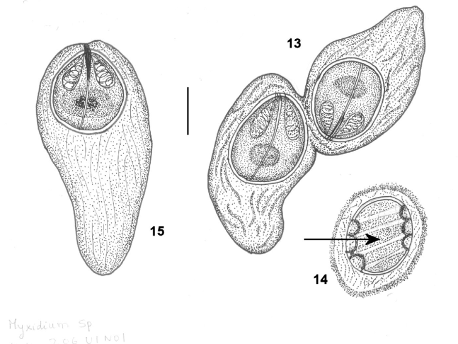
Host length range: 25 – 42 cm. Collection numbers: NHMUK 2012.3.19.2. Morphological description. Sporoblast oval or irregularly shaped, disporic (Fig. 13). Dimensions, based on 4 fixed specimens: 28.4 – 38.4 x 28.0 – 36.0. Developing sporoblasts show deeply staining cords (the origins of the membranous veil) twisted around the body (Fig. 14). These are clearly visible only when stained with Indian ink, but are indistinct and lightly stained in Giemsa preparations. Spore subspherical or broadly oval (Fig. 15). A smooth membranous veil originating anteriorly extends beyond the spore body posteriorly, enveloping the entire spore. Sutural ridge prominent anteriorly but thinner posteriorly. Keel-like appendages situated along the sutural ridge join together at the posterior extremity. Sporoplasm deeply staining and binucleate. Spore valves thin and smooth. Polar capsules pyriform, subterminal, one on either side of sutural line. Polar filament with 3 – 4 coils, not clearly visible. Dimensions, based on 15 fixed spores, as ranges with means and ± SD in parentheses: spore length 9.6 – 19.2 (13.64 ± 3.67); spore width 10.2 – 22.4 (19.03 ± 3.64); spore thickness 14.0 – 20.0 (16.09 ± 1.92); spore veil 32.0 – 48.0 (40.43 ± 6.47); polar capsule length 6.4 – 8.0 (6.93 ± 0.64); polar capsule width 3.2 – 5.4 (4.0 ± 0.80); spore length: spore width = 1: 0.8 – 0.96; polar capsule length: spore length = 1: 1.5 – 2.4.
Kalavati, Chaganti, Mackenzie, Ken, Collins, Catherine, Hemmingsen, Willy, Brickle, Paul (2013): Two new species of myxosporean parasites (Myxosporea: Bivalvulida) from gall bladders of Macruronus magellanicus Lönnberg, 1907 (Teleostei: Merlucciidae). Zootaxa 3647 (4): 541-554, DOI: 10.11646/zootaxa.3647.4.4
Site of infection: gall bladder Locality, date and depth: (1) Off Chiloe Island, Chile, 43 ºS, 73 ºW, June 2007, 300 m.
Kalavati, Chaganti, Mackenzie, Ken, Collins, Catherine, Hemmingsen, Willy, Brickle, Paul (2013): Two new species of myxosporean parasites (Myxosporea: Bivalvulida) from gall bladders of Macruronus magellanicus Lönnberg, 1907 (Teleostei: Merlucciidae). Zootaxa 3647 (4): 541-554, DOI: 10.11646/zootaxa.3647.4.4
Discussion. This species was not observed during the initial examinations of gall bladders, but was discovered later as a double infection with Pseudalataspora kovalevae in a formalin-preserved sample from a fish originally identified as infected only with the latter species. We therefore have no molecular sequence and no photographs of fresh material. Only five species of Palliatus have been previously described from marine fishes, four from the gall bladder and one from the pancreas (Shulman et al., 1979; Padma Dorothy & Kalavati, 1998; Aseeva, 2003). The new species differs considerably in morphology from all of these and the host and locality are both new for the genus Palliatus (Table 3).
Kalavati, Chaganti, Mackenzie, Ken, Collins, Catherine, Hemmingsen, Willy, Brickle, Paul (2013): Two new species of myxosporean parasites (Myxosporea: Bivalvulida) from gall bladders of Macruronus magellanicus Lönnberg, 1907 (Teleostei: Merlucciidae). Zootaxa 3647 (4): 541-554, DOI: 10.11646/zootaxa.3647.4.4
Material studied Host: Macruronus magellanicus Lönnberg, 1907
Kalavati, Chaganti, Mackenzie, Ken, Collins, Catherine, Hemmingsen, Willy, Brickle, Paul (2013): Two new species of myxosporean parasites (Myxosporea: Bivalvulida) from gall bladders of Macruronus magellanicus Lönnberg, 1907 (Teleostei: Merlucciidae). Zootaxa 3647 (4): 541-554, DOI: 10.11646/zootaxa.3647.4.4
Type locality: (1) Prevalence: 4 % (1 of 25).
Kalavati, Chaganti, Mackenzie, Ken, Collins, Catherine, Hemmingsen, Willy, Brickle, Paul (2013): Two new species of myxosporean parasites (Myxosporea: Bivalvulida) from gall bladders of Macruronus magellanicus Lönnberg, 1907 (Teleostei: Merlucciidae). Zootaxa 3647 (4): 541-554, DOI: 10.11646/zootaxa.3647.4.4
Export occurrence data
Darwin Core Archive (ZIP)
CLASSIFICATION
Taxonomic Classification Tree

MULTIMEDIA
Media Files(1)
FIGURES 13 – 15. Line drawings of Palliatus magellanicus n. sp. 13. Disporic sporoblast, stained with Indian ink. 14. Developing sporoblast, with deeply staining cords arrowed. 15. Spore stained with Giemsa. Scale bar: 10 μm.
Imageimage/png© Kalavati, Chaganti;Mackenzie, Ken;Collins, Catherine;Hemmingsen, Willy;Brickle, PaulKalavati, Chaganti;Mackenzie, Ken;Collins, Catherine;Hemmingsen, Willy;Brickle, Paul
IMAGES