AnimaliaNot EvaluatedacceptedspeciesAccepted
Ceratomyxa herouardi

Ceratomyxa herouardi

Georgevitch, 1916

GBIF:119594200

0year

ABOUT

Descriptions(1)

Type host: Sarpa salpa Linnaeus, 1758 goldline sea bream (Perciformes: Sparidae) Type localities: Mediterranean off Tunisia: Location 1: Gulf of Tunis (36 ° 45 ’ N, 10 ° 15 ’ E); Location 2: Bay of Bizerte (37 ° 20 ’ N, 9 ° 53 ’ E). Site of infection: Within gall bladder Prevalence: The overall prevalence is 28.2 % (93 / 330) (Fig. 9). At location 1, the prevalence of infection was 33.3 % (70 / 210) distributed as following, 03 / 2012: 36.7 % (11 / 30); 04 / 2012: 30 % (9 / 30); 05 / 2012: 40 % (12 / 30); 06 / 2012: 46.7 % (14 / 30); 07 / 2012: 23.3 % (7 / 30); 08 / 2012: 30 % (9 / 30); 05 / 2013: 25 % (5 / 20); 06 / 2013: 30 % (3 / 10). At location 2, the prevalence of infection is 19.2 % (23 / 120) distributed as following, 03 / 2013: 13.3 % (4 / 30); 04 / 2013: 13.3 % (4 / 30); 05 / 2013: 26.7 % (8 / 30); 06 / 2013: 23.3 % (7 / 30) (see Table 4). Mean intensity: 211.2 ± 50.6 spores / infected fish (++++++) (Fig. 9) (see Table 4). Type-material: Digitized photos of syntype spores were deposited in the parasitological collection of the Museum National d’Histoire Naturelle (MNHN), Paris, Coll. No. ZS 117. Description Vegetative stages. Trophozoites (n = 100) were freely floating in bile of the gallbladder in different stages of maturation and some seen attached to gall bladder epithelium. Polymorphous with great variety of shape and size, elongated with same breadth or tapering to one end or club-shaped with roundish enlargements (Fig. 3 B). Young trophozoïtes were spherical or pyriform (Figs. 3 E – F). Trophozoïtes were attached to each other by their pseudopodia while other possessed a long pseudopodia-like filopodia probably used for their motility (Fig. 3 A). Protoplasm homogeneous with fine refractile granules and inner generative cells. Subspherical to spherical plasmodia measuring 30.23 ± 5.5 (22 – 40) µm in length and 31.8 ± 5.1 (27 – 44.5) µm in width (n = 50). Disporous, each plasmodium contained ordinarily two identical spores (Figs. 3 C – E), or polysporous, with formation of numerous disporic plasmodia attached to each other within the trophozoïte mother (Figs. 3 C – D). Spores (n = 100 fresh spores). Mature spores were elliptic to hemispherical broadly with round ends in sutural view (Figs. 3 I – K, M, 8 C), measuring 10.5 ± 1.2 (8 – 12) Μm in length and 21.6 ± 1.6 (20 – 24) Μm in thickness. Posterior angle was flattened to straight 172.5 ± 6.8 (165 – 180 °). The suture line divided the spore in two equal valves smoothly ovoid in lateral view (Fig. 3 J). Polar capsules were nearly spherical to sub-spherical 3.91 ± 0.25 (3.5 – 4.5) Μm in length and 3.89 ± 0.27 (3.5 – 4.5) Μm in width (n = 100). The polar filament formed five to six turns arranged along the longitudinal axis of the capsule. A binucleate sporoplasm almost filled the spore cavity and was generally distributed symmetrically. Occasionally, aberrant spores with 3 polar capsules and 3 valves were observed (Figs. 3 F, L). Remarks The first description of C. herouardi was illustrated by Georgévitch (1916 a, 1916 b, 1916 c, 1917) from S. salpa captured in Mediterranean coast of Monaco, France. The description was focused on the huge variety of the vegetative forms. Georgévitch (1916) declared that C. herouardi has been especially deserved the most attention from all the Ceratomyxa spp. identified in that time by its extraordinary vegetative forms that were very diverse (Figs. 3 A – F): round, elliptical, pyriform, elongated and very unequal in size and by its mature spores that could show dissimilar shape in different views. In result of that, Jameson (1913) mentioned the same forms and variety of trophozoïtes in the same host, organ and locality (Monaco) without recognized the spores. He couldn’t determined the species and he declared in his study that the form of parasite found in the gall bladder of S. salpa has something of the appearance of a Lepthoteca (Kudo 1920). Georgévitch (1917) believed that the variety of the trophozoites and the complexity of it developmental cycle according to the process of endogenous or exogenous budding, were an important characteristics to identify C. herouardi from the other species of the same genus. This author made a traditional demonstration about the life cycle of C. herouardi based to observations in light microscopy and some erroneous hypothesis that attached to myxosporeans described many years ago. In recent paper, the different vegetative forms of C. herouardi are recognized and the first measurements of the sporogonic stages and as well as mature spore are given (see Table 5). Ecological notes During the sampling period, the overall rate of infection is 28.2 % and this myxosporean has a parasitic status as less frequent species even it has a great mean of intensity with 211.2 ± 50.6 spores per individual infected host (Fig. 10). Infection by C. herouardi was detected during the whole period of investigation. In Gulf of Tunis, the infection started from March to August with maximum prevalence in June 46.7 % while the infection commenced from March to June in Bay of Bizerte and maximum prevalence was recorded in May 26.7 %. During examination of goldline sea bream’s gall bladder, the intensity of infection was important and showed no significant variation with months (see Table 4).
Laamiri, Sayef (2014): New observations on Myxozoa of the goldline sea bream Sarpa salpa L. 1758 (Teleostei: Sparidae) from the Mediterranean coast of Tunisia. Zootaxa 3887 (2): 157-190, DOI: 10.11646/zootaxa.3887.2.3

Export occurrence data

Darwin Core Archive (ZIP)

CLASSIFICATION

Taxonomic Classification Tree

MULTIMEDIA

Media Files(5)

FIGURE 3 A – F. Photomicrographs of Ceratomyxa herouardi from the gall bladder of Sarpa salpa. (A) Round trophozoite (RT) attached to other polymorphous, notice the presence of inner generative cells and long filopodia (F). (B) Fresh smear of infested bile with the presence of elongated trophozoites (ET) contained pseudoplasmodi (pp), disporic plasmodia (P) and mature spores (ms). (C) Big rounded polysporic trophozoïtes (RT) contained several pseudoplasmodia (pp). (D) Part of trophozoite showing in Fig. C, presented the pseudoplasmodia (pp) and disporic plasmodia (P) containing immature spores (is). (E – F) Subspherical and pyriform trophozoites (PT) unequal in size attached to each other with formation of roundish disporic plasmodia (P) according to mechanism of endogenous or exogenous budding, notice of a disporic plasmodium of atypical spores (P *) in F. Scale bar = 50 µm in A – C; 20 µm in D – F;

Imageimage/png© Laamiri, SayefLaamiri, Sayef

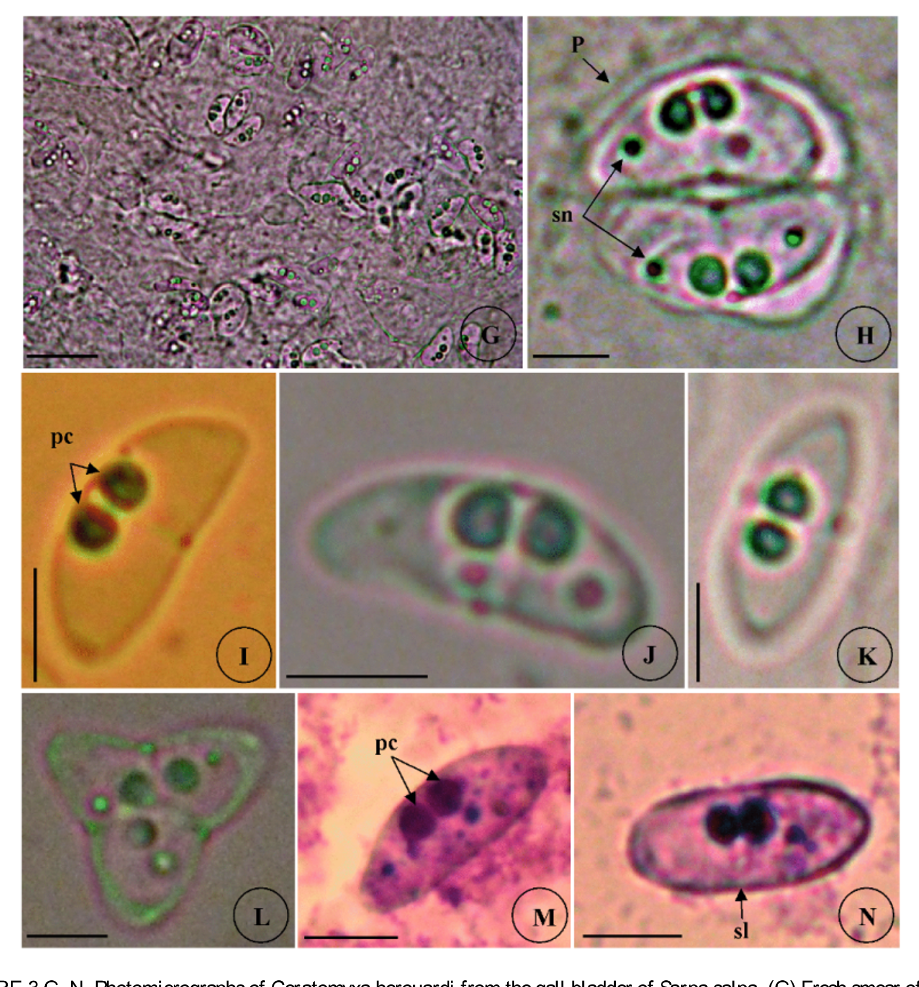
FIGURE 3 G – N. Photomicrographs of Ceratomyxa herouardi from the gall bladder of Sarpa salpa. (G) Fresh smear of heavy infested bile with sporogonic stages and mature spores. (H) Plasmodium (P) of premature spores showing the sporoplasm nuclei (sn). (I) Mature spore in sutural view showing the sub-spherical polar capsules (pc). (J) Mature spore in lateral view. (K) Mature spore in apical view. (L) Atypical spore with three polar capsules and three shell valves. (M – N) Different view of mature spores stained with Giemsa showing the distinct polar capsule (pc) and the suture line (sl). M, sutural view; N, apical view. Scale bar = 40 µm in G; 10 µm in H-N.

Imageimage/png© Laamiri, SayefLaamiri, Sayef

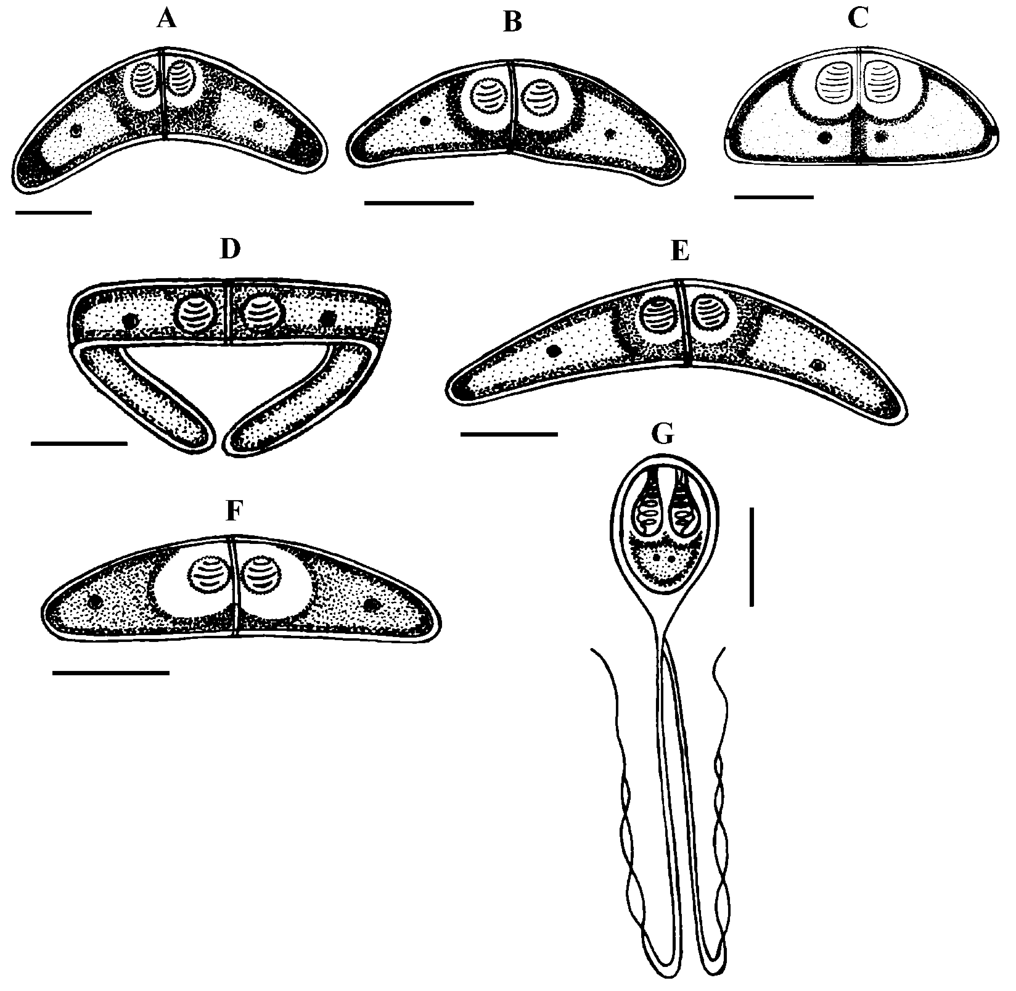
FIGURE 8. Line drawing of Myxozoan species infecting the goldline sea bream Sarpa salpa from the North coast of Tunisia. (A) Ceratomyxa arcuata Thélohan, 1892; (B) Ceratomyxa pallida Thélohan, 1895; (C) Ceratomyxa herouardi Georgévitch, 1916; (D) Ceratomyxa sp. 1; (E) Ceratomyxa sp. 2; (F) Ceratomyxa sp. 3; (G) Henneguya sp. Scale bar = 10 µm.

Imageimage/png© Laamiri, SayefLaamiri, Sayef

FIGURE 9. Prevalence of species of myxozoans from 2 localities, Gulf of Tunis and Bay of Bizerte. The total prevalence determined within the pooled sample of 330 goldline sea bream (Sarpa salpa).

Imageimage/png© Laamiri, SayefLaamiri, Sayef

FIGURE 10. Mean intensity ± standard deviation of species of myxozoans infecting Sarpa Salpa.

Imageimage/png© Laamiri, SayefLaamiri, Sayef

IMAGES

Gallery(5)

See Gallery

Occurrences with images

Source Information

New observations on Myxozoa of the goldline sea bream Sarpa salpa L. 1758 (Teleostei: Sparidae) from the Mediterranean coast of Tunisia

checklist

This dataset contains the digitized treatments in Plazi based on the original journal article Laamiri, Sayef (2014): New observations on Myxozoa of the goldline sea bream Sarpa salpa L. 1758 (Teleostei: Sparidae) from the Mediterranean coast of Tunisia. Zootaxa 3887 (2): 157-190, DOI: 10.11646/zootaxa.3887.2.3

Abstract

A member of the Sparidae family, the goldline sea bream Sarpa salpa (Linnaeus, 1758) collected from the Gulf of Tunis and the Bay of Bizerte in Northeast Tunisia, Western Mediterranean, were examined for the myxozoan parasites. During the parasitological exposure, a total of 7 myxosporean are found including 6 coelozoic species belong to the genus Ceratomyxa Thélohan, 1892 infected the gallbladder of their host of which 3 known species have been previously described, C. arcuata Thélohan, 1892, C. pallida Thélohan, 1895 and C. herouardi Georgévitch, 1916 and 3 species seem different in morphology to Ceratomyxa spp already known from Mediterranean Sea or from other localities in the wide world. These species are Ceratomyxa sp. 1, Ceratomyxa sp. 2 and Ceratomyxa sp. 3. Only one histozoic species belongs to the genus Henneguya Thélohan, 1892, Henneguya sp. identified for the first time infecting the mesentery vessels of S. salpa. The myxosporean parasite C. arcuata Thélohan, 1892 is reported for the first time in Tunisian waters from the goldline sea bream which represents as new host records. In addition to the Monoparasitism, the phenomenon of Polyparasitism was observed between the current species in both sampling sites with two types: Biparasitism and Triparasitism. The most frequent Polyparasitism was a Biparasitism-type between C. herouardi and C. pallida with frequency 16.97%. For all the species, no serious pathogenic changes have been recorded in the host organ or in the outward appearance of the fish. Morphological features, site of infection into the host, parasite prevalence and mean intensity of each myxosporean found during this survey are determined and their taxonomic affinities to other species are discussed.

Key words: Myxozoan fauna, Ceratomyxa, Henneguya, Morphology, Taxonomy, Biparasitism, Triparasitism, Sarpa salpa, Sparidae, Mediterranean coast, Tunisia

Laamiri S, plazi (2014). New observations on Myxozoa of the goldline sea bream Sarpa salpa L. 1758 (Teleostei: Sparidae) from the Mediterranean coast of Tunisia. Plazi.org taxonomic treatments database. Checklist dataset https://doi.org/10.11646/zootaxa.3887.2.3 accessed via GBIF.org on 2026-06-16.

CC0Published 12/31/2014View dataset
GBIF Usage Key
119594200
Dataset Key
190e4917-516a-4032-809a-42471bf7a52c
Origin
source
Backbone Key
4346268
Taxon ID
038887C4FFC3FFD3B3BBFF1580F042E5.taxon
Last Crawled
6/11/2026
Last Interpreted
6/11/2026