Amazhomidia
GBIF:119632591
0
Synonyms
ABOUT
Descriptions(4)
Export occurrence data
Darwin Core Archive (ZIP)
CLASSIFICATION
Taxonomic Classification Tree

NOMENCLATURE
Synonyms(1)
MULTIMEDIA
Media Files(6)
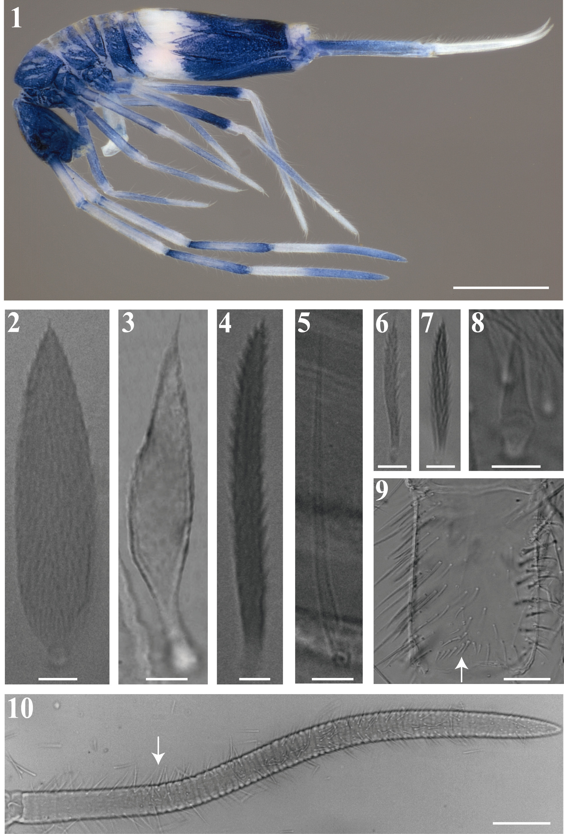
FIGURES 1 – 10. Amazhomidia ducke gen nov. sp. nov.: 1, habitus of a fixed specimen in ethanol (lateral view); 2 – 8, different shaped of scales and chaetae: 2, pointed scale, 3, scale-like chaetae of cephalic groove, 4, ciliated chaeta, 5, sens type II on Abd. IV, 6, ciliated microchaeta, 7, accessory chaeta of bothriotricha, 8, dental spine; 9, trochanter chaetotaxy, arrow indicates the extra spine-like chaetae of anterior face; 10, Ant. IV segment, arrow indicates the beginning of annulations. Scale bars: 1 (0.5 mm), 2 – 8 (0.005 mm), 9 (0.05 mm), 10 (0.1 mm).
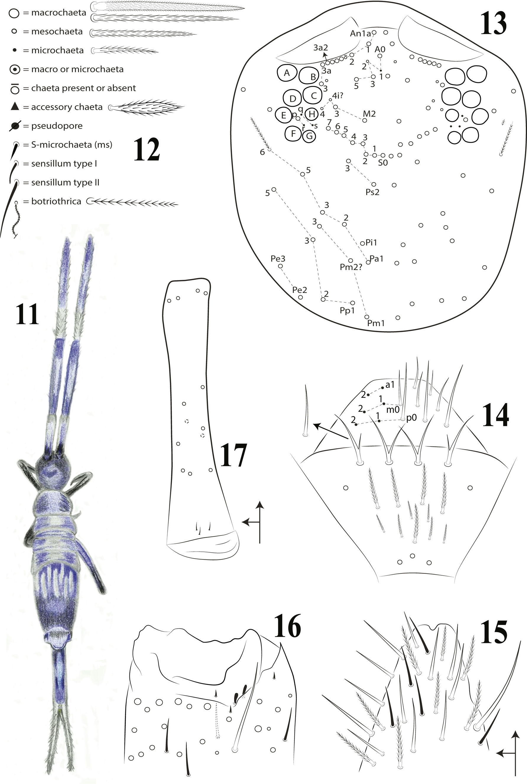
FIGURES 11 – 17. Amazhomidia ducke gen nov. sp. nov.: 11, habitus (dorsal view); 12, symbols used in detailed chaetotaxy schemes; 13, dorsal cephalic chaetotaxy; 14, chaetotaxy of the clypeus, prelabrum and labrum; 15, apex of Ant. IV; 16, Ant. III organ and associated chaetae and sensilla; 17, chaetotaxy of Ant. I (dorsal view).
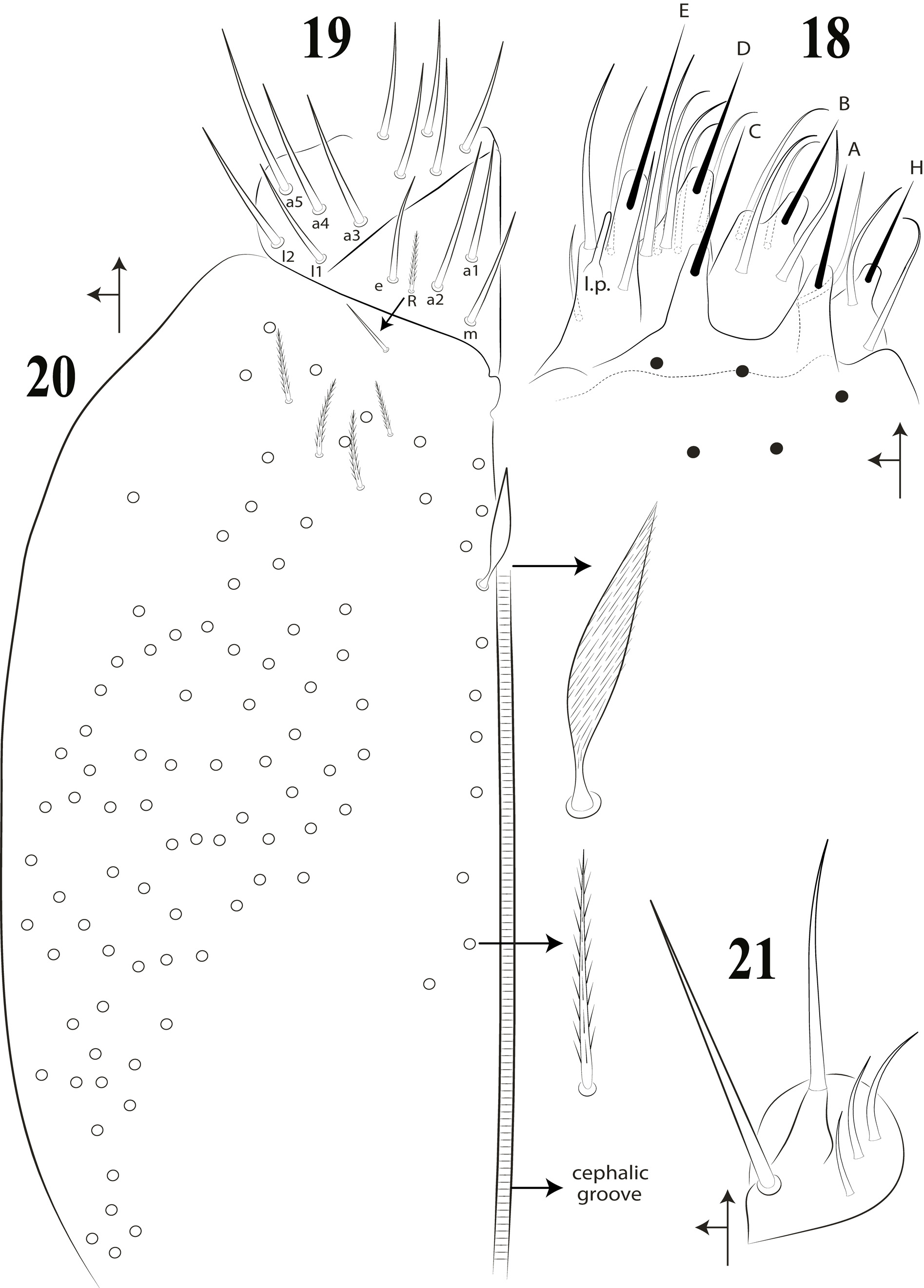
FIGURES 18 – 21. Amazhomidia ducke gen nov. sp. nov.: ventral head: 18, labial papillae and proximal chaetae; 19, labial region and proximal chaetae; 20, posterior labial chaetotaxy; 21, maxillary outer lobe and sublobal plate.
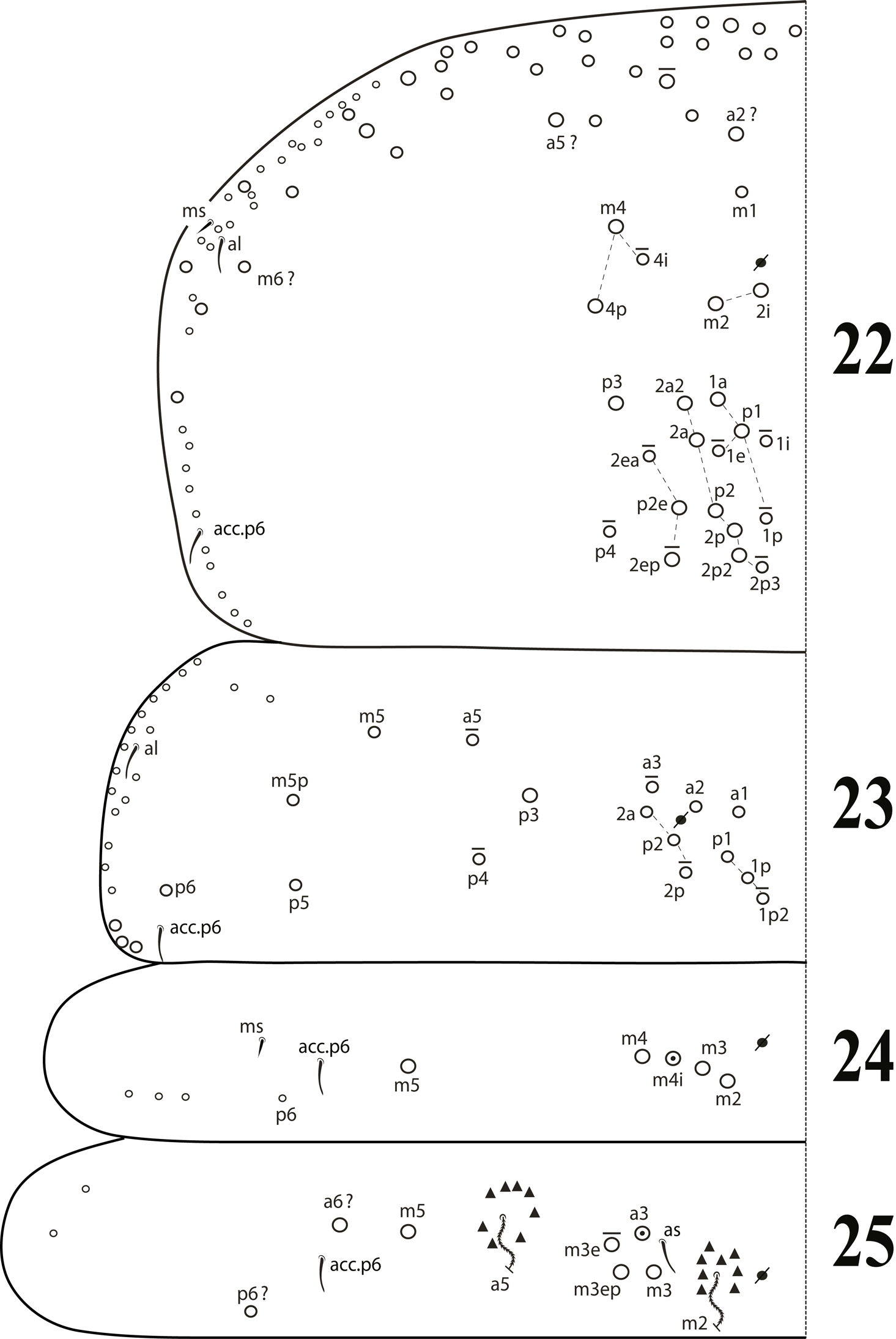
FIGURES 22 – 25. Amazhomidia ducke gen nov. sp. nov.: dorsal chaetotaxy: 22, Th. II; 23, Th. III; 24, Abd. I; 25, Abd. II.
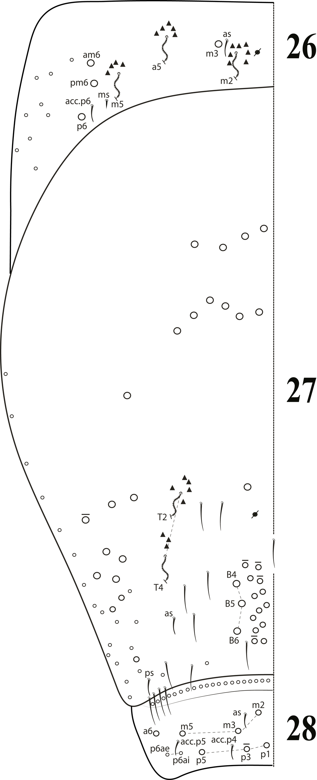
FIGURES 26 – 28. Amazhomidia ducke gen nov. sp. nov.: dorsal chaetotaxy: 26, Abd. III; 27, Abd. IV; 28, Abd. V.
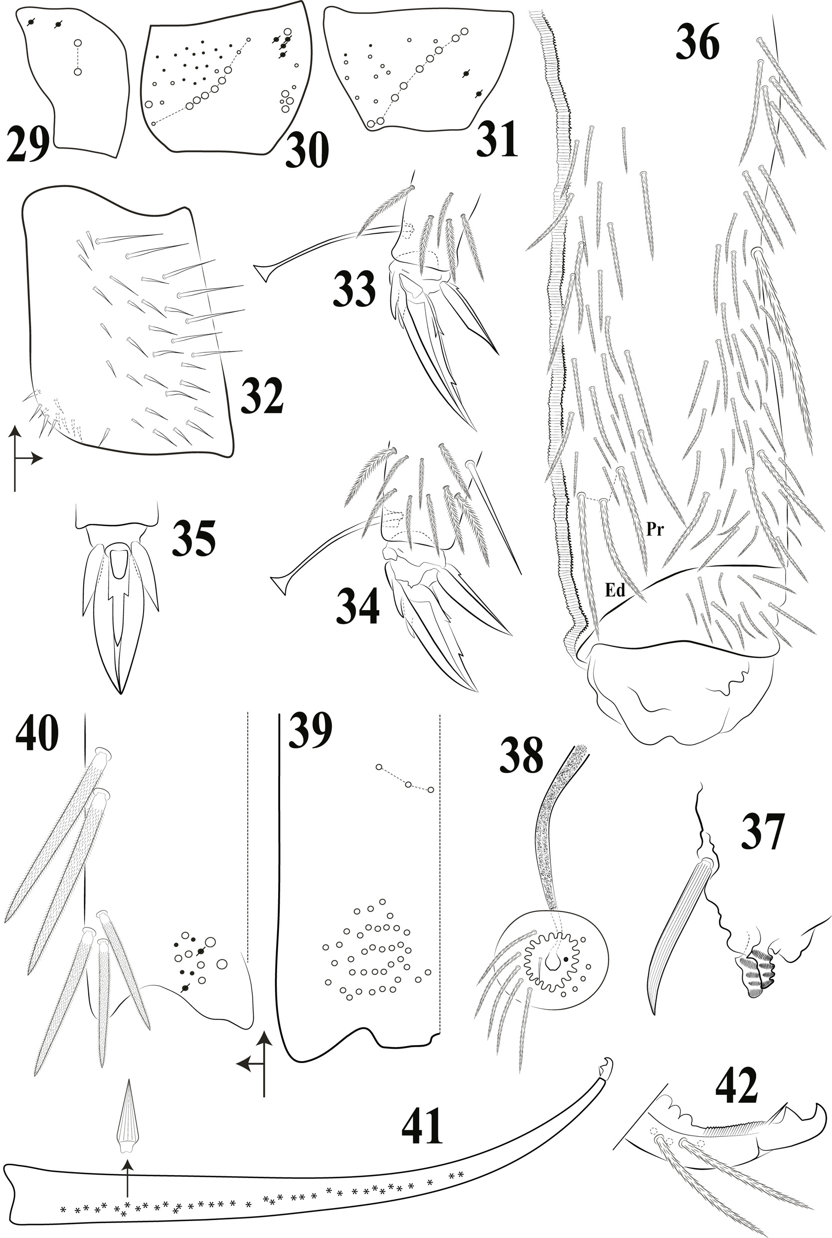
FIGURES 29 – 42. Amazhomidia ducke gen nov. sp. nov.: 29, subcoxa I; 30, subcoxa II; 31, subcoxa III; 32, trochanteral organ; 33 – 34, distal tibiotarsus and empodial complex (lateral view): 33, fore leg; 34, hind leg; 35, ventral view of hind empodial complex III; 36, collophore chaetotaxy (lateral view); 37, tenaculum (lateral view); 38, male genital plate and sperm duct; 39, distal manubrium (ventral view); 40, distal manubrium (dorso-lateral view); 41, dens and mucro (lateral view, showing dental spines distribution); 42, distal dens and mucro (lateral view).
IMAGES