Nephtyidae
GBIF:119637367
ABOUT
Descriptions(1)
Key to species of Nephtyidae of Lizard Island
1. Parapodia with branchiae (Figs 1 C, 2C, 3D)................................................................ 2
- Parapodia without branchiae (Figs 5 C, 6F), or if present, reduced in size and straight..................3 ( Micronephthys)
2. (1) Lyrate chaetae present (Fig. 2 G); notopodial interramal branchiae involuted (Fig. 2 C, E); proximal region of pharynx with ver- rucae............................................................................................... 6
- Lyrate chaetae absent; notopodial interramal branchiae recurved, start on chaetiger 5 and continue to posterior chaetigers; proximal region of pharynx smooth.......................................................... Nephtys inornata
3. (1) Notopodia of chaetiger 1 with specialised dentate chaetae (margin with teeth fused to form knobs, Fig. 11 D); eyespots present between chaetigers 2 and 3..................................................................... M. stammeri
- Notopodia of chaetiger 1 without specialised dentate chaetae................................................... 4
4. (3) Eyes externally visible (Figs 4 A, 8A)...................................................................... 5
- Eyes not visible externally.............................................................. M. platycephala n.sp.
5. (4) Two pairs of lensed overlapping eyes present on prostomium (Fig. 4 A).................................. M. oculifera
- One pair of eyes present at level of chaetiger 2 (Fig. 8 A)...................................... M. cf. sphaerocirrata
6. (2) Involute interramal notopodial branchiae present from chaetiger 7, interramal neuropodial branchiae present from chaetiger 8– 10 (Fig. 3 D); middorsal subdistal pharyngeal papilla absent.................................... Aglaophamus verrilli
- Involute interramal notopodial branchiae present from chaetiger 3, neuropodial branchiae absent (Fig.1 C, E); elongate middor- sal subdistal pharyngeal papilla present................................................. Aglaophamus cf. lobatus
Export occurrence data
Darwin Core Archive (ZIP)
CLASSIFICATION
Taxonomic Classification Tree

MULTIMEDIA
Media Files(7)
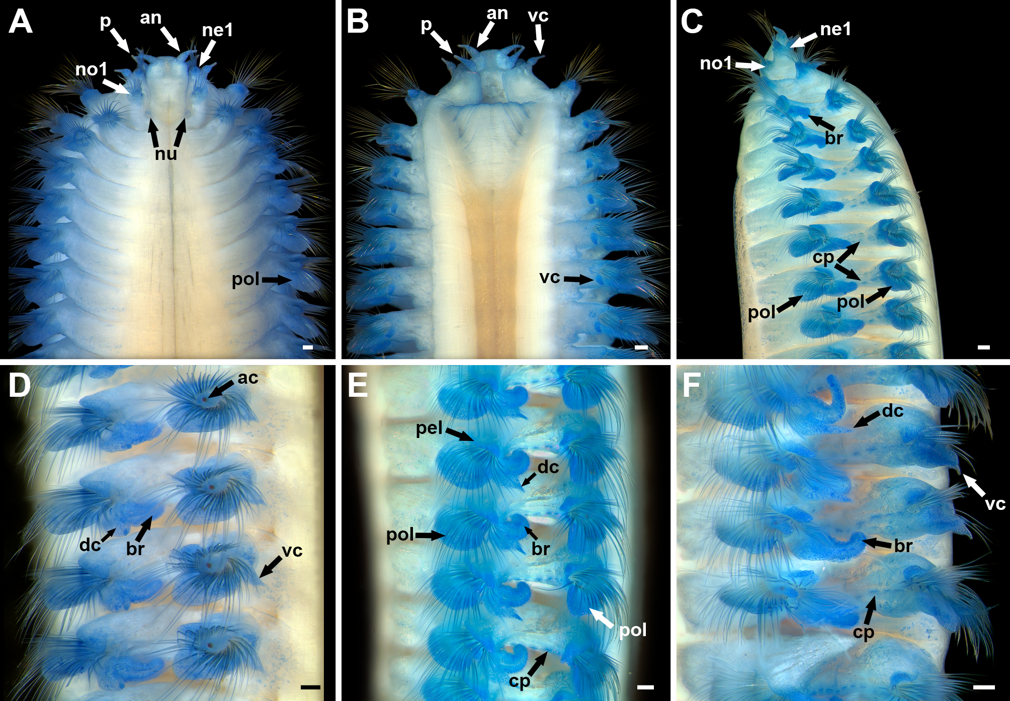
FIGURE 1. Aglaophamus cf. lobatus, specimen stained with methyl blue, AM W. 46976. A. Anterior end, dorsal view; B. Anterior end, ventral view; C. Anterior end, lateral view, branchiae starting from chaetiger 3; D. Anterior chaetigers, lateral view; E. Mid-body chaetigers, lateral view; F. Posterior chaetigers, lateral view. Abbreviations: ac = acicula, an = antenna, br = interramal branchia, cp = cilia patch, dc = dorsal cirrus, ne 1 = neuropodium 1, no 1 = notopodium 1, nu = nuchal pit, p = palp, pel = preacicular parapodial lobe, pol = postacicular parapodial lobe, vc = ventral cirrus. Scale bars: A – F = 0.1 mm.
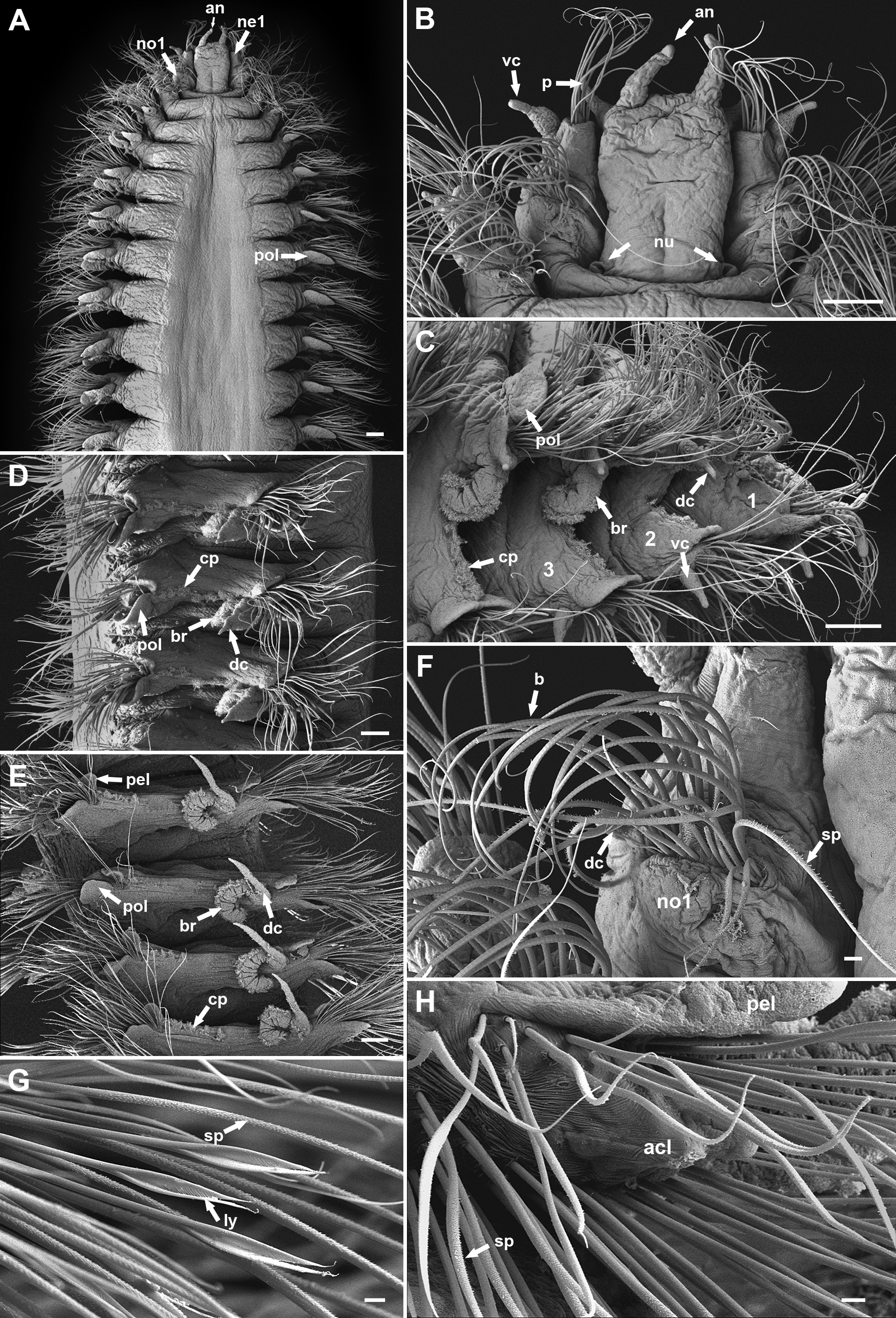
FIGURE 2. Aglaophamus cf. lobatus, SEM photos of AM W. 46973. A. Anterior end, dorsal view; B. Prostomium and chaetigers 1 – 2, dorsal view; C. Chaetigers 1 – 4, lateral view; D. Chaetigers 10 – 12, lateral view; E. Posterior chaetigers, lateral view; F. Notopodium 1, right side, dorsal view, showing barred and spinose chaetae and dorsal cirrus; G. Lyrate and spinose chaetae; H. Notochaetae of parapodium 20. Abbreviations: an = antenna, acl = acicular lobe, b = barred chaeta, br = interramal branchia, cp = cilia patch, dc = dorsal cirrus, ly = lyrate chaeta, ne 1 = neuropodium 1, no 1 = notopodium 1, nu = nuchal pit, p = palp, pel = preacicular parapodial lobe, pol = postacicular parapodial lobe, sp = spinose chaeta, vc = ventral cirrus. Scale bars: A – F = 0.1 mm; G – H = 0.01 mm.

FIGURE 3. Aglaophamus verrilli, specimen stained with methyl blue, AM W. 46971. A. Anterior end, dorsal view; B. Anterior end, ventral view, pharynx everted; C. Anterior end, lateral view, pharynx everted; D. Anterior segments, lateral view. Abbreviations: ac = acicula, an = antenna, db = dorsal interramal branchiae, dc = dorsal cirrus, e = eye, p = palp, pol = postacicular parapodial lobe, pp = pharyngeal papillae, vb = ventral interramal branchiae, vc = ventral cirrus. Scale bars: A – D = 0.1 mm.
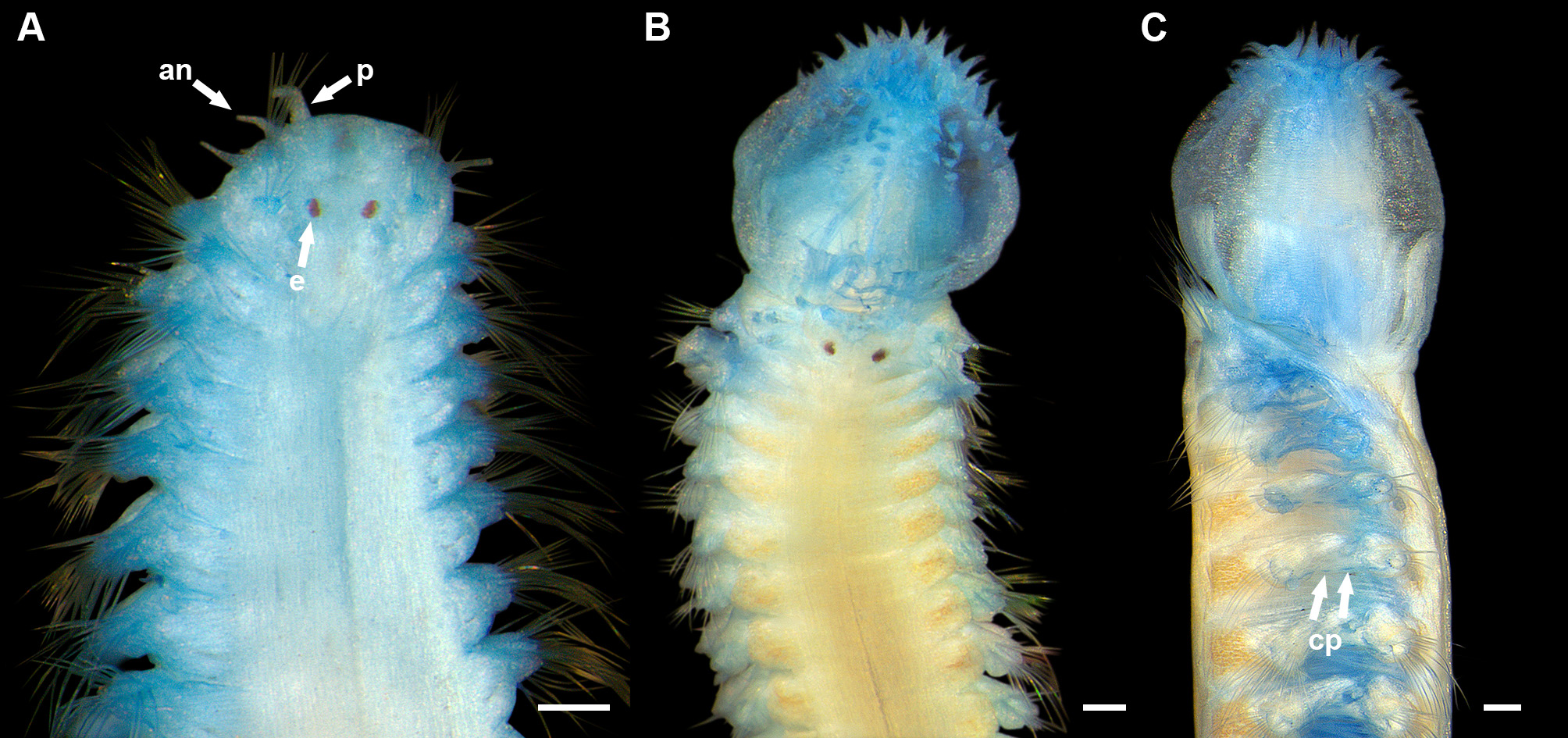
FIGURE 4. Micronephthys oculifera, specimens stained with methyl blue. A. Anterior end, dorsal view, AM W. 41544; B. Anterior end, dorsal view, pharynx everted, AM W. 41511; C. Anterior end, lateral view, AM W. 41578. Abbreviations: an = antenna, e = eye, cp = ciliated patches, p = palp. Scale bars: A – C = 0.1 mm.
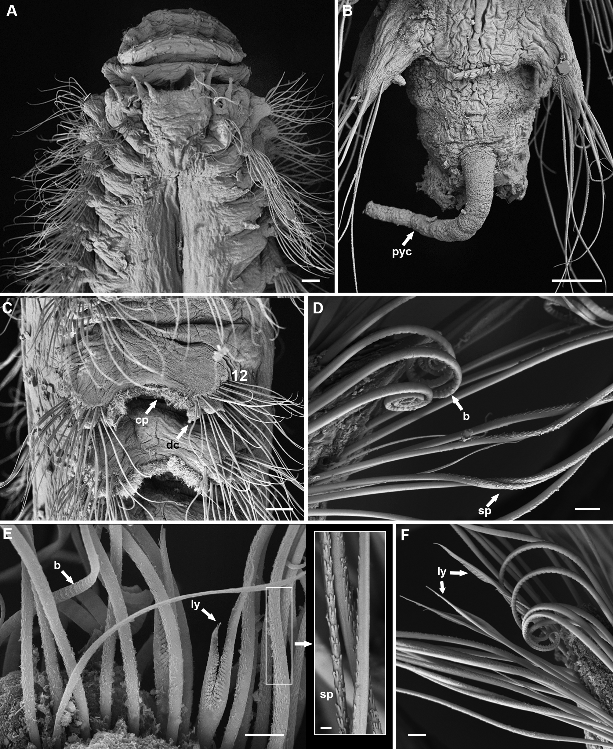
FIGURE 5. Micronephthys oculifera, SEM photos, AM W. 43653 (A – C), AM W. 41518 (D – F). A. Anterior end, dorsal view; B. Posterior end, ventral view; C. Chaetigers 12 – 13, lateral view; D. Barred and spinose chaetae, chaetiger 36; E. Barred and lyrate chaetae, inset with magnification of spinose chaetae, chaetiger 20; F. Postacicular lyrate chaetae, chaetiger 21. Abbreviations: b = barred chaeta, cp = cilia patch, dc = dorsal cirrus, ly = lyrate chaeta, pyc = pygidial cirrus, sp = spinose chaeta. Scale bars: A = 0.1 mm, B – C = 0.05 mm, D – F = 0.01 mm except for enlarged image 2 µm.
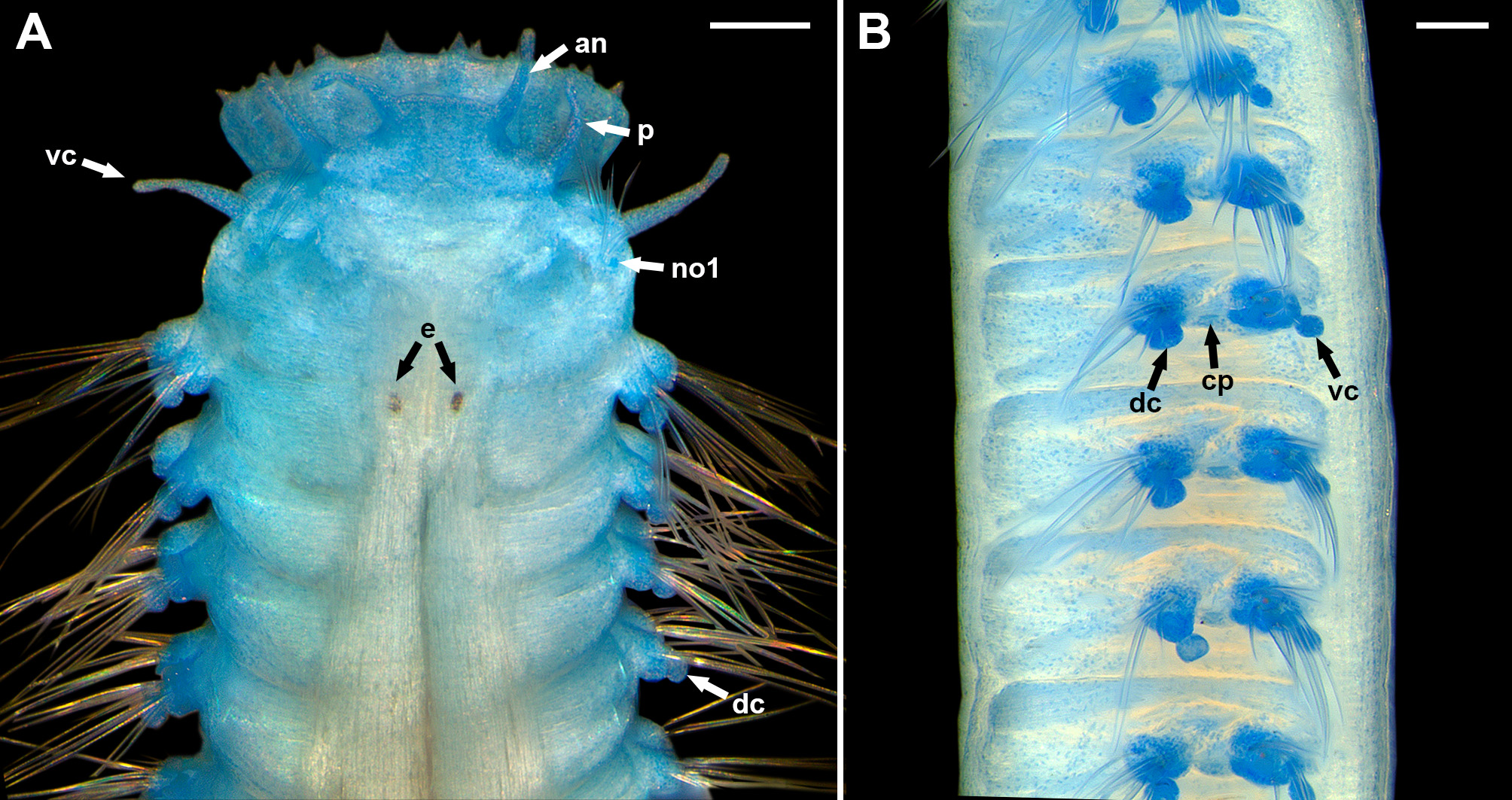
FIGURE 8. Micronephthys cf. sphaerocirrata, specimen stained with methyl blue, AM W. 46983. A. Anterior end, dorsal view; B. Chaetigers 2 – 8, lateral view. Abbreviations: an = antenna, cp = cilia patch, dc = dorsal cirrus, e = eye, no 1 = notopodium 1, p = palp, vc = ventral cirrus. Scale bars: A – B = 0.1 mm.
IMAGES