AnimaliaNot EvaluatedacceptedspeciesAccepted
Eugyra glutinans
(Möller, 1842) Moller, 1842
GBIF:120692787
0year
ABOUT
Descriptions(1)
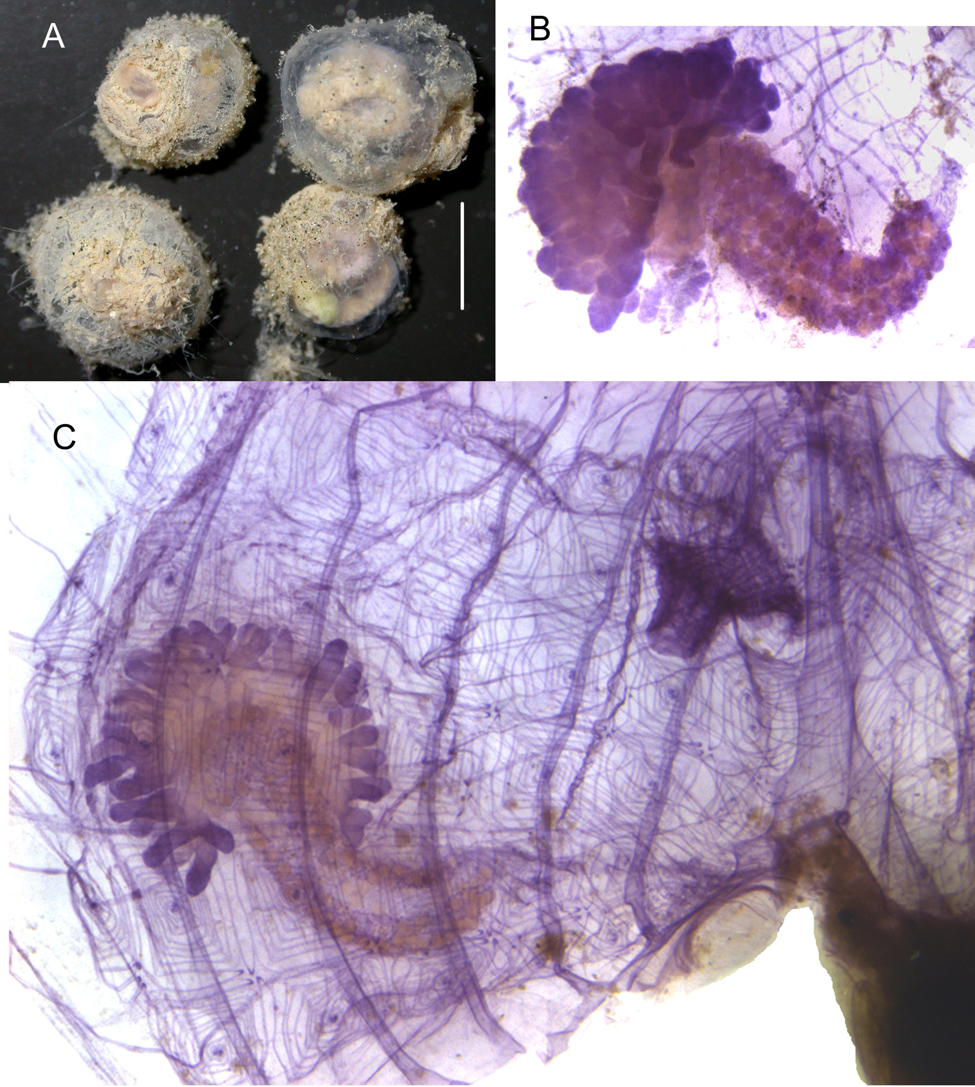
Stations. CP 4349; CP 4355; CP 4356; CP 4357; CP 4378; CP 4379; CP 4381; CP 4383; CP 4387; CP 4399; CP 4409; CP 4410. More than a hundred specimens were collected from 30 m to 130 m depth. All are spherical, 8 mm maximum in diameter (Fig. 30 A). The tunic is thin, resistant and bears thin processes and sand. The siphons are close together and not protruding. When removed from the tunic the internal organs are visible through the thin body wall. The muscular sphincters of the siphons are strong. On the body well- separated longitudinal muscular fibres start from the oral part and from the atrial siphon (Fig. 31 A). They are crossed by transverse fibres essentially in the anterior part of the body. Rare muscular ribbons lie on the ventral side (Fig. 31 A). There are 12 oral tentacles with short ramifications of one or 2 orders intercalated with buttons. The pre-pharyngeal band is double and almost straight dorsally. The dorsal tubercle has a vertical slit. The dorsal lamina is double along its entire length (Fig. 31 B) prolonged around the oesophagus entrance. There are 5 longitudinal vessels on each side (Fig. 31 B), each erect above a vertical row of infundibula. In addition on each body side there is ventrally and dorsally another row of infundibula without a longitudinal vessel. Two imbricate stigmata in many turns form the infundibulum (Figs 30 A, 31 BB). The gut occupies almost all the left body side (Fig. 31 A). The oesophagus is wide and short. The stomach is deeply pigmented, 4 to 5 low longitudinal folds can be seen on the cardiac side. The intestine is wide with a long closed primary loop and a deep secondary curve. The anus has a smooth edge. The hepatic gland covers the stomach in small flat dark papillae. The kidney (Fig. 30 C; 31 A) is a round transparent vesicle without inclusion located on the right ventral side near the oesophagus below the gonad. There is one gonad on each side with the same shape. The left one has the male part inside the primary gut loop but the distal part of the ovary crosses the intestine (Fig. 31 A). Each gonad has separate male and female elements but closely applied. The testis is made of several oval lobes, more or les divided, pressed to each other to make a crescent (Figs 30 C, 31 A). Their short sperm ducts converge, some of them fuse but not all and they open in separate papillae against the blind extremity of the ovary. The ovary is a wide S-shape tube ending in a wide papilla near the dorsal line. This species corresponds in all points to the description of E. glutinans by Hartmeyer (1923) and Van name (1945) with a typical structure of the branchial sac having 5 longitudinal vessels but 7 vertical rows of infundibula on each side. Records of this species after 1945 are not sufficiently detailed to be taken into account. It is likely that a confusion may occur with E. arenosa (Alder & Hancock, 1848) which has the same gonad shape but 7 longitudinal branchial vessels per side. Considering that E. glutinans was recorded from the peri-Antarctic area only (except one record off California. in Van Name 1945) its presence in Guiana is surprising.
Monniot, Françoise (2016): Ascidians (Tunicata) of the French Guiana Expedition. Zootaxa 4114 (3): 201-245, DOI: 10.11646/zootaxa.4114.3.1
Export occurrence data
Darwin Core Archive (ZIP)
CLASSIFICATION
Taxonomic Classification Tree


IMAGES