AnimaliaNot EvaluatedacceptedspeciesAccepted
Symplegma rubra
Monniot C., 1972
GBIF:120692791
0year
ABOUT
Descriptions(2)
The most part of the Guiana material is in poor condition and no coloration is retained. Most of the colonies encircle polychaete tubes. The zooid anatomy is that described from Bermuda (Monniot C. 1972). The gonads either male or female are located in different zooids. This species is recorded from diverse areas with variations in the number of stigmatal rows and stomach folds. In the western Atlantic it is known from Guadeloupe (Monniot C. 1983 b), Jamaica (Goodbody 1993), Gulf of Mexico (Lambert et al. 2005) and Brazil (Rocha & Costa 2005; Rodrigues et al. 1998). It is also present in the Indian and Pacific Oceans. Cnemidocarpa captiva n. sp. Figures: 18, 19, 20. Stations. CP 4395 (Type MNHN S 1 CNE 238); CP 4396.
Monniot, Françoise (2016): Ascidians (Tunicata) of the French Guiana Expedition. Zootaxa 4114 (3): 201-245, DOI: 10.11646/zootaxa.4114.3.1
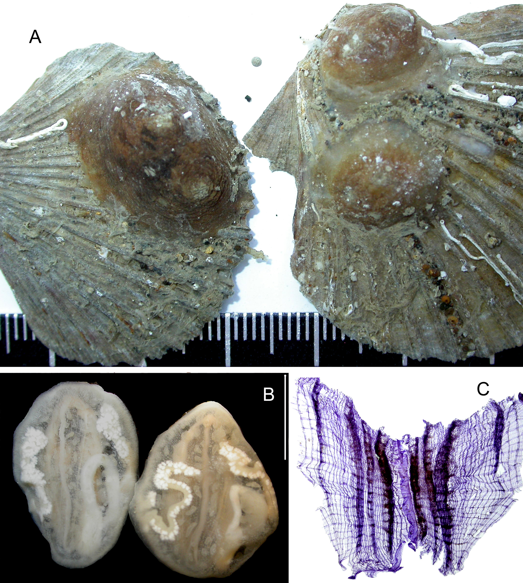
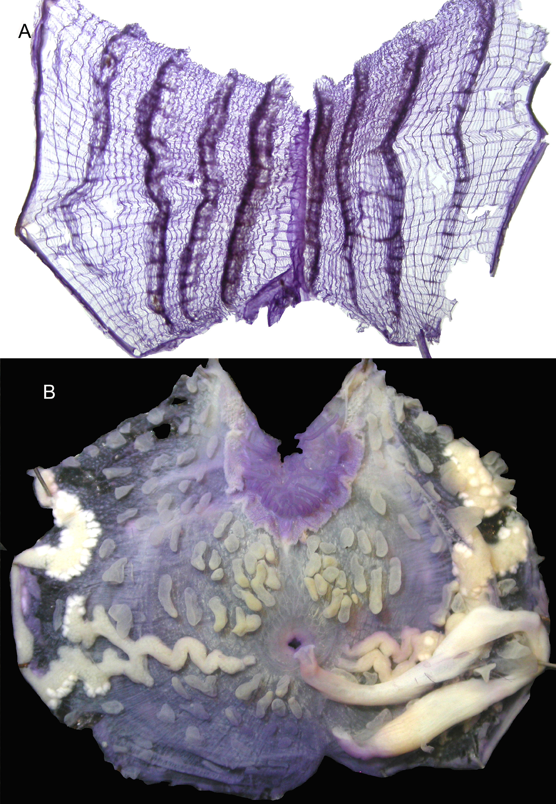
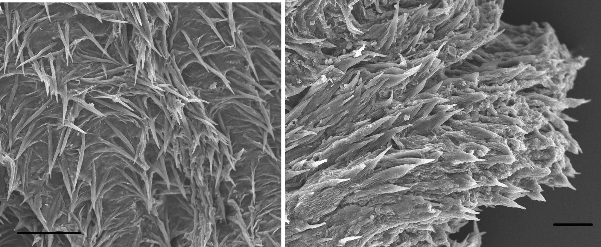
Etymology. captiva refers to the prison in the Iles du Salut which was formerly a prison. Most of the specimens are strongly attached on Pectinidae shells (Fig. 18 A) or some others on stones. Among 20 specimens collected the largest are 2 cm in diameter. The body is hemispherical and brown; the siphons on the upper side are distant of 1 / 3 of the body width. The tunic is thick and corrugated superficially but thinner and colourless and a little expanded on the fixation surface. The body wall only adheres to the tunic by the siphons. It is colourless except on the siphons having 4 brown longitudinal lines. The reflex tunic inside the siphons bears acicular spinules (Fig. 20 A, B). The musculature forms a dense uniform coating of thin fibres on the whole body wall. The oral tentacles are numerous in 3 orders of size behind a velum. The prepharyngeal band curves dorsally to include the dorsal tubercle which opens anteriorly in a U. The dorsal lamina is a high blade increasing in height posteriorly. There are 4 low branchial folds on each side (Fig. 18 C, 19 A) not recovering each other and the most ventral one becomes thinner posterioly to almost disappear near the ventral line (Fig. 19 A). The formula in one specimen is RE- 4 (12) 4 (14) 5 (12) 5 (15) 2 DL (17) 5 (13) 6 (12) 5 (10) 4 EL There are 2 to 3 long stigmata per mesh between the folds crossed by parastigmatic vessels except ventrally where the meshes are wider. The gut occupies the posterior part of the left side (Fig. 18 B, 19 B) It is closely attached to the body wall but not included in it. The oesophagus is narrow and curved. The stomach is long and spindle-like with internal folds and no caecum (Fig. 19 B). The intestine forms a closed long loop ending in a scalloped anus. There is one long gonad on each side (Fig. 18 B, 19 B). (The left gonad was missing in one specimen). The gonads are made of a long sinuous ovary with one or few lateral branches of varied design in different zooids (Fig. 18 B, 19 B). The female papillae open near the atrial siphon; in one specimen the ovary has its extremity divided into 2 branches ending each ending in a separate papilla (Fig. 19 B). The testis lobes form irregular masses of vesicles applied to the ovary but scattered along it. The sperm duct follows the ovary. The internal layer of the body wall is spotted with multiple clear vesicles. Numerous foliated endocarps are present on the body wall, not evenly distributed but in two groups: some around the gonads and the gut loop and some others gathered in the middle but dorsal part of each body side (Fig. 19 B). The entrance of the atrial aperture is fringed with a line of thread-like papillae in half circle posteriorly and extended dorsally in a loop up to the neural area. A large atrial velum closes the atrial siphon. The deep and tropical Cnemidocarpa captiva n. sp. has a surprising likeness to two other Cnemidocarpa recorded from the Antarctic: C. drygalskii (Hartmeyer, 1911) and C. nordenskjoldi (Michaelsen, 1898). These two species only differ from each other by the orientation of the gut loop parallel to the endostyle in the former and crossing the ventral line in the later. They have in common with C. captiva the dome shaped body, 4 branchial folds / side, a single very long sinuous gonad on each side and a closed gut loop with a long stomach. C. captiva differs from both Antarctic species by the number and arrangement of the endocarps, an important distinctive character among the Styelidae. As in C. drygalskii C. captiva has the gut entirely on the left side but differs by the absence of a stomach caecum and its dorsal lamina is smooth. C. nordenskjoldi is the only one having numerous cloacal tentacules over the whole atrial velum. The choice to include the Guiana species into the genus Cnemidocarpa instead of Styela is based on the relative position of the male and female elements in the gonads. In Styela the testis lobes are at some distance from the ovary and only the sperm ducts join to it but in Cnemidocarpa the testis is included in the membrane covering the ovary. This distinction is sometimes difficult to interpret.
Monniot, Françoise (2016): Ascidians (Tunicata) of the French Guiana Expedition. Zootaxa 4114 (3): 201-245, DOI: 10.11646/zootaxa.4114.3.1
Export occurrence data
Darwin Core Archive (ZIP)
CLASSIFICATION
Taxonomic Classification Tree

MULTIMEDIA
Media Files(3)
FIGURE 18. Cnemidocarpa captiva n. sp. A, individuals on scallop shells; B, ventral side of 2 specimens, scale bar = 1 cm; C, stained branchial tissue.
Imageimage/png© Monniot, FrançoiseMonniot, Françoise
IMAGES