AnimaliaNot EvaluatedacceptedspeciesAccepted
Diazona gigantea
Sluiter, 1919
GBIF:120692826
0year
0
Synonyms
ABOUT
Descriptions(4)
E. concklini Berrill 1932, redescribed in Monniot C. (1972) and in Goodbody & Cole (2006) differs in having large zooids sometimes exceeding 2 cm, and stolons creeping on solid substrate surfaces. They have usually 20 rows of stigmata and 2 to 3 stigmata per mesh. The primary gut loop is closed but the secondary loop is wider with a long rectum. E. minuta Berrill, 1932 has zooids attached to the substrate by their ventral part and the stolons are also linked to the substrate. The zooids are only 5 to 6 mm long. The oral siphon is apical and the atrial one at mid distance of the body side. The branchial sac has an average of 15 stigmata rows. The primary gut loop is closed; the intestine has 4 longitudinal swellings (Monniot C. 1972 Fig. 2 D) and the rectum lies horizontally before a sharp bend. The anus opens at the level of the top of the gut loop. The gonad differs from all other Ecteinascidia with a mass of testis lobes well separated from the nearby ovary.
Monniot, Françoise (2016): Ascidians (Tunicata) of the French Guiana Expedition. Zootaxa 4114 (3): 201-245, DOI: 10.11646/zootaxa.4114.3.1
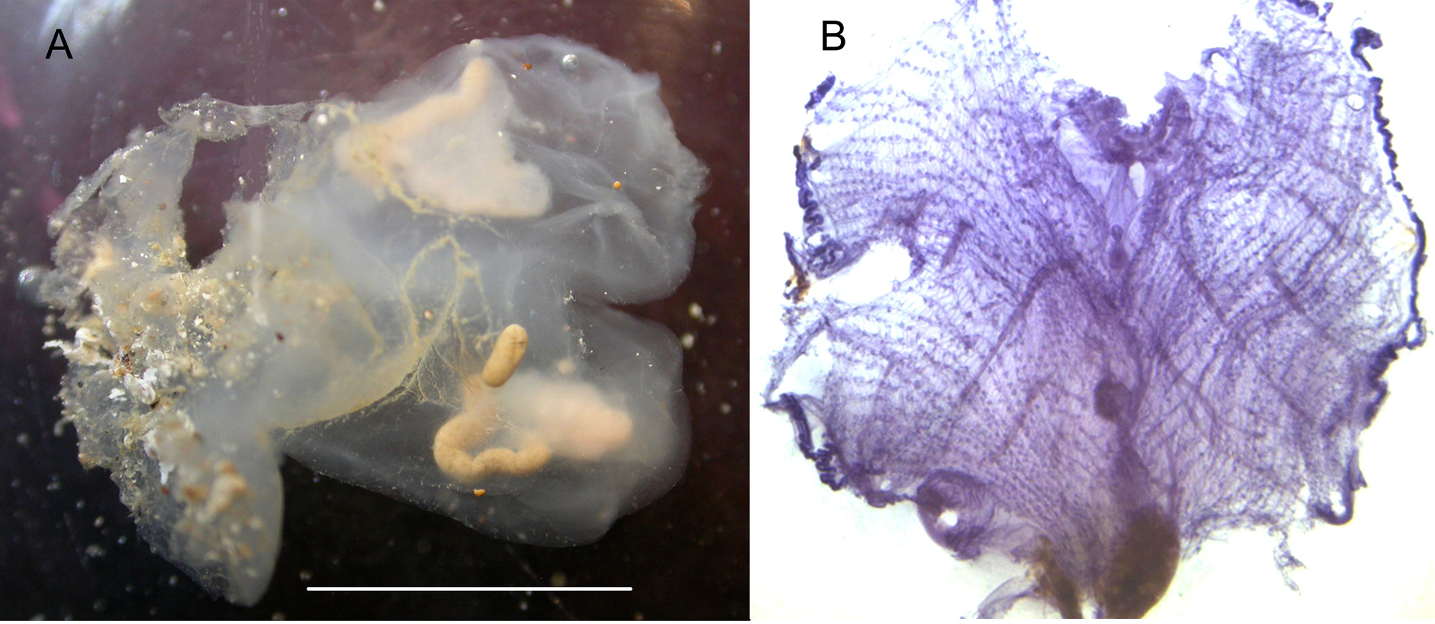
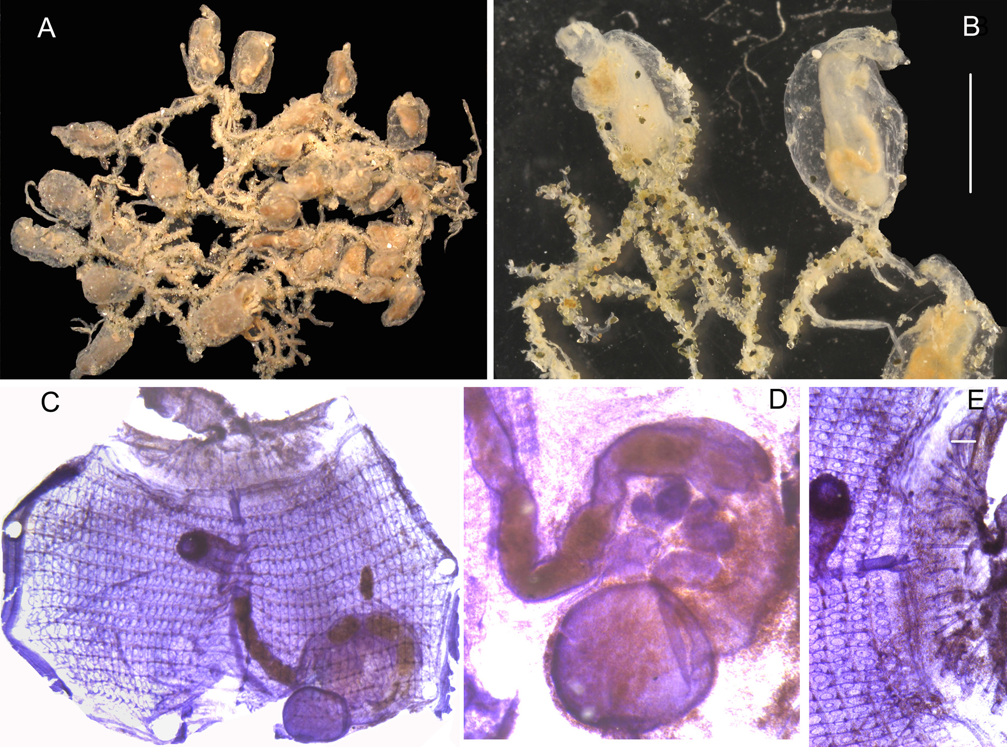
Non: Diazona gigantea: Monniot C. 1970 = Stomozoa roseola. Two colonies dredged at 145 m depth are not in good condition and provisionally placed in D. gigantea (Sluiter, 1919). One colony contained an isolated thorax, the other 15 mm x 12 mm is partially divided into 2 lobes (Fig. 12 A). In each lobe was a deeply retracted thorax but the abdomens were missing. The tunic is glassy, cartilaginous and naked except some sand at the fixation base. The shape of the siphon edges is not clear. There are 8 long oral tentacles on a ring and smaller ones intercalated. The dorsal tubercle opens in a vertical slit anterior to the oval neural ganglion. The branchial sac is wide (Fig. 12 B) with numerous straight stigmata in at least 35 rows. The high transverse vessels wear large papillae united in some places by longitudinal vessels. There is an average of 2 stigmata per mesh. The thoracic musculature includes the siphonal sphincters and only 6 to 8 strong longitudinal ribbons on each side of the thorax. No transverse musculature was detected. A wide rectum full of mud is at the base of the thorax but no other parts of the gut remain. The tunic structure and the shape of the thorax correspond to Sluiter’s original description completed by Ärnback-Christie-Linde (1925) who re-examined Sluiter’s type collected without precision from the West Indies. Caullery (1914) shortly described Diazona geayi from Guiana. Pieces of this species, cut in thin slices are present and labelled “ type ” in the MNHN collection. These pieces contain few abdomens and regenerating thoraces in very bad condition, but in one of them no papillae were present on the transverse thoracic vessels in high membranes. Considering the absence of branchial papillae this specimen cannot belong to the genus Diazona. No other species of the genus Diazona are recorded from the western part of the Atlantic Ocean. Ecteinascidia aranea n. sp. Figure 13.
Monniot, Françoise (2016): Ascidians (Tunicata) of the French Guiana Expedition. Zootaxa 4114 (3): 201-245, DOI: 10.11646/zootaxa.4114.3.1
Station. CP 4393
Monniot, Françoise (2016): Ascidians (Tunicata) of the French Guiana Expedition. Zootaxa 4114 (3): 201-245, DOI: 10.11646/zootaxa.4114.3.1
Etymology. aranea refers to a spider web Station. CP 4408 (Type: MNHN P 2 ECT 112). The colonies form a loose net of stolons several cm across (Fig. 13 A). Erect zooids are attached by a narrow base on thin ramified stolons incrusted with sand. The glassy tunic of the zooids is less impregnated than the stolons and allows seeing the internal organs (Fig. 13 B). The body is longer than wide with a terminal long oral siphon. The atrial tubular siphon opens at ¼ of the body length. The oral aperture has 6 petal-like lobes. The body wall is thin but with a more or less extended network of blood sinuses. The sphincters are well developed at the base of the siphons. Few longitudinal muscular fibres extend from the oral side to the base of the atrial aperture on the dorsal side. The transverse muscular fibres are spaced and only lying over the dorsal part of the body lacking on each side of the endostyle. About 45 oral tentacles form a circle in alternating sizes. The dorsal tubercle is urnshaped above an elongated neural ganglion (Fig. 13 D). The branchial sac has 15 rows of stigmata and an average of 22 longitudinal vessels on each side (Fig. 13 C) but this number could be counted only in one less contracted colony. There is only one stigma per branchial mesh. The digestive tract occupies ¼ of the posterior left body side (Fig. 13 C). The stomach is spherical with a smooth wall (Fig. 13 D). The first part of the intestine shows 2 constrictions. The rectum curves to reach the oesophagus and then follows the dorsal line to the atrial aperture. The anus has 2 short lobes. The testis comprises piriform lobes arranged in a crescent inside the gut loop, not overpassing the intestine. The sperm duct follows the rectum and opens close to the anus. The ovary is placed near the centre of the testis half circle. No brood pouch was present in any zooid. Several Ecteinascidia species occur in the western Atlantic and are compared below.
Monniot, Françoise (2016): Ascidians (Tunicata) of the French Guiana Expedition. Zootaxa 4114 (3): 201-245, DOI: 10.11646/zootaxa.4114.3.1
Export occurrence data
Darwin Core Archive (ZIP)
CLASSIFICATION
Taxonomic Classification Tree

MULTIMEDIA
Media Files(4)
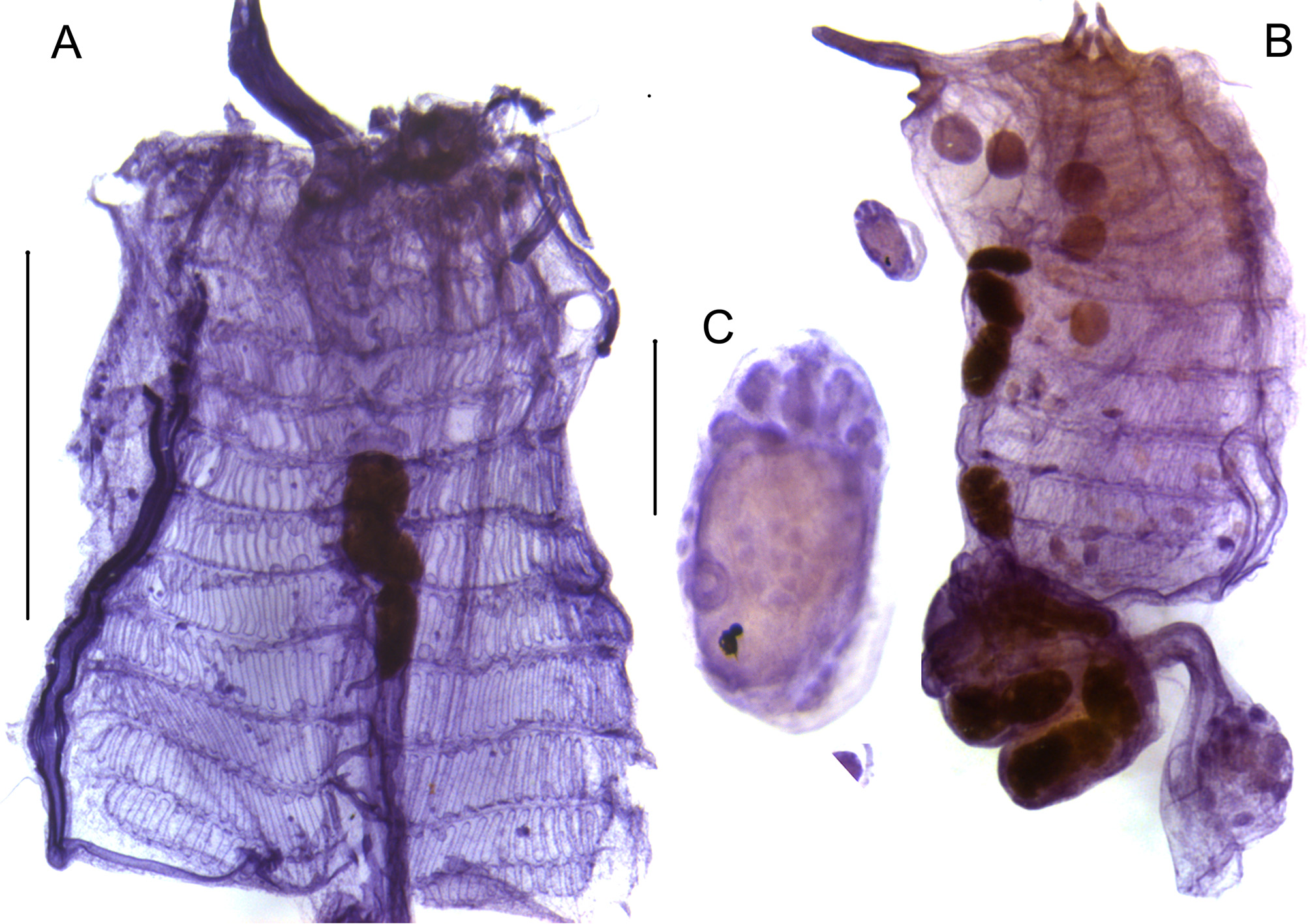
FIGURE 2. Polyclinum constellatum (stained with hemalum). A: branchial sac; B, zooid; C, larva. Scale bars: A – B = 2.5 mm; C = 0.25 mm.
Imageimage/png© Monniot, FrançoiseMonniot, Françoise
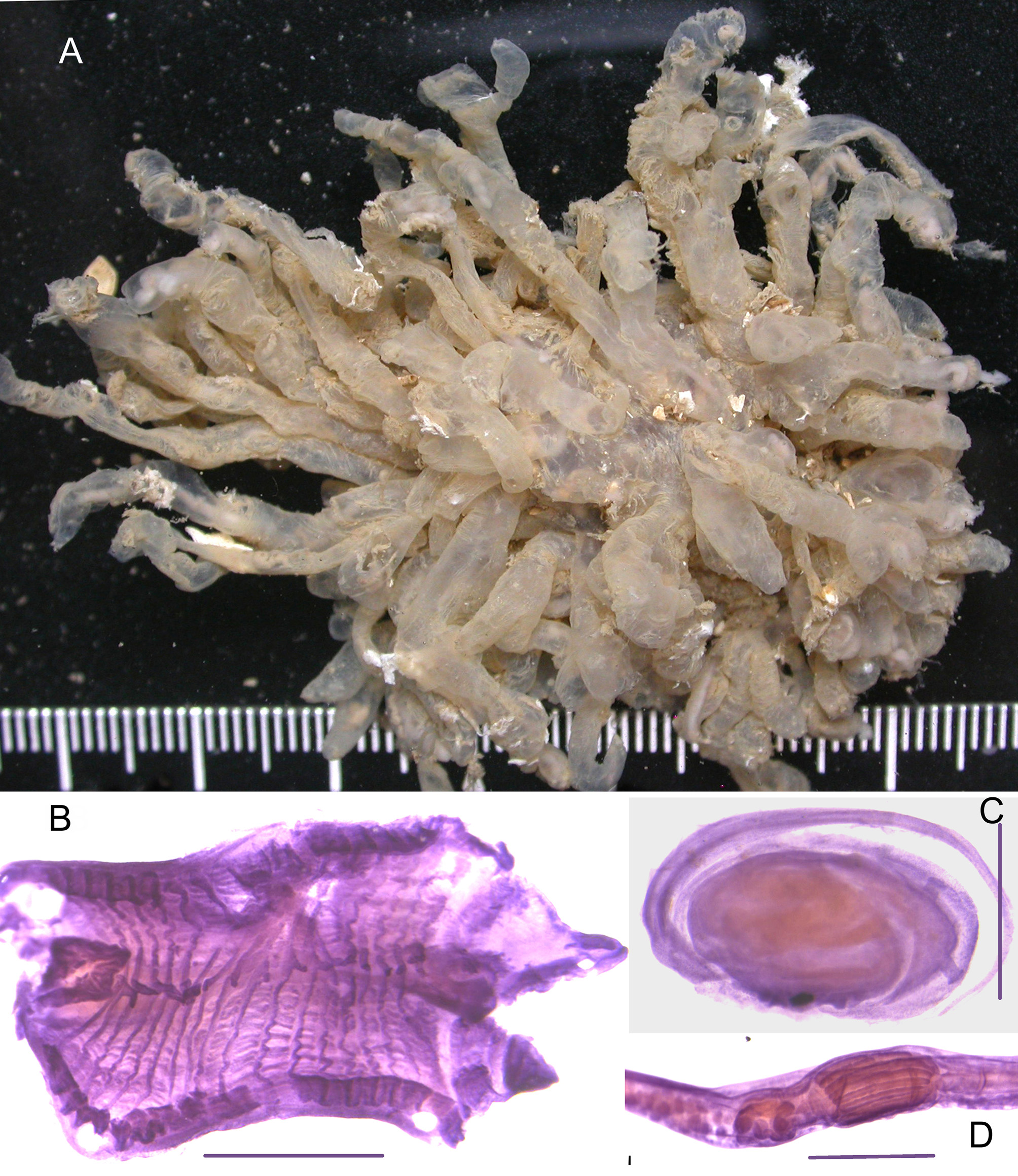
FIGURE 3. Euherdmania fasciculata. A, colony; B, branchial sac; C, larva; D, stomach. Scale bars B = 1 mm; C = 0.5 mm; D = 1 mm.
Imageimage/png© Monniot, FrançoiseMonniot, Françoise
IMAGES