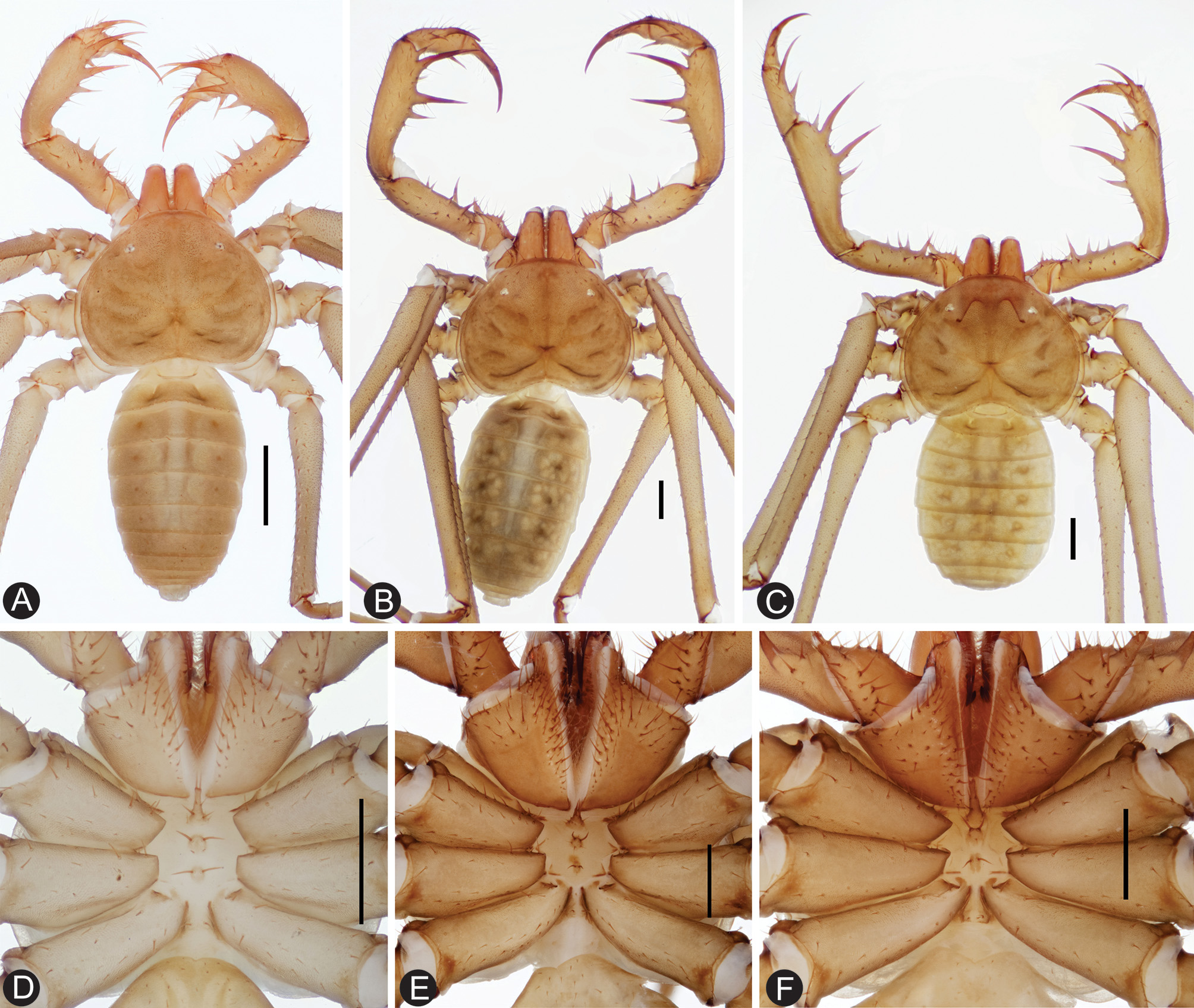
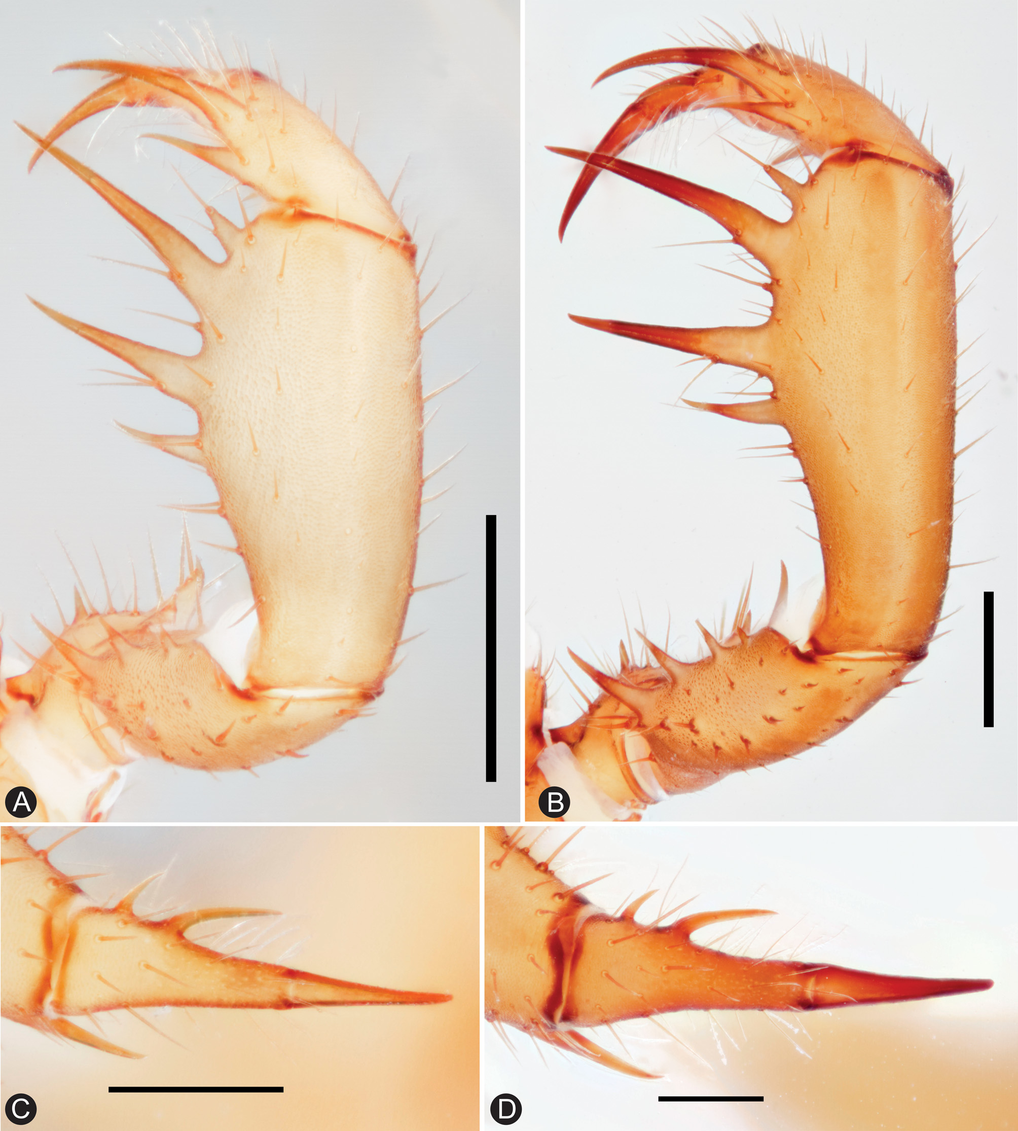
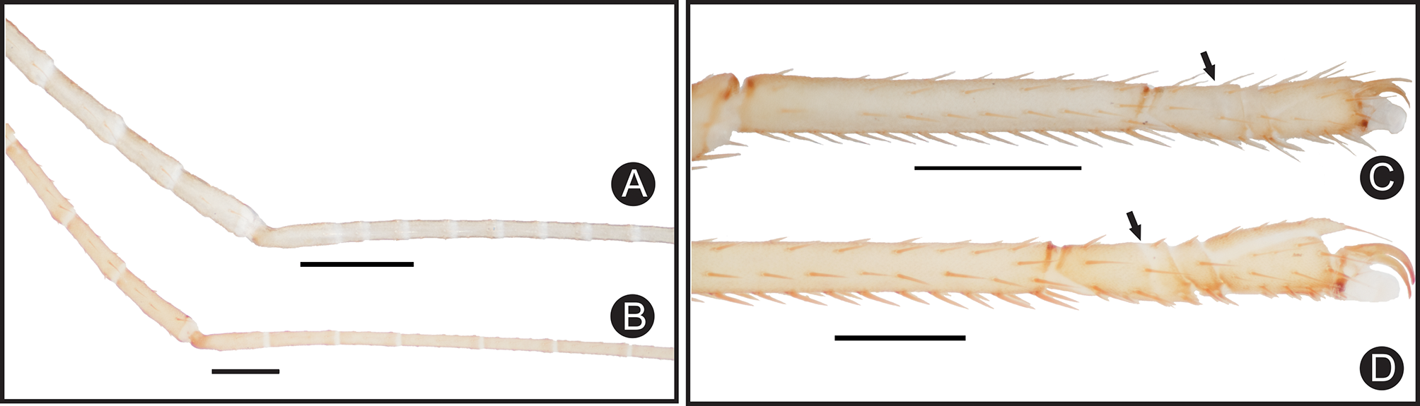
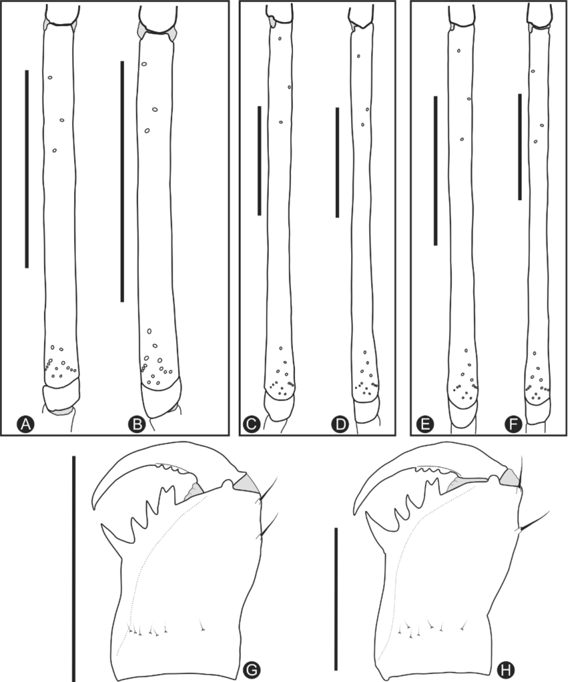
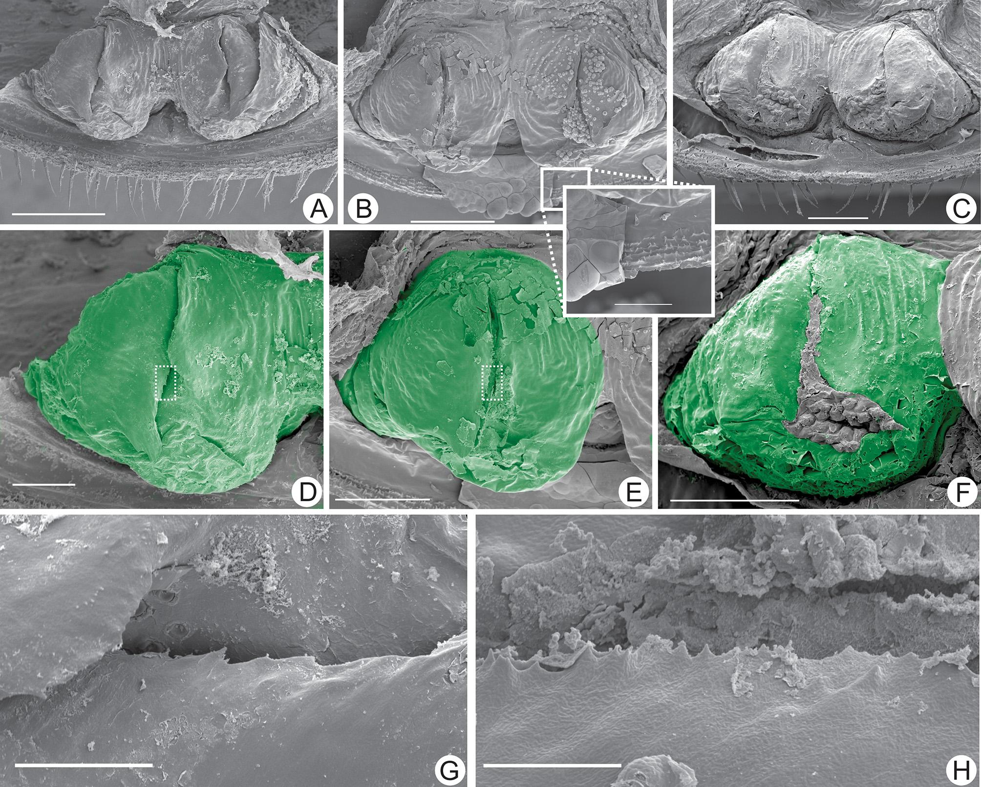
Description of the male holotype (variations found in the paratypes are indicated in brackets; description of the chelicerae and gonopod are based on paratypes): Carapace (Fig. 1 A). Carapace flattened, wider than long (1.6 times), slightly bent downwards below lateral eyes; a thin median furrow reaches the fovea starting from the depression that replaces the median eye and tubercle. Anterior margin straight, with six frontal setae. Frontal process large, triangular, not visible from above, with a rounded tip. Three pairs of shallow furrows in the lateral of the carapace, and an oval and deep fovea. First pair of furrows placed just behind the lateral boss behind the lateral eyes; any of the furrows reaches the middle line. Median eyes and tubercle completely absent, a deep depression instead; no setae present in the depression. Lateral eyes well developed, pale, one large seta behind each triad; lenses directed upwards and slightly anteriorly. Sternum (Fig. 1 D): tetra-segmented, all pieces well sclerotized. Tritosternum with a round basis and projected anteriorly in a small blunt tubercle, reaching the base of the pedipalp coxae, with two apical, two median and two basal setae, with smaller ones on the base. Middle piece (tetrasternum) in one convex piece, with a pair of large setae in its apex, and a pair of small setae in its base. Third piece (pentasternum) formed by one convex piece, smaller than the middle piece, with two long setae at its top and with no small setae on its base. Sternites separated from each other by length of the third piece. Metasternum not paired (i. e., one single piece), with one pair of setae on an elevation at the posterior region of the plaque. Abdomen (Fig. 1 A): oblong, with almost indistinguishable punctuations. Ventral sacs not developed. Chelicera (Fig. 5 G): Cheliceral furrow with four internal teeth; first tooth (upper) bifid, proximal cusp of the same size as distal cusp. Third tooth slightly thinner and shorter than second tooth. Fourth tooth one third larger than the third. No tooth in the external row of the basal segment. Mesal face with a longitudinal row of seven setae. Claw with four denticles. Pedipalp: Trochanter (Fig. 1 A, 2 A, B): large ventral apophysis, located in the posterior border of the trochanter, spiniform, bearing 11 large setae, and with a blunt tip pointed forward; two subequal spines, one in about the center of the anterior row of setiferous tubercles (three setae on each side), the other at the external border, below the apophysis, a bit curved inwards. Femur (Fig. 1 A, 2 A, B, 3 A): three dorsal spines (I> II> III) with two prominent setiferous tubercle before the first spine; three ventral spines (I> II> III) with one small setiferous tubercle before the first spine [one female paratype have two spines]. Tibia (Fig. 1 A, 2 A, B, 3 A): three dorsal spines (I> II> III); one spine distal to I (about one third the size of I); one small setiferous tubercle proximal to spine III; spine II two thirds spine I and spine III one third spine I; spine I and II with two setiferous tubercle on its basal third; spine III with one setiferous tubercle in its half. Two ventral spines; second spine half size of the first (I> II). Basitarsus (Fig. 1 A, 2 A, B): two dorsal spines, the basal 2 / 3 the size of the distal. One ventral spine at the distal half, 2 / 3 the basal spine dorsal. Distitarsus (Fig. 3 C): two large curved spines, the distal half the size of the article and pointed forward; the proximal half the size of the distal and pointed upward. Cleaning organ about half of the article length. Claw (Fig. 1 A, 2 A, B, 3 C): long, with an acute, curved tip. Legs: All setose. Ventral corner of the prolateral face of femora II – IV projecting in a distinct spiniform process. Femur length: I> III> II> IV. Tibia I with 23 articles; distal segments (Fig. 4 A) with two small trichobothria, one on the dorsal and one in the lateral (ectal) side of the segment; one trichobothria in the second and fourth (from distal to proximal) segments, close to the distal border, one more lateral and the other more dorsal, respectively; no trichobothria on the other segments. Tarsus (basitarsus + distitarsus) I with 41 articles; proximal segment 3.3 times longer than the next (Fig. 4 A). Leg IV: Basitibia: divided into three pseudo-articles, with one trichobothrium on the first third of the last pseudo-segments (trichobothrium bt). Distitibia (Fig. 5 A, B): three proximal and 13 distal trichobothria (total of 16); trichobothrium bc midway to bf and sbf [in the paratypes, bc is closer to sbf than to bf]; sf and sc with five trichobothria. Basitibia-distitibia length DT> BT 1> BT 4> BT 3> BT 2. Tarsus: with very weak mark of the white ring in the distal part of the second segment of distitarsus IV (Fig. 4 C). Measurements (in mm): Female (n = 2): Carapace: Length: 1.97, Width: 2.94. Pedipalp: Femur 1.5, Tibia 1.55, Basitarsus 0.88, Distitarsus 0.63, Tarsal claw 0.45. Leg I: Femur 4.35, Tibia 6.80, Tarsus 6.80. Leg II: Femur 3.20, Basitibia 1.63, Distitibia 1.38, Basitarsus 0.75, other tarsal articles 0.50. Leg III: Femur 3.60, Basitibia 2.0, Distitibia 1.6, Basitarsus 0.88, Other tarsal articles 0.76. Leg IV: Femur 3.20, Basitibia I 1.56, Basitibia II 0.41, Basitibia III 0.72, Distitibia 1.97, Basitarsus 1.96, Other tarsal articles 0.51. Measurements (in mm): Male holotype: Carapace: Length: 1.72, Width: 2.78. Pedipalp: Femur 1.58, Tibia 1.56, Basitarsus 0.91, Distitarsus 0.66, Tarsal claw 0.51. Leg I: Femur 4.63, Tibia 8.00, Tarsus 8.50. Leg II: Femur 3.50, Basitibia 2.25, Distitibia 1.95, Basitarsus 1.00, Other tarsal articles 0.60. Leg III: Femur 3.80, Basitibia 2.68, Distitibia 1.72, Basitarsus 1.08, Other tarsal articles 1.00. Leg IV: Femur 3.20, Basitibia I 1.52, Basitibia II 0.40, Basitibia III 0.76, Distitibia 1.80, Basitarsus 1.04, Other tarsal articles 0.60. Color Pattern (in alcohol): Chelicerae, pedipalps, carapace and abdomen yellowish-brown. Legs tibia and tarsus lighter colored. Color in live animals is similar, except for the chelicerae that are burgundy. Genitalia: Female gonopod (Fig. 6 A, D, G): posterior margin of genital operculum straight, with few setae along its margin and on its surface. Gonopods oval, cushion-like, placed close to the border of the genital operculum, with a soft projection in the shape of a claw-like flap that covers the genital operculum. Internal border of the flap serrated, with few and spaced cusps. Male gonopod with distal border of fistula sclerotized; PI straight; Lol 1 short and fimbriated. Natural history: C. belizensis sp. nov. inhabits decomposing parts of fallen tree logs and deserted termite galleries in the broadleaf forest. It shares this habitat with several other arthropods, and occasionally it is found together with Diplocentrus maya Francke, 1977 (Scorpiones: Scorpionidae) and millipedes of the order Platydesmida. More than one individual of C. belizensis can be found using the same log cavity, which suggests a degree of tolerance towards conspecifics. It is unknown whether C. belizensis leaves the log to forage. It was often recorded feeding on small spiders and insects inside the log.
Miranda, Gustavo Silva De, Giupponi, Alessandro Ponce De Leão, Wizen, Gil (2016): Two new species of whip spider (Amblypygi): an epigean and a cave dwelling Charinus Simon, 1892 from Belize. Zootaxa 4098 (3): 545-559, DOI: 10.11646/zootaxa.4098.3.7