AnimaliaNot EvaluatedacceptedspeciesAccepted
Williamsium parviflorum

Williamsium parviflorum

(Thomson, 1916) Moore, Alderslade & Miller, 2017

GBIF:132631042

0year

0

Synonyms

ABOUT

Descriptions(6)

Description:
Moore, Kirrily M., Alderslade, Philip, Miller, Karen J. (2017): A taxonomic revision of Anthothela (Octocorallia: Scleraxonia: Anthothelidae) and related genera, with the addition of new taxa, using morphological and molecular data. Zootaxa 4304 (1): 1-212, DOI: 10.11646/zootaxa.4304.1.1
Polyps and calyces: Calyces are sparsely arranged on all sides of the branches, approximately 3.5 – 5 mm apart. Terminal clumps of calyces are not large or overly crowded (Fig. 90 C). The calyces are tall and cylindrical, with eight distinct longitudinal furrows. Most calyces are approximately 3 – 4.5 mm tall and 1.5 – 2.0 mm wide although there are rare calyces only 2 – 2.5 mm tall. The exsert part of the polyps are approximately 1 – 2.3 mm tall but none are fully extended — most have the polyp head resting on the lip of the calyx (Fig. 90 D). Polyp heads are approximately 1.3 – 1.6 mm in diameter and some are fully retracted into the calyx (Fig. 91 A). The tentacles are often crumpled in a haphazard way over the polyp mouth (Fig. 91 B), although there are occasionally polyps with the tentacles folded in, across the polyp mouth, giving those polyps a mounded, starred apex to the polyp head. There is a single row of 10 very long and narrow pinnules along each side of the tentacles. The pinnules taper to a sharp tip and often twist and curl. Medulla and Cortex: The medulla is constructed of tightly packed sclerites, arranged longitudinally in general, and is surrounded by a cortex, similarly consisting of crowded sclerites. The cortex is distinctly separated from the medulla by a ring of relatively large and well-defined boundary canals (Fig. 92 A). These canals do not obviously anastomose to form a boundary space but run longitudinally, adjacent to each other, the length of the fragments. This is similar to that seen in specimens of Anthothela although here the canals are much larger relative to the diameter of the branches; within the 1 – 2 mm branch diameter the boundary canals have approximate diameters of 0.1 – 0.15 mm. The cortex is approximately 0.1 – 0.2 mm thick while the medulla is approximately 1 - 1.5 mm in diameter. There are no coelenteric canals within the medulla. Thomson (1916) mentions “ a few small canals ” in the medulla but there was no indication of such found here. Due to the scarcity of material, no investigation was conducted on the canal arrangement at the branch tips. Sclerites: Sclerites cover the calyces and polyps. On the polyp head, straight or very slightly curved, tuberculate sticks and spindles are arranged in eight points (Fig. 92 B, C). These sclerites are 0.18 – 0.38 mm long and are arranged longitudinally on the central ridge of the points, and en chevron to obliquely on the sides of the points. A collaret is not present. There are intermediate sclerites arranged longitudinally between the points, with clumps of up to five proximally, reducing to only one or two distally (Fig. 92 Ba). They are similar in form to the sclerites of the points. In the distal part of the points, the longitudinally arranged sticks and spindles cease and are replaced by transversely arranged, small, lightly tuberculate, mostly flattened rods (Fig. 93 A, B) along the back and side of the rachis. They continue to the tip of the tentacle, decreasing in size distad, and range from 0.11 – 0.21 mm in length. The pinnules have numerous small, very lightly tuberculated scales (Fig. 93 C). Some are butterflyshaped or have a waist but most or straight with slightly crenulated edges. They are arranged transversely in the pinnules (Fig. 93 A), and although numerous, are not overly crowded. Their length ranges from 0.05 – 0.12 mm. The polyps have an irregular arrangement of sclerites spread over the neck region, mostly short rods with simple tubercles (Fig. 94 A). Sizes range from 0.07 – 0.19 mm long. The pharynx is fleshy and thick, and when contracted shows rounded, transverse ridges which are possible muscle bands (Fig. 94 B). Sclerites are rare or absent proximally but distally are arranged in indistinct longitudinal groups. They are short rods with few tubercles (Fig. 94 C), similar to those in the neck, and differ slightly from those pharynx sclerites found in the other genera included herein. They are approximately 0.08 – 0.13 mm long. On the calyces, the sclerites are arranged mostly longitudinally or slightly obliquely and are quite crowded together. They are visible as a silvery layer with individual, large sclerites discernible for most of the calyx, but at the lip they overlap so only the sclerite tips are visible. Almost all are narrow to stout spindles with simple to relatively crowded tubercles (Fig. 95) and they range from 0.06 – 0.32 mm long. In the cortex, the sclerites are similar to those from the calyx, that is, sticks and spindles with simple tubercles through to complex warts (Fig. 96) and they are 0.06 – 0.34 mm long. There tends to be a higher percentage of warty spindles, particularly short, stout ones (Fig. 96 a), in the surface than in the calyx but this may be an artefact of sampling or patchiness. The medulla contains similar warty spindles as found in the cortex, as well as simple, lightly tuberculate sticks and spindles, short, mostly smooth spindles and rare warty crosses (Fig. 97). The tuberculate sticks and spindles are easily damaged or broken during the sampling process so maximum length is an underestimate, but they appear to be 0.11 – 0.4 mm long. The warty spindles are relatively consistent in length (0.18 – 0.25 mm) while the mostly smooth spindles are only 0.06 – 0.1 mm long. All sclerites are universally transparent and colourless under transmitted light.
Moore, Kirrily M., Alderslade, Philip, Miller, Karen J. (2017): A taxonomic revision of Anthothela (Octocorallia: Scleraxonia: Anthothelidae) and related genera, with the addition of new taxa, using morphological and molecular data. Zootaxa 4304 (1): 1-212, DOI: 10.11646/zootaxa.4304.1.1
Remarks: This species was originally placed in the genus Anthothela by Thomson on the then justifiable grounds of a similar scleraxonian medulla and mostly comparable polyp and sclerite form. Based on the redefinition of Anthothela herein, this species can no longer be considered an example of the genus due to the distinct differences in the sclerites of the tentacles and pinnules and in the colony form. Anthothela species have tuberculate sticks and spindles arranged longitudinally along the rachis of the tentacle, while W. parviflorum n. comb. has short, flat rods arranged transversely along the tentacle. Similarly, Anthothela species have long, narrow-handled spatulate clubs crowded longitudinally in the pinnules where W. parviflorum n. comb. has short scales arranged transversely. Differences in colony form are also noteworthy — specimens of Anthothela have no single trunk or main stem, having instead a tangled, anastomosing colony form with little consistent structure and crowded polyps, while specimens of W. parviflorum have single trunks with no noted examples of anastomoses and only sparse branching and relatively isolated polyps. In his discussion comparing his new species with Anthothela grandiflora, Thomson (1916) mentioned that A. parviflora has “ long, thin spindles or rods with few processes ” in the tentacles and that these are similar to those found in A. grandiflora “ but apparently in some cases at least are much longer ”. This description does not correspond to the short, flat transverse rods found in the tentacles in this study. In his re-description of the species where the lectotype was designated, Williams (1992 a) does not document the placement of sclerites on the polyp, mentioning only that there are “ numerous needle-like spindles or a few stout rods ”. Just before publication there was an opportunity to examine the lectotype (South African Museum) and there was good congruence between the fragment examined and the paralectotype described and figured here. Thus it appears likely that Thomson was erroneously referring to the sclerites from the points. The combination of a sparsely branching, arborescent colony with no coelenteric canals in the central medulla, widely dispersed, tall calyces, short, flat, transverse rods and scales in the tentacles and a predominance of broad, warty spindles in the calyx distinguishes this colony from all other genera in the family Anthothelidae.
Moore, Kirrily M., Alderslade, Philip, Miller, Karen J. (2017): A taxonomic revision of Anthothela (Octocorallia: Scleraxonia: Anthothelidae) and related genera, with the addition of new taxa, using morphological and molecular data. Zootaxa 4304 (1): 1-212, DOI: 10.11646/zootaxa.4304.1.1
Distribution: This species has rarely been reported and it is assumed it is restricted to the waters around South Africa (Williams 1992 b). Considering the report by Williams (1992 a) on four full colonies in one trawl in an area outside the type locality, the species may be locally common. Depth: 180 – 500 metres.
Moore, Kirrily M., Alderslade, Philip, Miller, Karen J. (2017): A taxonomic revision of Anthothela (Octocorallia: Scleraxonia: Anthothelidae) and related genera, with the addition of new taxa, using morphological and molecular data. Zootaxa 4304 (1): 1-212, DOI: 10.11646/zootaxa.4304.1.1
Colony form: The fragments examined here are from one of three syntypes, which was only briefly mentioned in Thomson’s (1916) original description. His description was based on the “ most complete example ” which was probably the specimen designated as the lectotype by Williams (1992 a). Unfortunately it was not possible to view the lectotype which is stored at the South African Museum. The material examined here is from a different location to the lectotype but corresponds with Thomson’s extensive description and strongly resembles the figures in Williams (1992 a). The complete lot consists of a small, incomplete colony plus the three fragments examined here (Fig. 90 A). They are all pieces of branches with tall calyces emanating at right angles or obliquely from the branch (Fig. 90 B). The largest fragment is 51.5 mm long with eleven polyps spread evenly along it; another fragment is 28 mm long with seven visible polyps (approximately half of the branch is surrounded by an encrusting sponge); and the smallest fragment is 14 mm long with four polyps. The two smallest pieces have intact branch tips which have small clumps of adult polyps (one has two juvenile polyps on the very apex of the branch). None of the fragments examined have any evidence of branching, however the largest portion of the colony pictured in Fig. 90 A is consistent with the species description by Thomson; that is, colonies with a spreading base, sparse, irregular branching, basically in one plane, bifurcation at approximately 45 degrees and branches which are twisted or curved. There are no anastomoses noted for the three specimens described by Thomson (1916) or the four colonies examined by Williams (1992 a), although Thomson does mention that colonies can be “ creeping ”, perhaps indicating this species can grow both membranous and branching forms. Williams, however, does not mention any such growth form from the four “ whole ” colonies included in his description. The three branch fragments are circular in cross-section and diameter varies little (from 1 – 2 mm). In the larger colony portion pictured, the major branches are up to 3 mm wide while the bulky basal stem is approximately 5 mm wide. All fragments are in good condition with mostly intact polyps and an undamaged colony surface. Colour: No mention is made of live colour by either Thomson or Williams, although Thomson mentions that the (presumably preserved) colony has a “ slightly silvery appearance ”. This is more likely due to the dense layer of sclerites than colony colour. The fragments are now cream in alcohol.
Moore, Kirrily M., Alderslade, Philip, Miller, Karen J. (2017): A taxonomic revision of Anthothela (Octocorallia: Scleraxonia: Anthothelidae) and related genera, with the addition of new taxa, using morphological and molecular data. Zootaxa 4304 (1): 1-212, DOI: 10.11646/zootaxa.4304.1.1
Material examined. Paralectotype: NHMUK 1962.7. 20.40, off Algoa Bay, South Africa, ‘ P. F. 524 ’, depth 183 m, 1 st November 1898, S. J. Hickson collection.
Moore, Kirrily M., Alderslade, Philip, Miller, Karen J. (2017): A taxonomic revision of Anthothela (Octocorallia: Scleraxonia: Anthothelidae) and related genera, with the addition of new taxa, using morphological and molecular data. Zootaxa 4304 (1): 1-212, DOI: 10.11646/zootaxa.4304.1.1

Export occurrence data

Darwin Core Archive (ZIP)

CLASSIFICATION

Taxonomic Classification Tree

MULTIMEDIA

Media Files(12)

FIGURE 2. A. Josephinae clubs found in the pinnules and tentacle rachis of Ƒictorgorgia and Lateothela n. gen.; B. Spindles (93 – 95), needles (86 – 87) and bars (88 – 89) from Bayer et al. 1983, Plate 16; C. Sticks.

Imageimage/png© Moore, Kirrily M.;Alderslade, Philip;Miller, Karen J.Moore, Kirrily M.;Alderslade, Philip;Miller, Karen J.

FIGURE 3. Sclerite forms found in the points, calyx and cortex of Anthothela and Ƒictorgorgia species.

Imageimage/png© Moore, Kirrily M.;Alderslade, Philip;Miller, Karen J.Moore, Kirrily M.;Alderslade, Philip;Miller, Karen J.

FIGURE 4. Anthothela grandiflora (Sars, 1856), holotype: A. Holotype fragments; B. Polyps and branches.

Imageimage/png© Moore, Kirrily M.;Alderslade, Philip;Miller, Karen J.Moore, Kirrily M.;Alderslade, Philip;Miller, Karen J.

FIGURE 5. Anthothela grandiflora (Sars, 1856), holotype: A. Terminal polyp bunch (a. fully retracted polyp); B. Partly retracted polyp; C. Cross-section of decalcified medulla; D. Cross-section of medulla.

Imageimage/png© Moore, Kirrily M.;Alderslade, Philip;Miller, Karen J.Moore, Kirrily M.;Alderslade, Philip;Miller, Karen J.

FIGURE 90. Williamsium parviflorum (Thomson, 1916) n. comb., paralectotype: A. Paralectotype lot; B. Branch fragment; C. Branch tip; D. Polyp. (A. Courtesy of NHMUK staff).

Imageimage/png© Moore, Kirrily M.;Alderslade, Philip;Miller, Karen J.Moore, Kirrily M.;Alderslade, Philip;Miller, Karen J.

FIGURE 91. Williamsium parviflorum (Thomson, 1916) n. comb., paralectotype: A. Retracted polyp; B. Crumpled tentacles.

Imageimage/png© Moore, Kirrily M.;Alderslade, Philip;Miller, Karen J.Moore, Kirrily M.;Alderslade, Philip;Miller, Karen J.

IMAGES

Gallery(12)

See Gallery

Occurrences with images

Source Information

A taxonomic revision of Anthothela (Octocorallia: Scleraxonia: Anthothelidae) and related genera, with the addition of new taxa, using morphological and molecular data

checklist

This dataset contains the digitized treatments in Plazi based on the original journal article Moore, Kirrily M., Alderslade, Philip, Miller, Karen J. (2017): A taxonomic revision of Anthothela (Octocorallia: Scleraxonia: Anthothelidae) and related genera, with the addition of new taxa, using morphological and molecular data. Zootaxa 4304 (1): 1-212, DOI: 10.11646/zootaxa.4304.1.1

Abstract

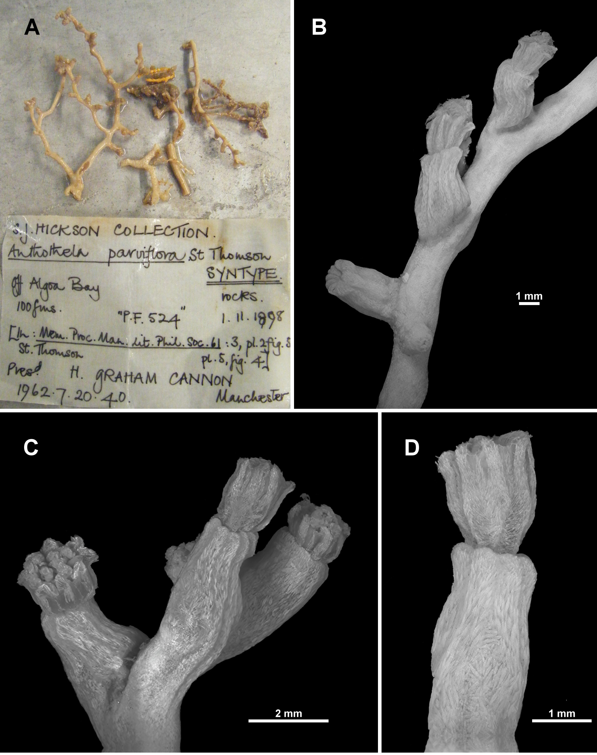
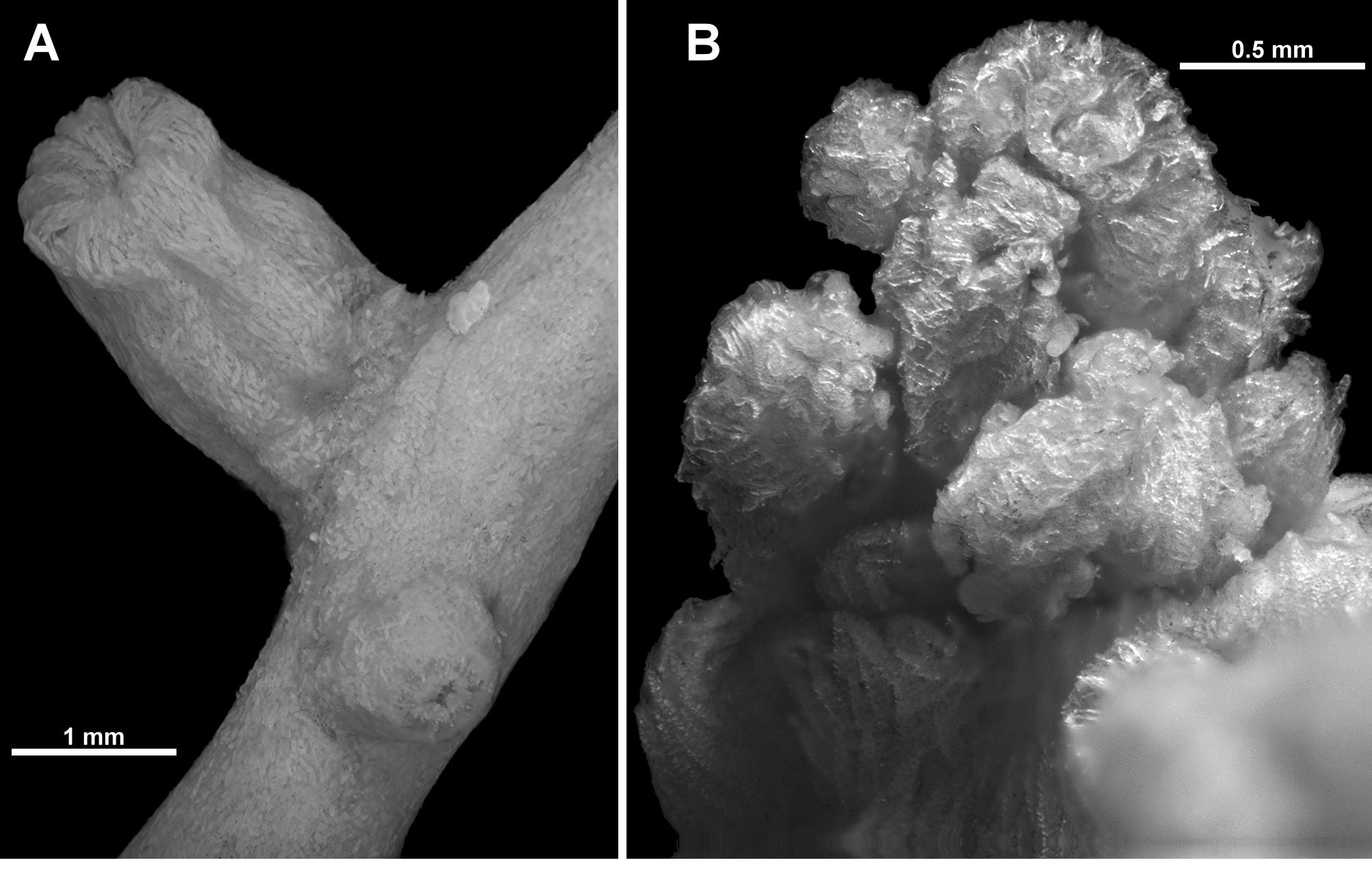
A complete taxonomic revision of the genus Anthothela (Anthothelidae) and closely related taxa is presented herein, based on original type material of nominal species and additional specimens from multiple deep-water surveys. A multi-disciplinary approach was used, combining morphological characteristics such as colonial branching patterns, polyp structure, and sclerite form and arrangement, together with phylogenetic reconstructions using two mitochondrial gene regions (mt- MutS and igr1– cox1). The genus Anthothela, with seven nominal species globally, is here divided into four genera, two of which are new. Three of the original species of Anthothela are validated (A. grandiflora Sars, 1856, A. pacifica Kükenthal, 1913 and A. tropicalis Bayer, 1961), Spongioderma (?) vickersi Benham, 1928 is reassigned to Anthothela and two new species, A. aldersladei and A. quattriniae, are described. Anthothela argentea Studer, 1894, A. macrocalyx (Nutting, 1911) and A. nuttingi Bayer, 1956 are reassigned to Victorgorgia López-González & Briand, 2002 and two new species of this genus, V. eminens and V. nyahae are described. A new family, Victorgorgiidae is described for Victorgorgia due to clear morphological and genetic differences from Anthothela, the type genus of Anthothelidae. A new genus, Williamsium (Anthothelidae), is described for A. parviflora Thomson, 1916 which is restricted to South African waters. A number of North Atlantic Ocean specimens that have traditionally been mistaken for Anthothela grandiflora were found to be synonymous with Alcyonium grandiflorum (Tixier-Durivault & d'Hondt, 1974) and a second new genus, Lateothela (Anthothelidae), is erected for these specimens based on morphological and molecular evidence that Alcyonium grandiflorum was incorrectly placed in the genus Alcyonium Linnaeus. There is good congruence between morphological characteristics and molecular data at a generic level but at a species level, the degree of congruence was inconclusive as morphological and genetic variation is very low. Anthothela and Lateothela n. gen. are found to be closely related to some nominal Alcyonium species, and the family Anthothelidae and subfamily Anthothelinae are shown to be paraphyletic. These are the first records of Anthothela and Victorgorgia from Australian waters.

Key words: Cnidaria, Southern Ocean, deep-sea, octocoral, soft coral, gorgonian, Alcyonacea

Moore K M, Alderslade P, Miller K J, plazi (2017). A taxonomic revision of Anthothela (Octocorallia: Scleraxonia: Anthothelidae) and related genera, with the addition of new taxa, using morphological and molecular data. Plazi.org taxonomic treatments database. Checklist dataset https://doi.org/10.11646/zootaxa.4304.1.1 accessed via GBIF.org on 2026-06-16.

CC0Published 12/31/2017View dataset
GBIF Usage Key
132631042
Dataset Key
3b9bd2c4-7dee-46a3-a36d-80077b88a78b
Origin
source
Backbone Key
9592348
Taxon ID
039B87ED3E36FF30FF4BE37E783EDB32.taxon
Last Crawled
6/11/2026
Last Interpreted
6/11/2026