Anthothela grandiflora
(Sars, 1856)
GBIF:132631046
0
Synonyms
ABOUT
Descriptions(6)
Export occurrence data
Darwin Core Archive (ZIP)
CLASSIFICATION
Taxonomic Classification Tree

MULTIMEDIA
Media Files(27)
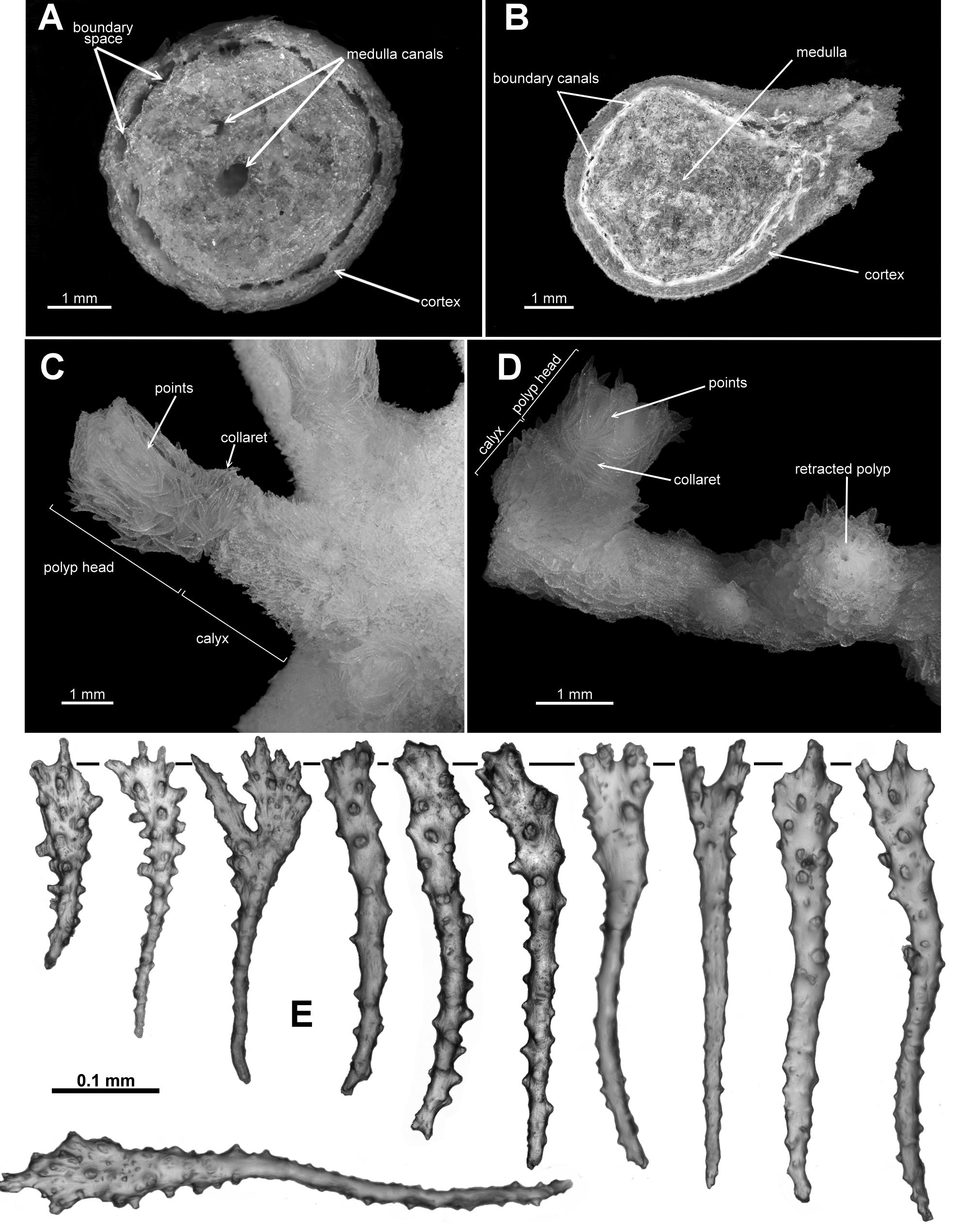
FIGURE 1. A. Cross-section of the holotype of Ƒictorgorgia eminens n. sp. showing extensive boundary canals which frequently anastomose and distinct central canals in the medulla; B. Cross-section of the holotype of Anthothela grandiflora showing boundary canals, adjacent but separate, and a medulla lacking any obvious central canals; C. Extended polyp of the holotype of A. aldersladei n. sp. showing polyp, calyx, points and collaret; D. Partly extended polyp of A. quattriniae n. sp. showing polyp head, calyx, points and collaret and, on right, a fully retracted polyp; E. Spatulate clubs found in Anthothela.
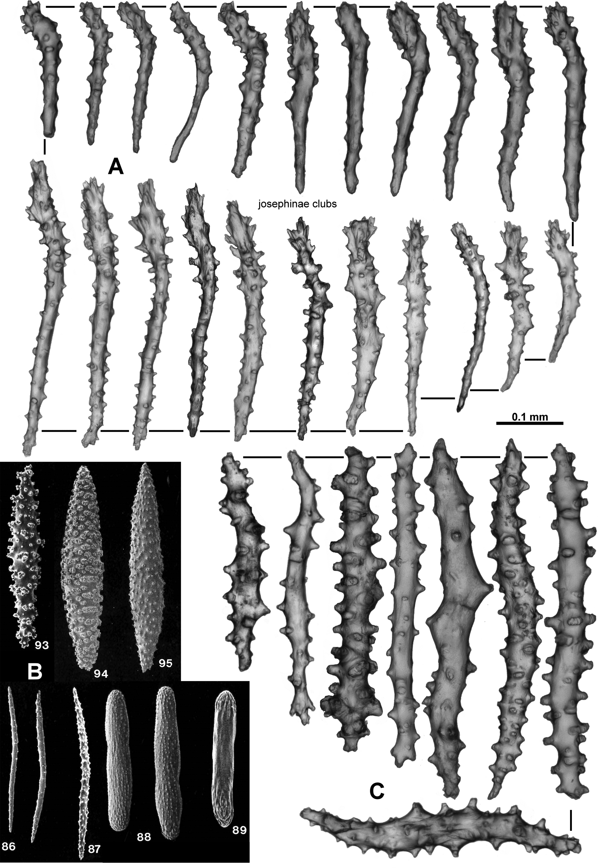
FIGURE 2. A. Josephinae clubs found in the pinnules and tentacle rachis of Ƒictorgorgia and Lateothela n. gen.; B. Spindles (93 – 95), needles (86 – 87) and bars (88 – 89) from Bayer et al. 1983, Plate 16; C. Sticks.
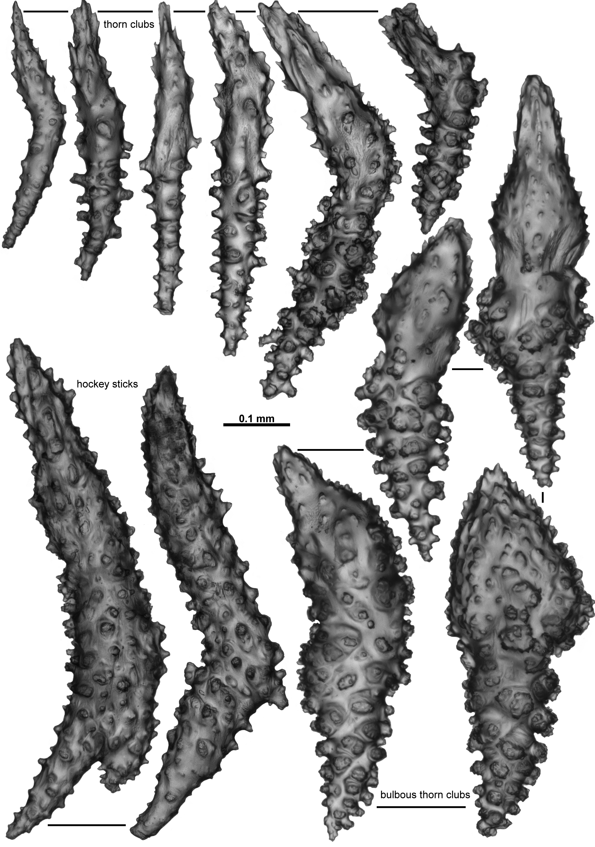
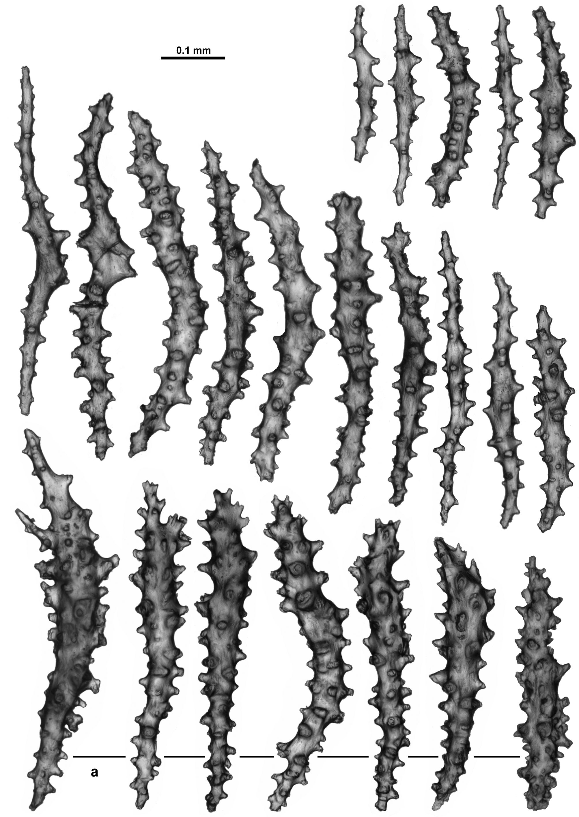
FIGURE 3. Sclerite forms found in the points, calyx and cortex of Anthothela and Ƒictorgorgia species.
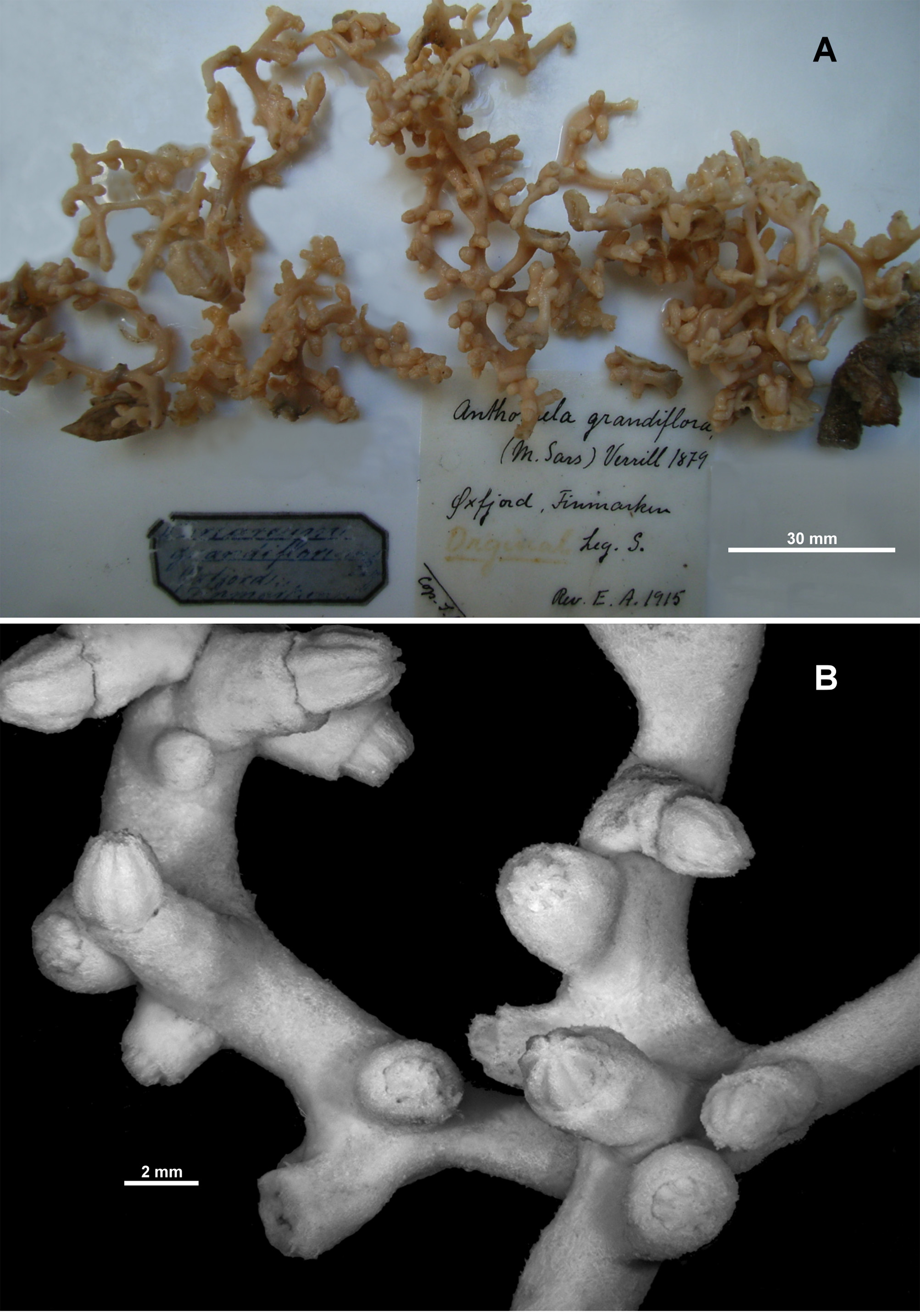
FIGURE 4. Anthothela grandiflora (Sars, 1856), holotype: A. Holotype fragments; B. Polyps and branches.
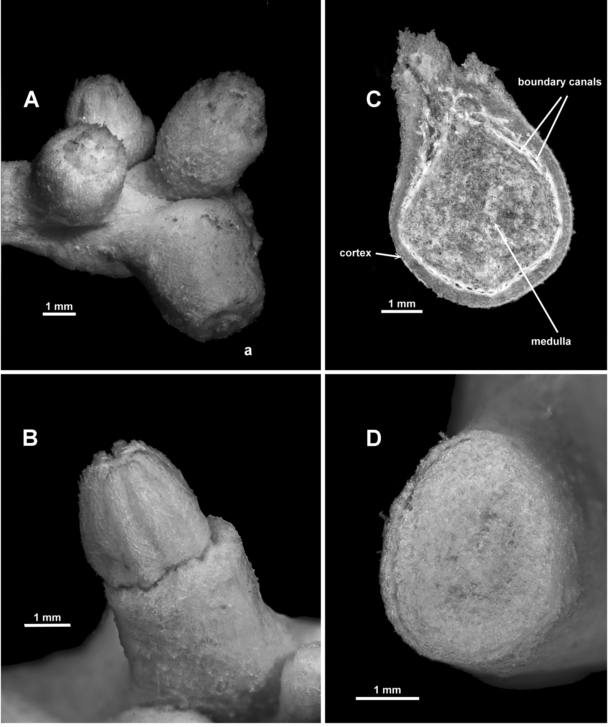
FIGURE 5. Anthothela grandiflora (Sars, 1856), holotype: A. Terminal polyp bunch (a. fully retracted polyp); B. Partly retracted polyp; C. Cross-section of decalcified medulla; D. Cross-section of medulla.
IMAGES