AnimaliaNot EvaluatedacceptedspeciesAccepted
Ascidia interrupta
Heller, 1898
GBIF:141263991
0year
ABOUT
Descriptions(2)
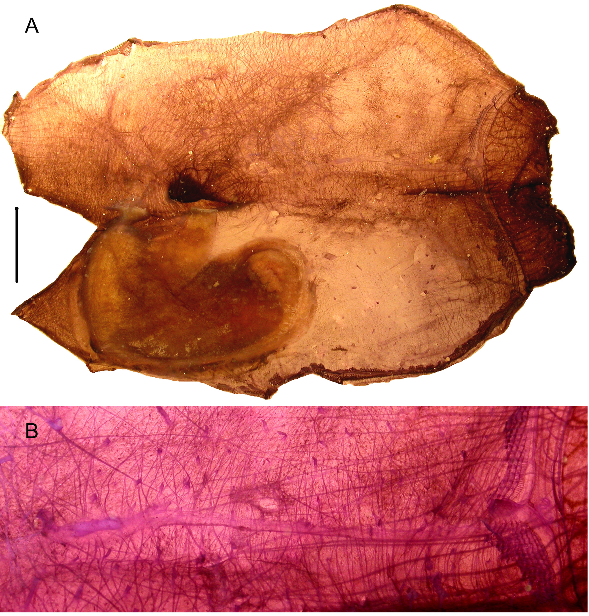
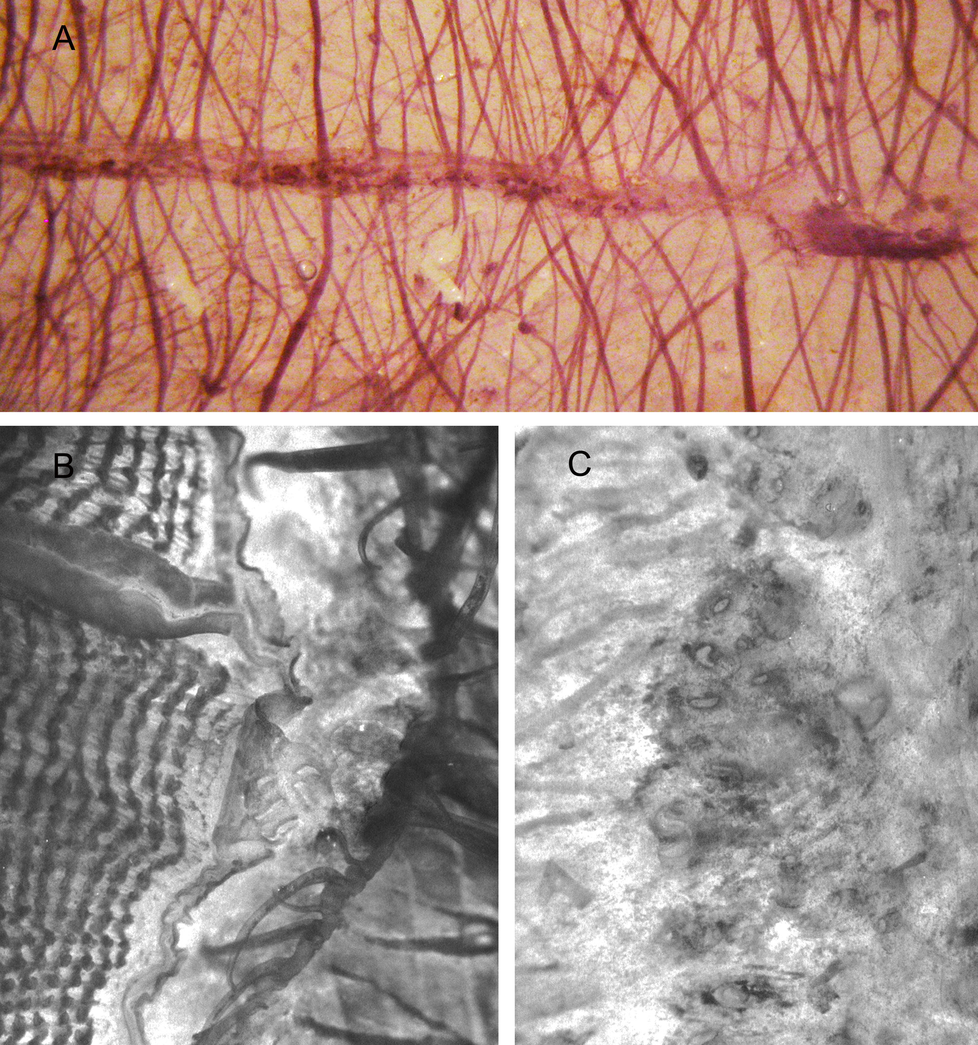
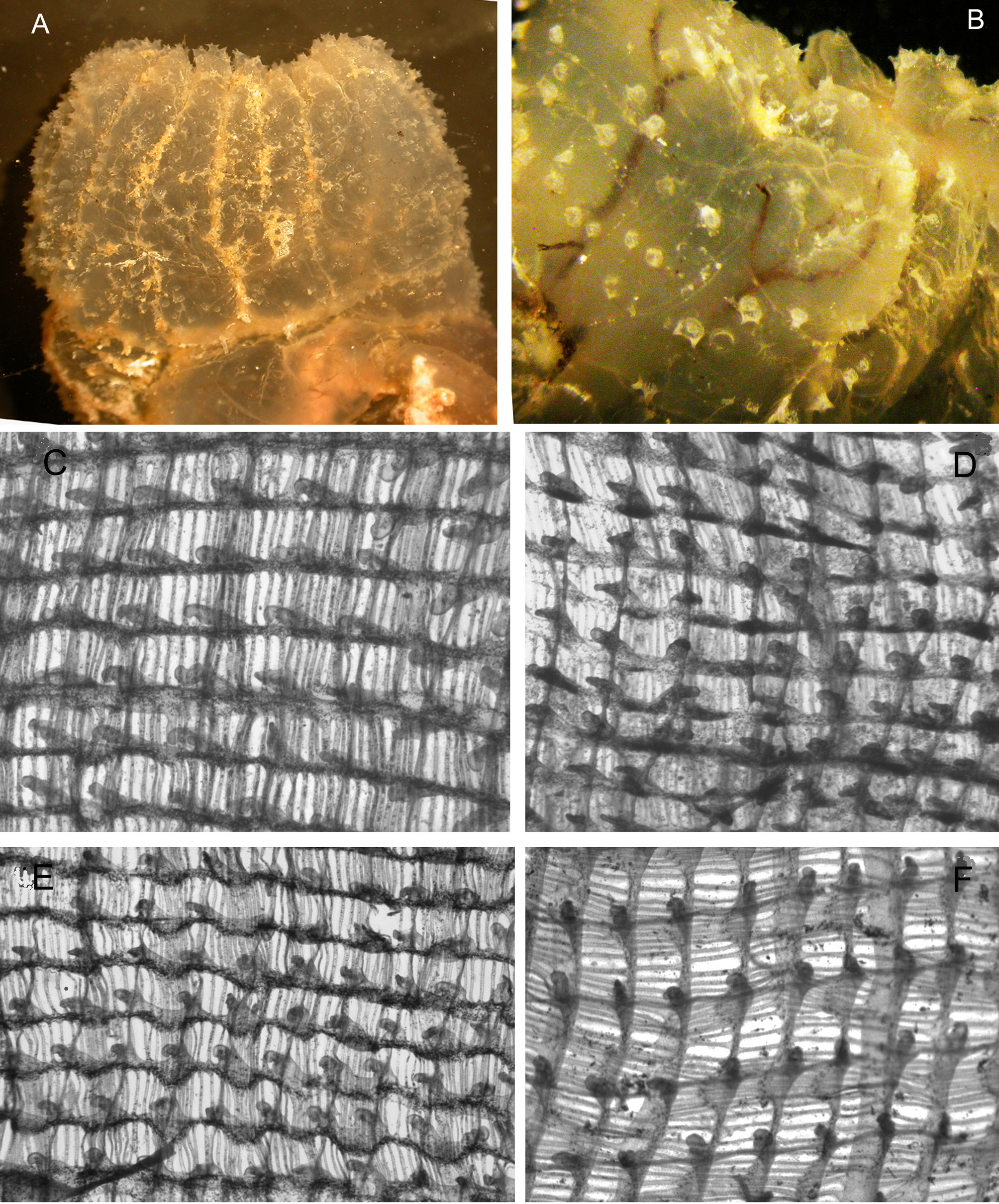
Stations: AB 120; AB 123; AB 149; AB 155; AB 157; AB 163; AB 169; AB 175; AB 187; AB 189; AB 195; AB 197; AR 63; AR 65; AR 69; AR 72; AR 101; AR 103; AR 107; AR 110; AR 118; AR 170; AR 172; AR 177; AR 194; AR 364; AR 415. (MNHN P 5 ASC. A 434 – 437) Synonymy and distribution in Bonnet & Rocha (2011) Numerous ascidians collected by the Madibenthos survey would be normally identified as Ascidia interrupta Heller, 1878 as their anatomy is that generally described in the Caribbean area for this species and its organ variability was emphasised by Van Name (1945). But in some Martinique specimens the structure of the neural gland has several openings along the duct and that would better correspond to the diagnosis of the genus Phallusia. So a question arises about the genus for the Martinique samples. The presence of more than one species also might be possible. In my opinion, for the Madibenthos specimens, the different neural gland structures are here considered as a variability inside the species A. interrupta as all other anatomical characters are more or less variable and particularly the tunic surface with or without papillae. These variations are not linked together among the different specimens, they can occur in the same station and their degree is progressive. The anatomical description below will detail the differences observed between 40 specimens dissected and stained with hemalum. Description. The body shape mostly depends on how the specimens were attached to the substrate, by the whole ventral side or the posterior end or if they were partially burrowed. The tunic is stiff, thick, cartilaginous with brown vessels in its thickness (Fig. 4 A). The surface has irregular grooves and swellings and often contains epibionts and sand. Some specimens are covered by large spiny papillae (Fig. 5 A; 6 A; 8 A, B), others have only sparse very small pointed papillae, many specimens have no papillae at all. The tunic papillae have 3 to 4 short soft spines at the top (Fig. 8 A, B). The oral siphon of fixed specimens remains wide open, it is apical and sometimes shows 8 external ridges but internally the rim is divided into 18 to 22 lobes with a black spot between them. The atrial siphon is sessile at mid distance of the body length. Extracted from the tunic the body has a yellowish colour with dark brown pigment on the siphons and irregular dark patches on the body wall (Figs. 4 A; 6 B, C) .. The thin and numerous oral tentacles are dark in 3 orders of size but vary in number. The prepharyngeal area is narrow and has no papillae. The prepharyngeal groove has 2 blades which do not curve on the dorsal line. The neural ganglion is placed near the atrial aperture (Fig. 6 B). The neural gland lies above the ganglion. In most of the specimens the duct of the neural gland is straight without secondary apertures (Fig. 4 B) and ends anteriorly in a U-shaped dorsal tubercle (Fig 5 C). In other specimens there are no accessory apertures along the duct but instead multiple grouped openings take the place of the dorsal tubercle (Fig; 7 B). In a few specimens some or numerous apertures are lined along the neural gland duct (Fig. 5 B; 6 B; 7 A, C). This variability of the neural duct is not linked to the animal size or to the differences in the tunic surface. The dorsal lamina begins by two long blades prolonged posteriorly as single membrane with ribs ending in small papillae. The branchial tissue has the same structure in all specimens (Fig. 8 C, D, E, F) with very numerous longitudinal vessels some of them oblique, wearing spoon-like papillae which irregularly have a button-like protuberance at the base. The meshes have an irregular size and contain an average of 5 to 8 stigmata or occasionally more. There are no intermediate papillae along the longitudinal vessels. The branchial tissue contains dark pigment irregularly distributed and particularly abundant in the wider transverse vessels. The branchial sac is prolonged far below the gut. The musculature has exactly the same design in all specimens (Figs 4 A; 6 B). The longitudinal fibres of the oral siphon are thin, not grouped in bundles; they are shortly prolonged on the left side but on the right they are longer. Dense transverse muscles extend the whole width of the right body side, the fibres being thicker dorsally and ventrally. Oblique fibres start from the base of the atrial siphon and spread in a fan on the half posterior right side and cross the transverse muscles (Fig. 6 B). On the left side close to the ventral line short transverse fibres form a narrow ribbon which does not reach the gut level. The atrial siphon has numerous but thin circular muscles and a few strong but short longitudinal fibres (Fig. 6 B). The gut occupies the lower half of the left body side (Figs. 4 A; 6, B, C). The stomach is olive shaped with a few indistinct longitudinal folds. The intestine makes two closed loops. The descending limb of the intestine is wide more often inflated in a large pouch densely pigmented in dark brown (Figs 6 B, C). The anus has 2 slightly undulated lobes or a smooth thin rim. The ovary is placed inside the gut loop. Small testis vesicles are spread on the whole intestine. The gonoducts follow the rectum and open together against the anus. A very large blood vessel starting from the oesophagus level (where it contains a round concretion (Fig. 6 B) encircles the gut, follows the ventral line and divides in several branches with one of them larger protruding externally from the body to reach the tunic.
Monniot, Françoise (2018): Ascidians collected during the Madibenthos expedition in Martinique: 1 - Phlebobranchia. Zootaxa 4387 (3): 451-472, DOI: 10.11646/zootaxa.4387.3.3
Remarks. The Madibenthos specimens are assigned here to A. interrupta often recorded in the tropical western Atlantic (references in Bonnet & Rocha 2011) even though some of the characters evoke similarities with Phallusia. All Phallusia known from the Atlantic Ocean differ by several linked characters, a dark brown pigment of all tissues, numerous siphonal lobes, the muscle design, and an inflated intestinal pouch. The other Phallusia species registered in the western Atlantic are: P. caguayensis (Millar & Goodbody, 1974); P. fragilis Bonnet & Rocha, 2011; P. nigra (Savigny, 1816) P. fumigata (Grube, 1864) and P. ingeria (Traustedt, 1883).
Monniot, Françoise (2018): Ascidians collected during the Madibenthos expedition in Martinique: 1 - Phlebobranchia. Zootaxa 4387 (3): 451-472, DOI: 10.11646/zootaxa.4387.3.3
Export occurrence data
Darwin Core Archive (ZIP)
CLASSIFICATION
Taxonomic Classification Tree

MULTIMEDIA
Media Files(5)
FIGURE 4. Ascidia interrupta (st. AB 175): A, body ventrally opened without stain (scale 1cm); B, duct of the neural gland (stained)
Imageimage/png© Monniot, FrançoiseMonniot, Françoise
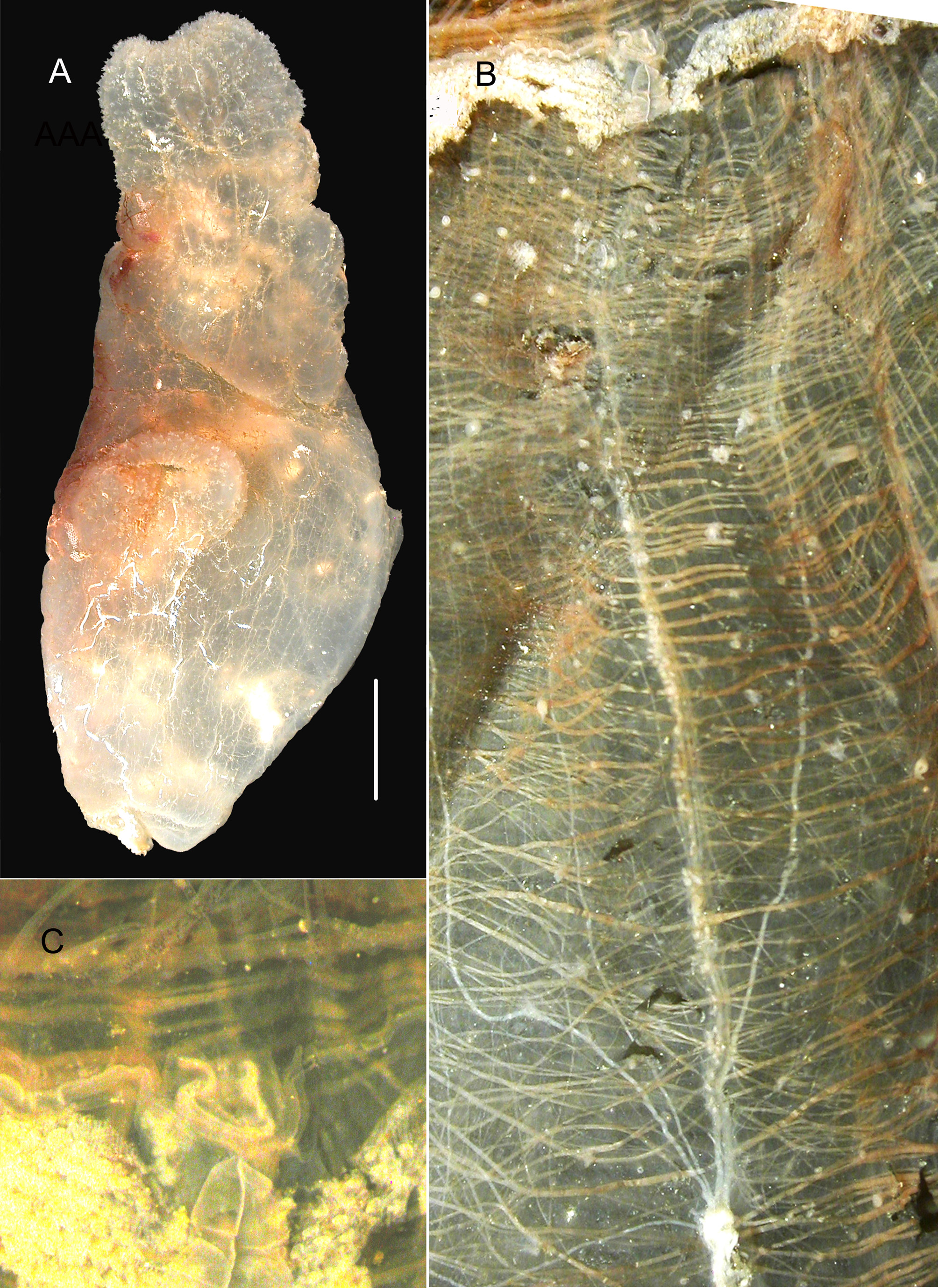
FIGURE 5. Ascidia interrupta (st. AR 69). A, body; B, apertures of the neural gland duct; C, dorsal tubercle.
Imageimage/png© Monniot, FrançoiseMonniot, Françoise
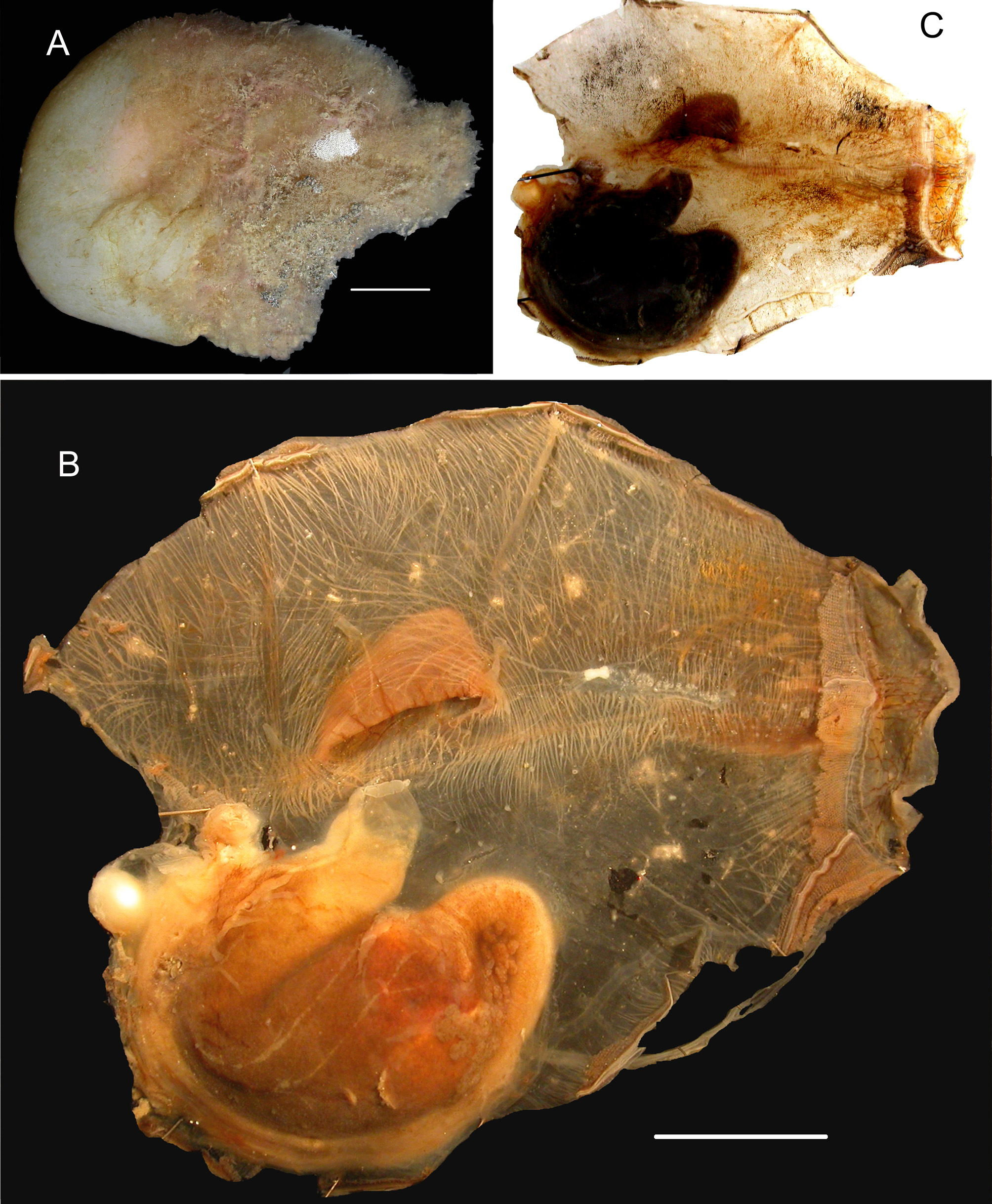
FIGURE 6. Ascida interrupta (st. AR 415): A body; B, neural area and muscles; C, other specimen ventrally opened. Scale bars = 1cm.
Imageimage/png© Monniot, FrançoiseMonniot, Françoise
IMAGES