AnimaliaNot EvaluatedacceptedspeciesAccepted
Ecteinascidia minuta
Traustedt, 1882
GBIF:141264000
0year
ABOUT
Descriptions(1)
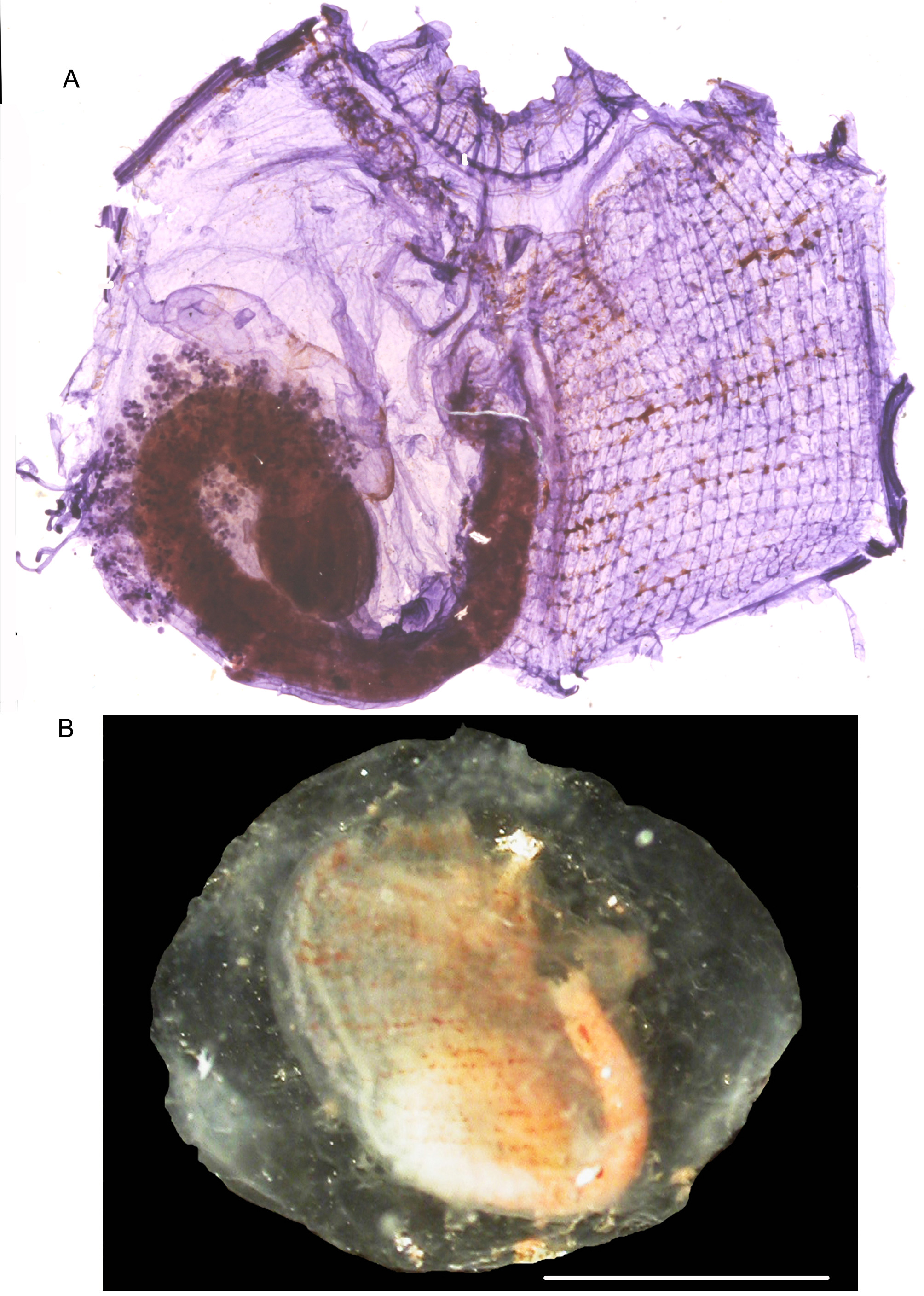
Stations: AS 66; AR 132; AR 309. (MNHN P 4 COR. A 72) Monniot F. 2016: and synonymy Attached by the right body side to algae or to corals this ascidian has a transparent cartilaginous tunic (Fig. 16 B) with siphons opening at 90 ° to each other. Some brown pigment remained in a specimen fixed with formalin on the siphons and the branchial sac. There are 8 oral lobes with ocelli. Twenty-eight tentacles of two orders of size are separated by small buttons. The prepharyngeal groove is not dorsally indented. The dorsal tubercle opens in a slit. The dorsal lamina has triangular languets of increasing length posteriorly. The branchial tissue (Fig. 16 A) contains 35 longitudinal vessels on the right side of the largest specimen of 12 mm. The spiral stigmata are very regular. The gut forms a wide loop on half of the right body side (Fig. 16 A). The stomach shows 5 folds seen by the internal side and has a caecum. The anus his smooth with 2 lips. The ovary consists of several lobes, some of them around the gut (Fig. 16 A) and others lie on the intestinal loop mixed with testis vesicles. The musculature is weak on the siphons; on the body it is reduced to a few longitudinal short fibres beginning at the prepharyngeal level and do not extend on the body sides. A few short muscles are on the sides of the atrial opening. All descriptive elements are the same for specimens recorded in the tropical Atlantic, Indian or Pacific Oceans (Monniot F. 2016) but the origin of this species is unknown.
Monniot, Françoise (2018): Ascidians collected during the Madibenthos expedition in Martinique: 1 - Phlebobranchia. Zootaxa 4387 (3): 451-472, DOI: 10.11646/zootaxa.4387.3.3
Export occurrence data
Darwin Core Archive (ZIP)
CLASSIFICATION
Taxonomic Classification Tree

IMAGES