
Frontohornera frontalis
Grischenko, Gordon & Melnik, 2018
GBIF:148403713
ABOUT
Descriptions(5)
Export occurrence data
Darwin Core Archive (ZIP)
CLASSIFICATION
Taxonomic Classification Tree

MULTIMEDIA
Media Files(4)
FIGURE 2. Colonies of some cyclostome bryozoans, in vivo, attached to polymetallic nodules. A–E, Pandanipora helix n. gen., n. sp.: A, specimen GLD4–09, Stn 190; B, specimen GLD4–12, Stn 262; C, specimen YMG4–07, Stn 143; D, specimen YMG4–13, Stn 295; E, specimen GLD 4–11, Stn 212. F, Tubuliporina sp. indet., specimen YMG18–01, Stn 7. G, H, Abyssoecia elevata n. gen., n. sp.: G, specimen GLD4–09, Stn 196; H, specimen GLD4–09, Stn 191. I, Discantenna metallica n. sp.: specimen GLD4–11, Stn 224. J, K, Frontohornera frontalis n. gen., n. sp.: J, specimen YMG4–07, Stn 124; K, specimen GLD4–11, Stn 210. L, Alyonushka hystricosa n. gen., n. sp.: specimen GLD4–09, Stn 199. M, Calyssopora volcano n. gen., n. sp.: specimen YMG18–01, Stn 33. N, O, Anyuta anastema n. gen., n. sp.: N, specimen GLD4–09, Stn 180; O, specimen YMG4–06, Stn 71. Scale bars: 1 mm.

FIGURE 16. Frontohornera frontalis n. gen., n. sp. Holotype specimen, ZIRAS 1/50707, respectively seen in A, frontal and B, abfrontal views. Scale bars: 1 mm.

FIGURE 17. Frontohornera frontalis n. gen., n. sp. Holotype, ZIRAS 1/50707, showing details of morphology. A, part of branch distal to gonozooid, showing cancelli and zooidal peristomes; B, part of infertile branch with fewer cancelli; C, D, parts of infertile branch showing peristomial dimorphism; E, part of branch near gonozooid showing numerous cancelli and peristomial dimorphism; F, zooids showing peristomial and apertural dimorphism; G, abfrontal part of branch in vicinity of gonozooid; H, J, K, gonozooid profiles; I, autozooidal peristome showing interior spinules; L, ooeciostome and ooeciopore. Scale bars: A, B, D, E, G, 300 µm; C, H, J, K, 200 µm; F, 150 µm; I, 100 µm; L, 50 µm.
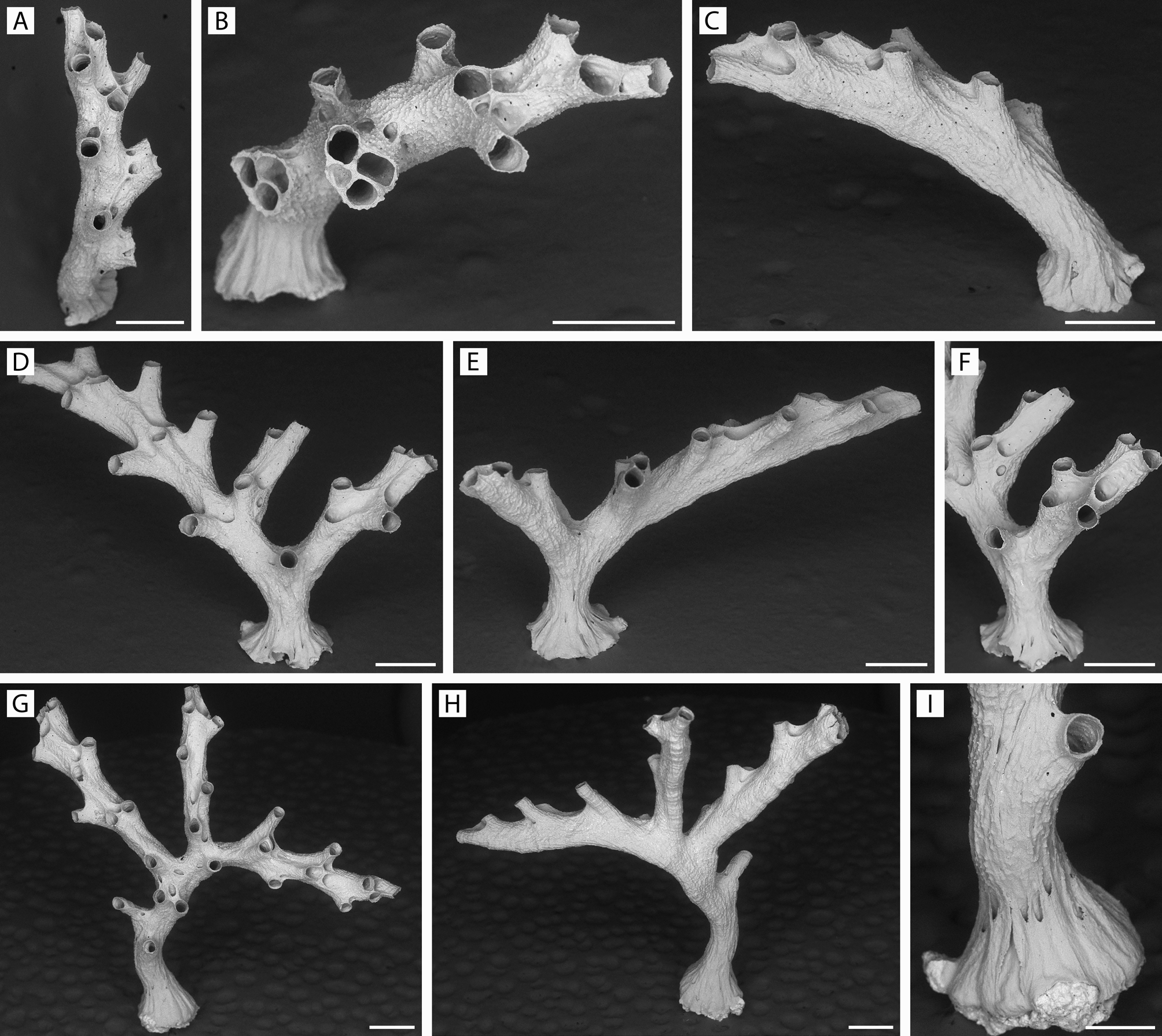
FIGURE 18. Frontohornera frontalis n. gen., n. sp. Progressive stages of development of young colonies. A–C, specimen GLD4–11, Stn 215, from different angles; D–F, specimen YMG4–14, Stn 334, from different angles; G–I, specimen GLD4–11, Stn 210, in frontal and abfrontal views and a close-up of the colony base with secondary calcification. Scale bars: A–H, 500 µm; I, 250 µm.
IMAGES