
Discantenna metallica
Grischenko, Gordon & Melnik, 2018
GBIF:148403720
ABOUT
Descriptions(5)
Export occurrence data
Darwin Core Archive (ZIP)
CLASSIFICATION
Taxonomic Classification Tree

MULTIMEDIA
Media Files(4)
FIGURE 2. Colonies of some cyclostome bryozoans, in vivo, attached to polymetallic nodules. A–E, Pandanipora helix n. gen., n. sp.: A, specimen GLD4–09, Stn 190; B, specimen GLD4–12, Stn 262; C, specimen YMG4–07, Stn 143; D, specimen YMG4–13, Stn 295; E, specimen GLD 4–11, Stn 212. F, Tubuliporina sp. indet., specimen YMG18–01, Stn 7. G, H, Abyssoecia elevata n. gen., n. sp.: G, specimen GLD4–09, Stn 196; H, specimen GLD4–09, Stn 191. I, Discantenna metallica n. sp.: specimen GLD4–11, Stn 224. J, K, Frontohornera frontalis n. gen., n. sp.: J, specimen YMG4–07, Stn 124; K, specimen GLD4–11, Stn 210. L, Alyonushka hystricosa n. gen., n. sp.: specimen GLD4–09, Stn 199. M, Calyssopora volcano n. gen., n. sp.: specimen YMG18–01, Stn 33. N, O, Anyuta anastema n. gen., n. sp.: N, specimen GLD4–09, Stn 180; O, specimen YMG4–06, Stn 71. Scale bars: 1 mm.
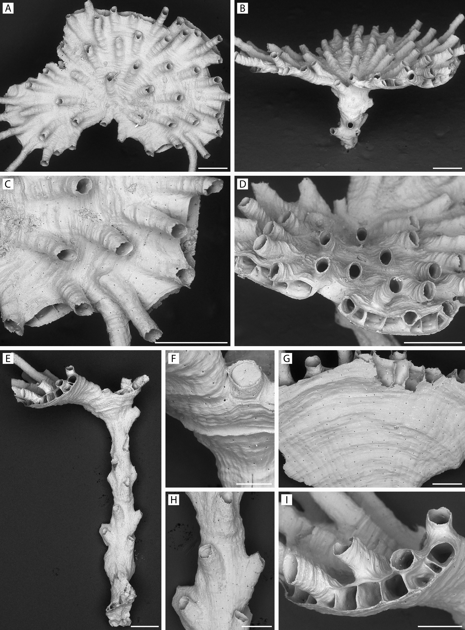
FIGURE 10. Discantenna metallica n. sp. Holotype and paratype colonies. A–D, holotype, ZIRAS 1/50675; E–I, paratype 1, ZIRAS 2/50676. A, apical and B, lateral views of colony; C, D, same, edge of disk, respectively showing peristomes and differentiating zooids; E, lateral view of colony showing numerous stalk zooids; F, autozooidal peristome near top of stalk with calcified terminal diaphragm; G, underside of part of capitulum seen in E; H, part of stalk with short peristomes and terminal diaphragms; I, margin of capitulum seen in E. Scale bars: A–E, 500 µm; F, 150 µm; G–I, 250 µm.
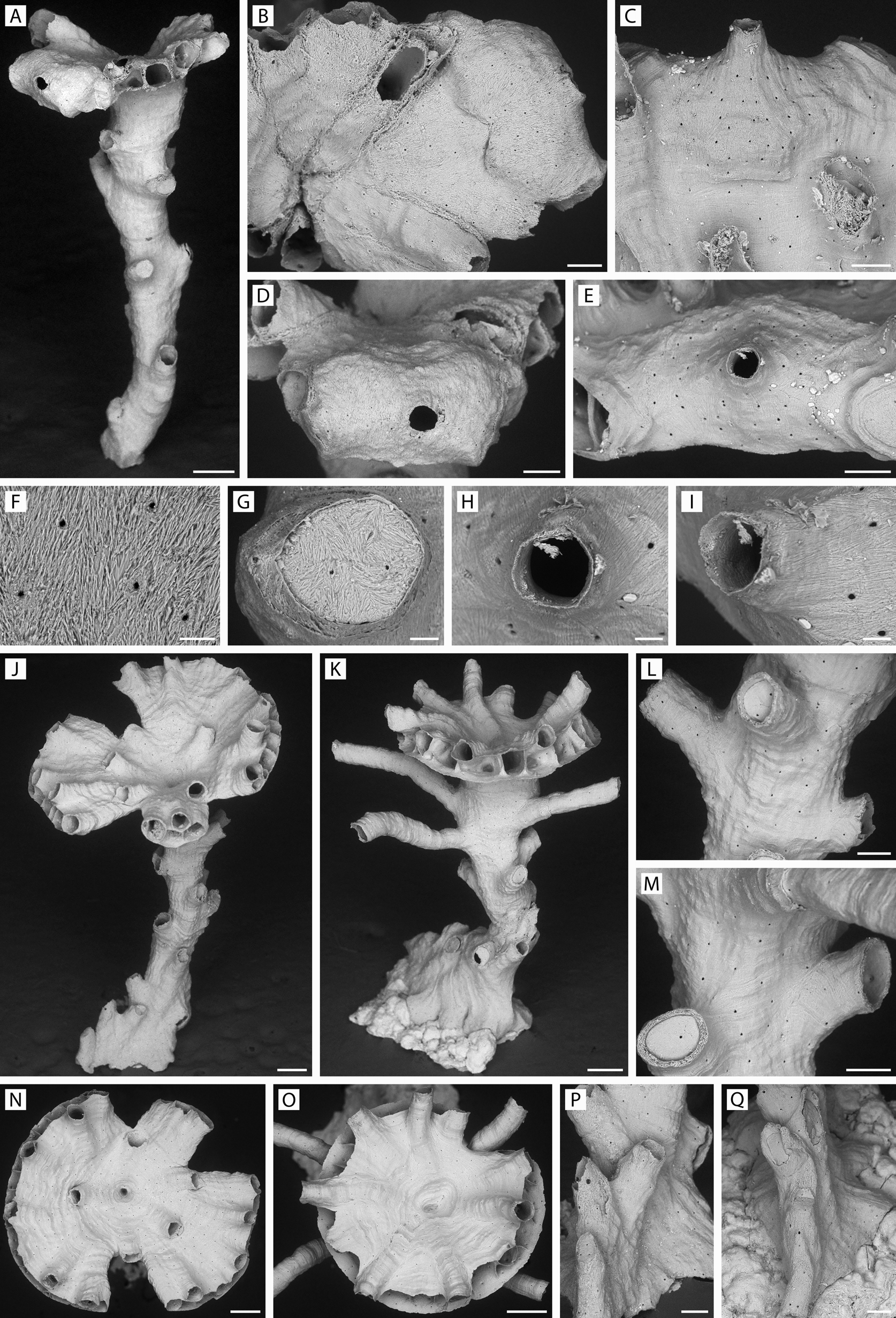
FIGURE 11. Discantenna metallica n. sp. Paratype and other colonies. A, B, D, F, G, paratype 2, ZIRAS 3/50702; C, E, H, I, paratype 1, ZIRAS 2/50676; J, L, N, P, specimen YMG4–06, Stn 69; K, M, O, Q, specimen GLD4–08, Stn 151. A, fertile colony with broken base and small capitulum; B, D, same, showing gonozooid in frontal and apertural views; C, E, a second gonozooid with longer ooeciostome; F, G, high- and lower-magnification views of terminal diaphragm, showing skeletal microstructure; H, I, ooeciopores of gonozooid in C and E; J, N, lateral and apical views of colony with small lobate capitulum; K, O, lateral and apical views of another colony with a small capitulum that is more symmetrical; L, M, parts of stalks with short autozooidal peristomes and terminal diaphragms; P, Q, proximal parts of two colonies in post-ancestrular region. Scale bars: A, J, K, N, O, 250 µm; B–E, L, M, P, Q, 100 µm; F–I, 25 µm.
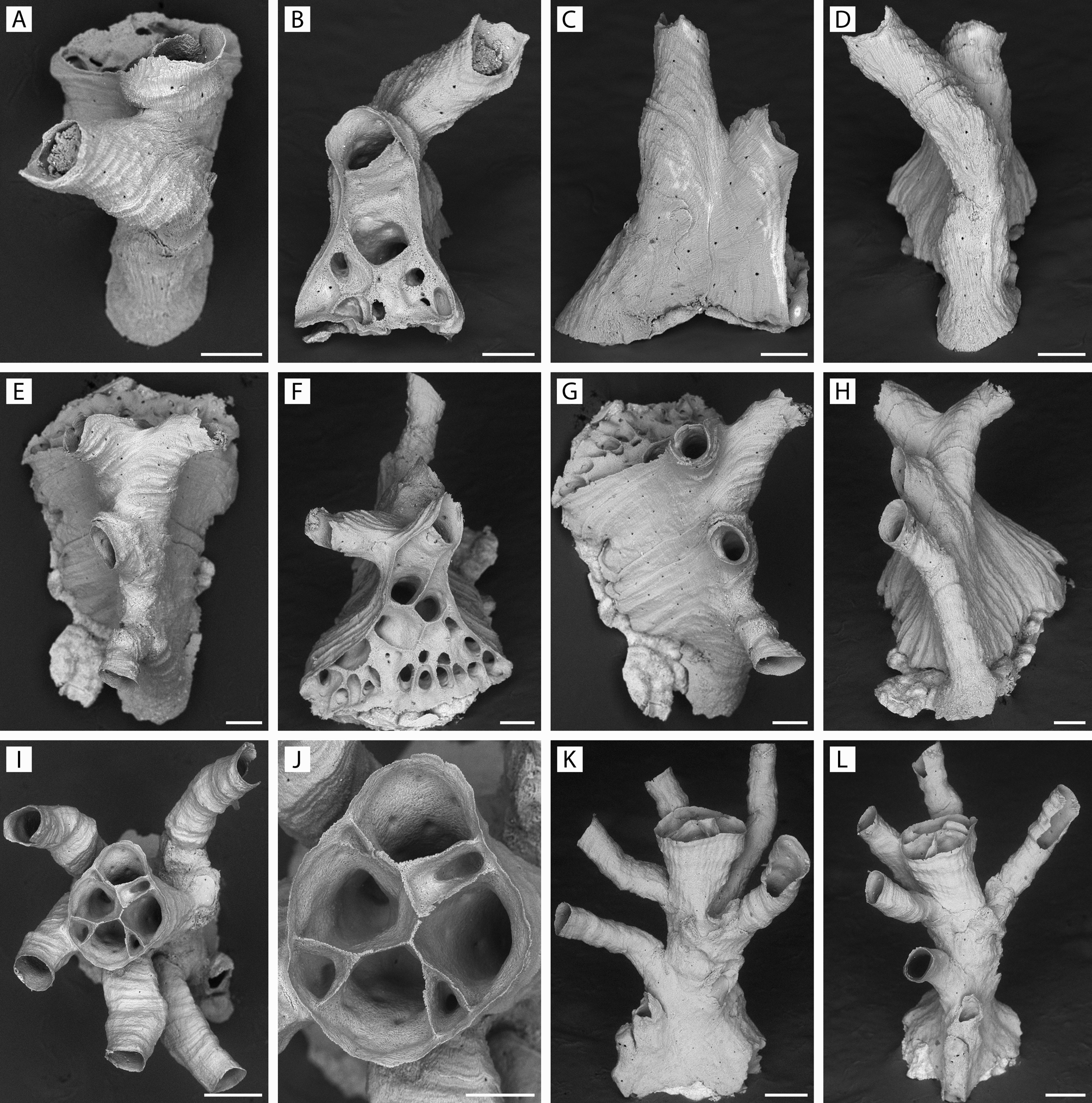
FIGURE 12. Discantenna metallica n. sp. Progressive stages of development of ancestrulate and young colonies. A–D, specimen YMG4–14, Stn 358; E–H, specimen GLD4–11, Stn 224; I–L, specimen GLD4–11, Stn 207. A–D, rotational views (respectively frontal, distolateral, lateral, proximal) of newly formed colony with ancestrular zooid and developing daughter peristome; note developing kenozooids in B; E–H, similar views of a slightly older colony with further kenozooidal development in F; I–L, apical and profile views of an older colony (ancestrular protoecium missing) with a definite stalk underway and peristomes opening all around it. Scale bars: A–H, J, 100 µm; I, K, L, 200 µm.
IMAGES