
Anyuta anastema
Grischenko, Gordon & Melnik, 2018
GBIF:148403736
ABOUT
Descriptions(5)
Export occurrence data
Darwin Core Archive (ZIP)
CLASSIFICATION
Taxonomic Classification Tree

MULTIMEDIA
Media Files(5)
FIGURE 2. Colonies of some cyclostome bryozoans, in vivo, attached to polymetallic nodules. A–E, Pandanipora helix n. gen., n. sp.: A, specimen GLD4–09, Stn 190; B, specimen GLD4–12, Stn 262; C, specimen YMG4–07, Stn 143; D, specimen YMG4–13, Stn 295; E, specimen GLD 4–11, Stn 212. F, Tubuliporina sp. indet., specimen YMG18–01, Stn 7. G, H, Abyssoecia elevata n. gen., n. sp.: G, specimen GLD4–09, Stn 196; H, specimen GLD4–09, Stn 191. I, Discantenna metallica n. sp.: specimen GLD4–11, Stn 224. J, K, Frontohornera frontalis n. gen., n. sp.: J, specimen YMG4–07, Stn 124; K, specimen GLD4–11, Stn 210. L, Alyonushka hystricosa n. gen., n. sp.: specimen GLD4–09, Stn 199. M, Calyssopora volcano n. gen., n. sp.: specimen YMG18–01, Stn 33. N, O, Anyuta anastema n. gen., n. sp.: N, specimen GLD4–09, Stn 180; O, specimen YMG4–06, Stn 71. Scale bars: 1 mm.

FIGURE 39. Anyuta anastema n. gen., n. sp. A–D, holotype, ZIRAS 1/50718, in apical and lateral profiles; note the clusters of fascicles, at least three of which have dimorphic orifices with terminal flanges (dimorphic orifices arrowed). Scale bars: 500 µm.
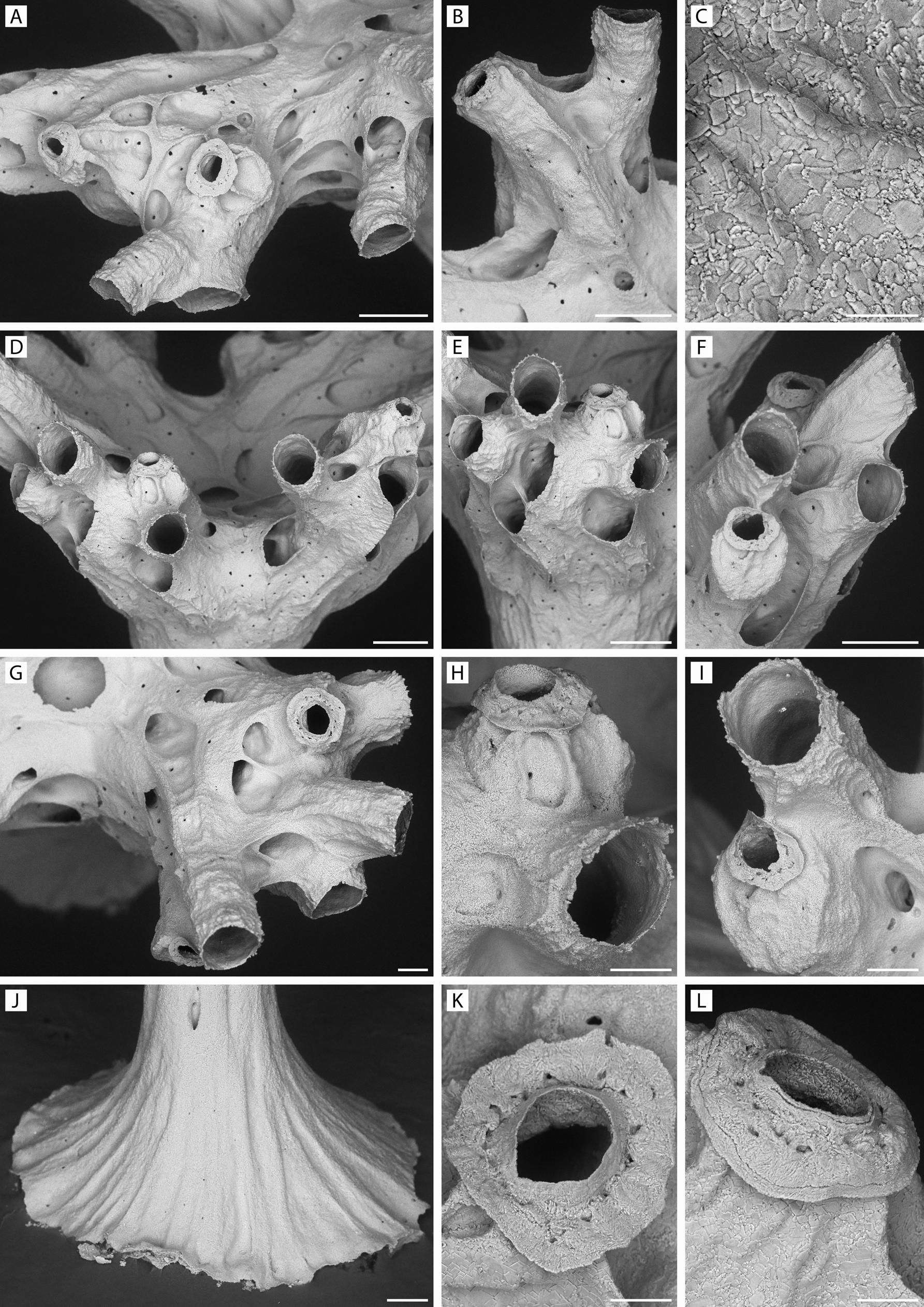
FIGURE 40. Anyuta anastema n. gen., n. sp. Holotype, ZIRAS 1/50718. A, part of fascicle with autozooidal peristomes and two dimorphic peristomes with flanges; note large shallow alveoli on sides of peristomes; B, two peristomes, left one dimorphic; C, skeletal microstructure of part of peristome in L; D–G, subfascicles with both autozooidal and flanged dimorphic peristomes; H, close-up of squat flanged dimorphic peristome in E; note small alveoli on flanks of peristome; I, close-up of lower dimorphic peristome in F, showing it to be derived from an autozooidal peristome by its partial closure; J, base of column; K, L, close-ups of perforated flanges of dimorphic peristomes. Scale bars: A, B, D, F, 250 µm; G–J, 100 µm; K, L, 50 µm; C, 25 µm.

FIGURE 41. Anyuta anastema n. gen., n. sp. Progressive stages of development of young colonies. A–D, specimen YMG4–14, Stn 360, three-zooid stage; E–H, specimen YMG4–14, Stn 338, five–six-zooid stage; I–L, specimen YMG4–13, Stn 281, and M–P, specimen YMG4–06, Stn 71, showing later stages in which trabecular walls define spaces destined to become either autozooids or alveoli. Scale bars: 200 µm.
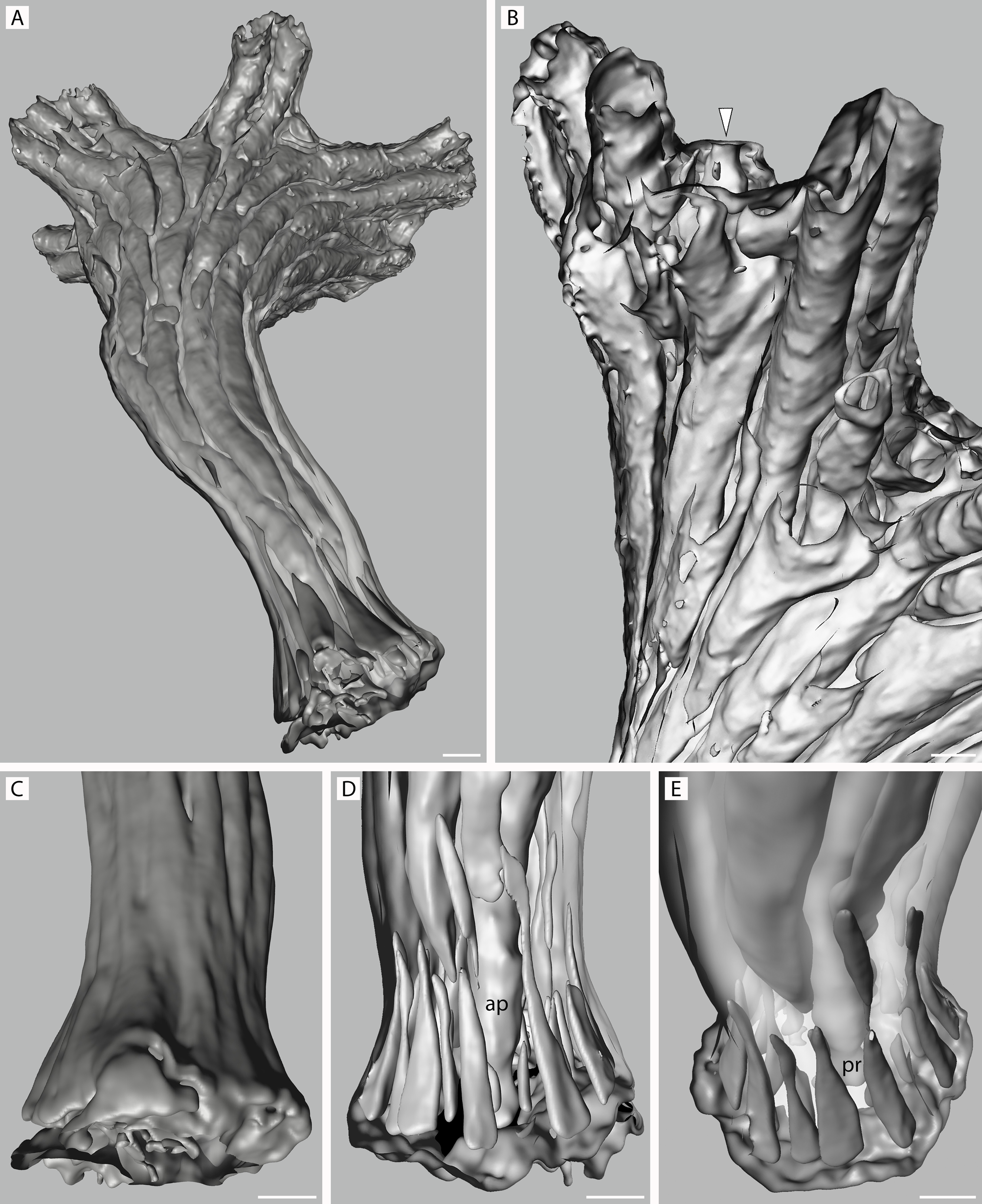
FIGURE 53. Anyuta anastema n. gen., n. sp. Micro-CT scans of paratype, NIWA 127725, as surface (C) and back-face isosurface renders showing colony interiors. A, view of colony from below; note absence of kenozooids from most of column; B, lateral abfrontal view of two small fascicles with entrance to dimorphic zooid indicated by arrowhead; note relative paucity of communication pores; C–E, exterior and interior views of colony base, showing lateral (C, D) and frontolateral (E) profiles with palisade of elongate-triangular kenozooidal chambers surrounding ancestrular protoecium (pr) and proximal part of its peristome (ap). Scale bars: 100 µm.
IMAGES