
Calyssopora clarionensis
Grischenko, Gordon & Melnik, 2018
GBIF:148403739
ABOUT
Descriptions(5)
Export occurrence data
Darwin Core Archive (ZIP)
CLASSIFICATION
Taxonomic Classification Tree

MULTIMEDIA
Media Files(3)
FIGURE 33. Calyssopora clarionensis n. gen., n. sp. General view of holotype and paratype colonies. A, holotype, ZIRAS 1/ 50719, with fully hooded ooeciostome; B, paratype 1, ZIRAS 2/50720, with mostly hooded ooecostome; C, paratype 2, ZIRAS 3/50721, with ooeciopore visible; D, paratype 3, ZIRAS 4/50722, with partially developed ooeciostome not yet concealing ooeciopore. Scale bars: 250 µm.
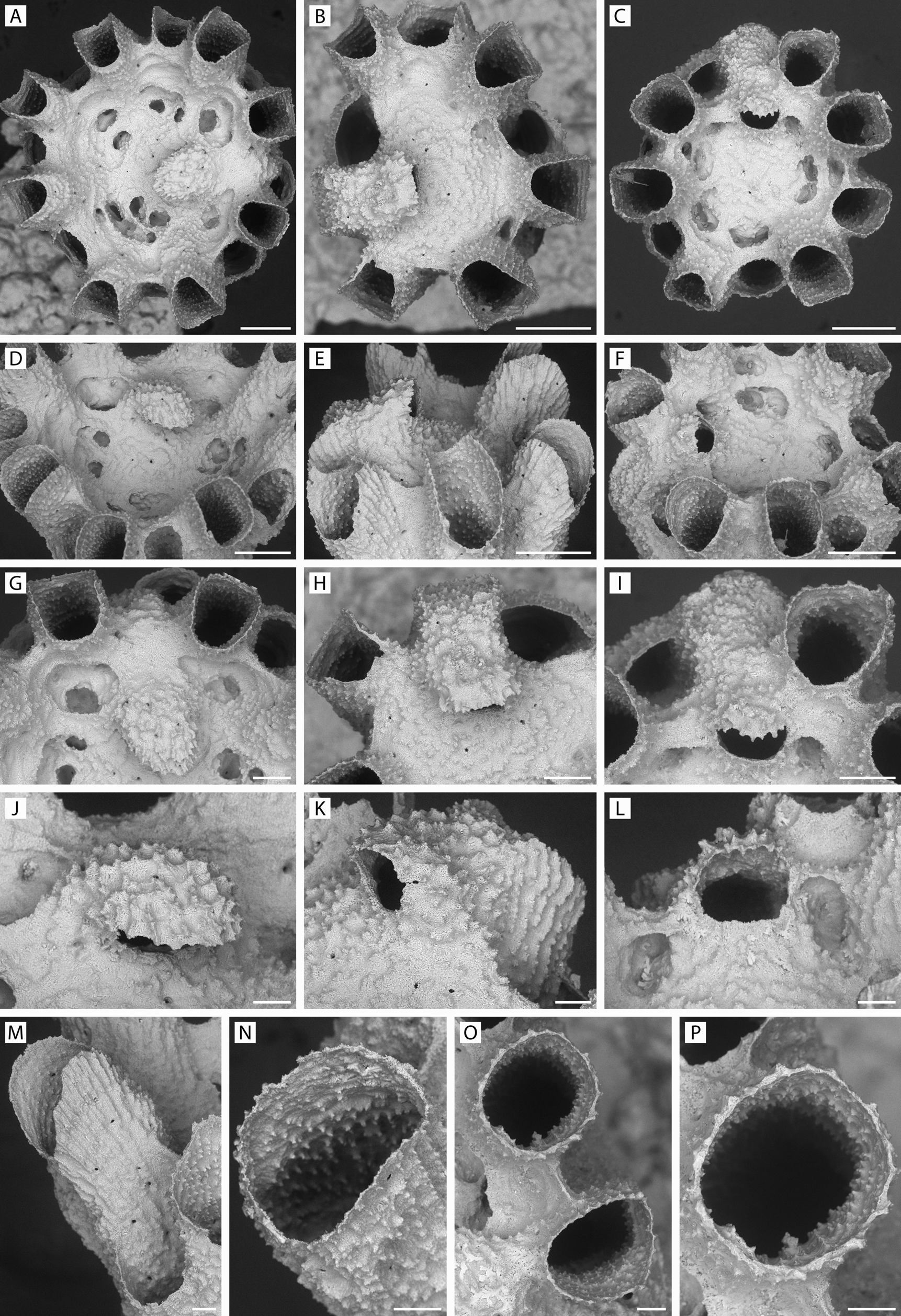
FIGURE 34. Calyssopora clarionensis n. gen., n. sp. A, D, G, J, holotype, ZIRAS 1/50719, respectively showing an apical view of the fertile colony and profiles of the hooded ooeciostome; B, E, H, K, paratype 2, ZIRAS 3/50721, respectively showing an apical view of the gonozooid and profiles of a more-truncated ooeciostome; C, F, I, L, paratype 3, ZIRAS 4/50722, similar views of a colony with a more-open ooeciostome; M, specimen YMG4–13, Stn 287, mitriform shape of autozooecial orifice when seen in oblique lateral view; N, specimen GLD4–11, Stn 209, oblique view of autozooidal peristome and aperture showing spinose granulation of interior and exterior surfaces; O, P, paratype 1, ZIRAS 2/50720, respectively showing trabecula joining two peristomes and vertical view of autozooidal aperture. Scale bars: A–F, 200 µm; G–I, 100 µm; J–P, 50 µm.
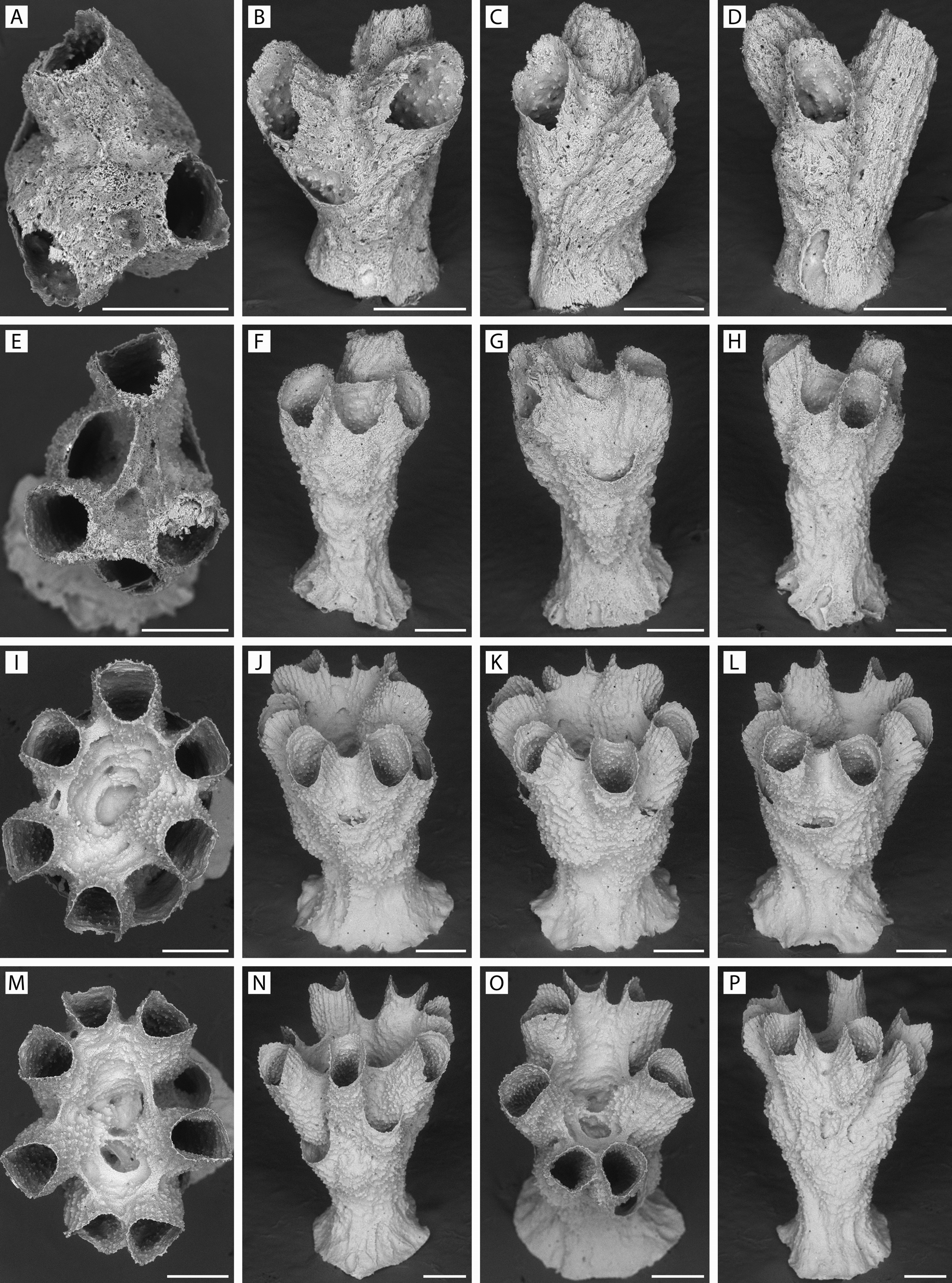
FIGURE 35. Calyssopora clarionensis n. gen., n. sp. Progressive stages of development of ancestrular and young colonies. A–D, specimen YMG18–01, Stn 24, three-zooid colony; E–H, specimen GLD4–08, Stn 144, four–five-zooid stage; I–L, specimen YMG4–07, Stn 134, seven-zooid stage with central cavity presumably representing incipient incubation chamber; M–P, specimen GLD4–11, Stn 209, eight-zooid stage with paired central cavities. Scale bars: 200 µm.
IMAGES