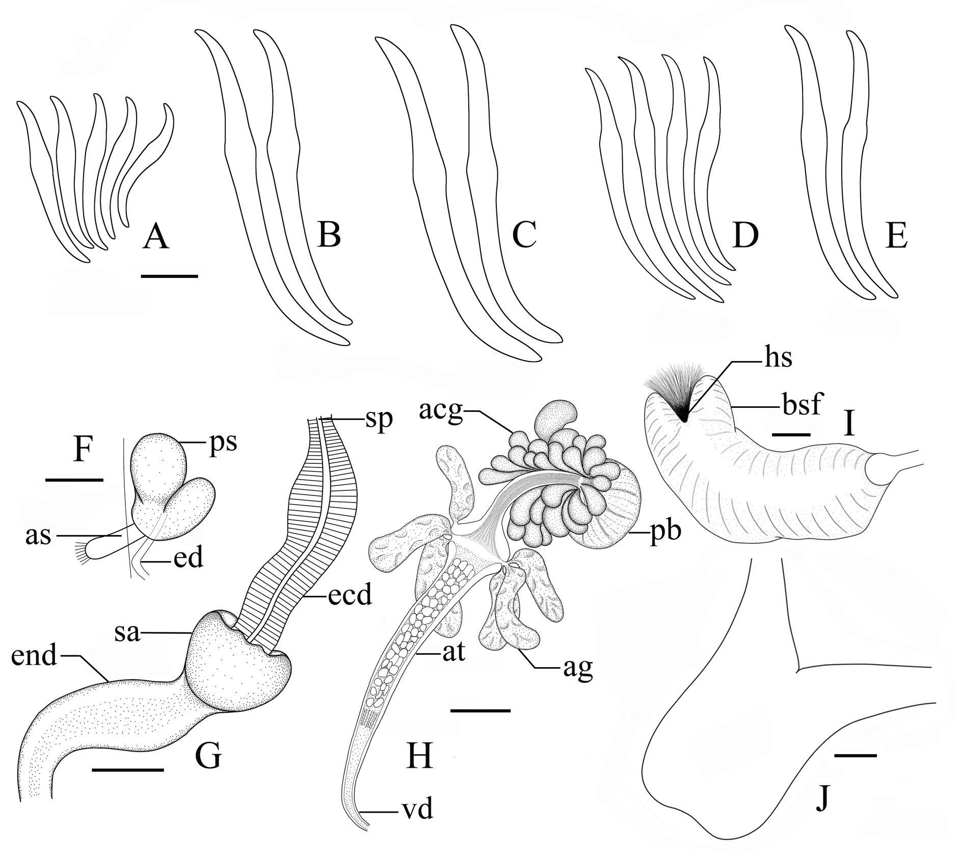
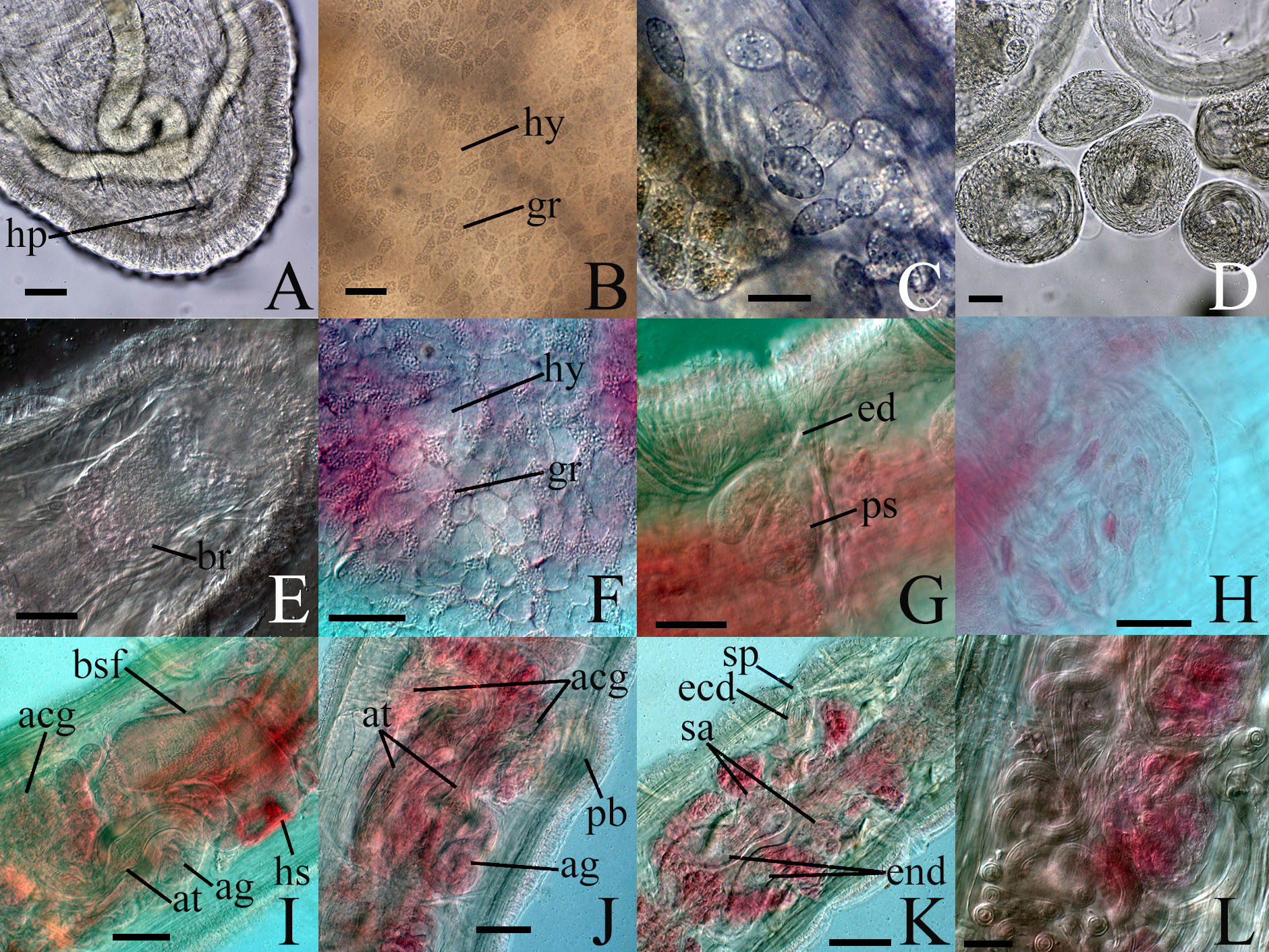
Description. Small worms, active, twisting when submerged in water, pale in vivo, body wall thin, transparent. Body length 6.5 – 11.5 mm (holotype 8.0 mm), 7.0 – 12.5 mm in vivo; body width 270 – 355 µm at V (330 – 450 µm in vivo), 470 – 575 µm at clitellum (320 – 625 µm in vivo). Segments 28 – 46 (holotype 34). Head pore transverse slit near the apex of prostomium (Fig. 2 A). Epidermal gland cells inconspicuous, almost absent. Chaetal formula: 2,3 - 3,4: 2 – 5 (6) - 2 – 5 (6), up to 6 per bundle in the last segments. Chaetae sigmoid, nodulated, distally thinner and single-pointed. Lateral chaetae 44 – 100 µm in length and 3.5 – 5 µm in maximal width, shortest in II (44 – 48 µm), gradually increasing in size from III backwards, maximal size in IX (80 – 100 µm) (Table 1). Ventral chaetae, except those in V, VI and XI, 63 – 120 µm in length and 4 – 7.5 µm in maximal width (Fig. 1 A, D). Ventral chaetae in V, VI and XI reduced to 2 per bundle (occasionally 3) (Fig. 1 B, C, E). Ventral chaetae in V and VI longer and stouter than the others even in immature specimens, 126 – 158 µm in length and 7.5 – 11 µm in maximal thickness (Fig. 1 B, C). Ventral chaetae in XI shorter and thinner than in V and VI, but thicker than the ordinary ventral ones, 6 – 8.5 µm in maximal thickness (Fig. 1 E). Chaetae of XII lacking in fully mature specimens. Clitellum extending from 1 / 2 XI to XIII, elevated conspicuously. Hyalocytes (12 – 24 µm in diameter) more abundant than granulocytes, irregularly arranged and in contact with each other. Granulocytes (13 – 22 µm in diameter) irregular in shape, having some connection with each other and forming a loose net (Fig. 2 B, F). Male pores distinct in the ventral middle of XII. Spermathecal pores paired, in the lateral line at IV / V (Fig. 2 K). Brain in I-II (Figs. 1 J, 2 E), trapezoidal, deeply concave anteriorly and slightly incised posteriorly, 137 – 170 µm long and 105 – 125 µm in maximal width. Blood colourless or slightly yellowish. Dorsal vessel arising from XIV – XVI. Two pairs of primary pharyngeal glands in IV and V, attached to septa of 4 / 5 and 5 / 6 respectively, and both not connected dorsally. Three pairs of secondary lobes ventrally in V – VII, largest in VI. No oesophageal appendages. Intestinal diverticula absent. Chloragogen cells developed, originating from VIII, 12 – 45 µm high, absent in clitellum. Coelomocytes abundant, disc-shaped with conspicuous nuclei, 7.5 – 15.0 µm in diameter, with uneven refractile granules (Fig. 2 C). Five pairs of nephridia in preclitellar segments from VI / VII to X / XI. Anteseptale stalked, containing nephrostome only; postseptale bilobed and with fewer interstitial tissue. Efferent duct arising between the two lobes (Figs. 1 F, 2 G). Sperm funnels cylindrical, occupying 1 / 3 – 1 / 2 body cavity in XI, 390 – 440 µm in length and 122 – 200 µm in maximal width. Their ental openings much narrower than funnel body, 43 – 73 µm wide. The heads of spermatozoa 17 – 20 µm in length (Figs. 1 I, 2 I). Vasa deferentia with conspicuous cilia in the canal, wound in irregular spirals in coelom from XII and posteriorly into XV – XXII, 18 – 30 µm in width, lumen of vasa deferentia 14 – 24 µm wide (Fig. 2 L). The transition between vas deferens and atrium gradual. Atria in XII – XIII, cylindrical, remarkably wider than vasa deferentia, 415 – 650 µm long and 65 – 85 µm in maximal width. No cilia observed in atrial canal. Each atrium possessing 4 – 6 tubular prostate-like glands (each 117 – 195 µm in length and 50 – 68 µm in width) (Figs. 1 H, 2 I, J). Each atrium divided into two distinct parts, an ental part (length: 217 – 400 µm; width: 27.5 – 58 µm) with thin muscle layer and thick epithelium, and an ectal part (length: 200 – 350 µm; width: 25 – 44 µm) with a muscular duct connecting with the penial bulb. Penial bulb large, compact in ventral of XII, consisting of muscle strands and surrounded by masses of accessory glands of different size (Figs. 1 H, 2 J). A pair of sperm sacs large from XI – XIV in different size, and backwards extending to XV – XXIV, each containing numerous ball-shaped sperm bundles (diameter: 50 – 110 µm), a large number of spermatozoa coiled inside in vivo but much more loose in fixed condition (Fig. 2 D, H). Egg sacs paired, large from XIV – XXII, continuing caudad to XVII – XXXIII, usually containing 2 – 11 mature eggs at a time. One pair of spermathecae in V – VII, without ectal glands. Ectal duct 215 – 385 µm long and 37 – 50 µm wide, projecting into ampulla. Ampulla spherical, 83 – 102 µm in diameter. No diverticula visible. Ampulla filled with spermatozoa in fully mature specimens. Ental ducts stout, 195 – 244 µm long and 62 – 67 µm wide, connecting to the dorsal side of oesophagus separately in the posterior of VI or anterior of VII (Figs. 1 G, 2 K).
Zhang, Junqian, Lu, Yajing, Xie, Zhicai (2018): Two new Mesenchytraeus species (Annelida: Clitellata: Enchytraeidae) from Changbai Mountain, China. Zootaxa 4496 (1): 382-394, DOI: 10.11646/zootaxa.4496.1.28