Seira
Lubbock, 1870
GBIF:148692409
ABOUT
Descriptions(5)
Export occurrence data
Darwin Core Archive (ZIP)
CLASSIFICATION
Taxonomic Classification Tree

MULTIMEDIA
Media Files(16)
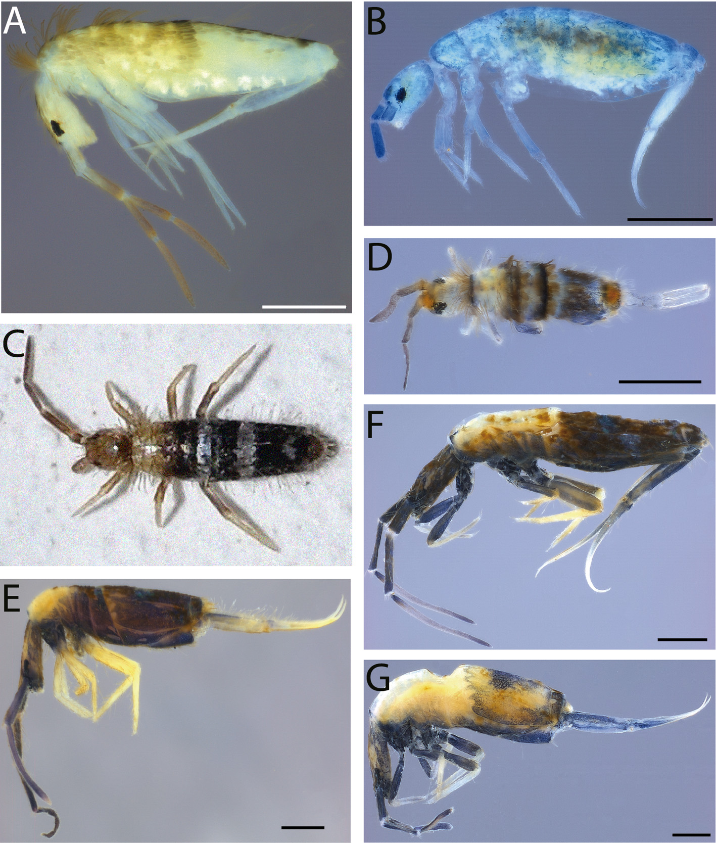
FIGURE 2A–G. Seira spp., habitus: A, S. domestica; B, S. dinizi; C‒D, S. ferrarii: C, in natural habitat, courtesy of Francisco Rodríguez (http://www.biodiversidadvirtual.org/insecta rium/Seira-ferrarii-img750580.html), D, in alcohol; E‒G, S. pini: E, paratype, F, specimen with pigmentation in legs from Monegros (Spain), G, specimen depigmented on Th II to Abd III and with one median spot on Abd IV from Monegros (Spain). Scale bars: 0.5mm.

FIGURE 3A–E. Seira spp., habitus (lateral view, except A in dorsal): A‒B, in natural habitat, A, S. betica sp. nov., courtesy of André Burgers (http://www.biodiversidadvirtual.org/insectar ium/Seira-betica-img961392.html), B, S. burgersi sp. nov., courtesy of André Burgers (http://www.biodiversidadvirtual.org/insectarium/Seira-burgersi-img961393.html); C‒E, preserved in alcohol: C, S. barrai sp. nov., D, S. burgersi sp. nov., E, S. betica sp. nov. Scale bars: 0.5mm.
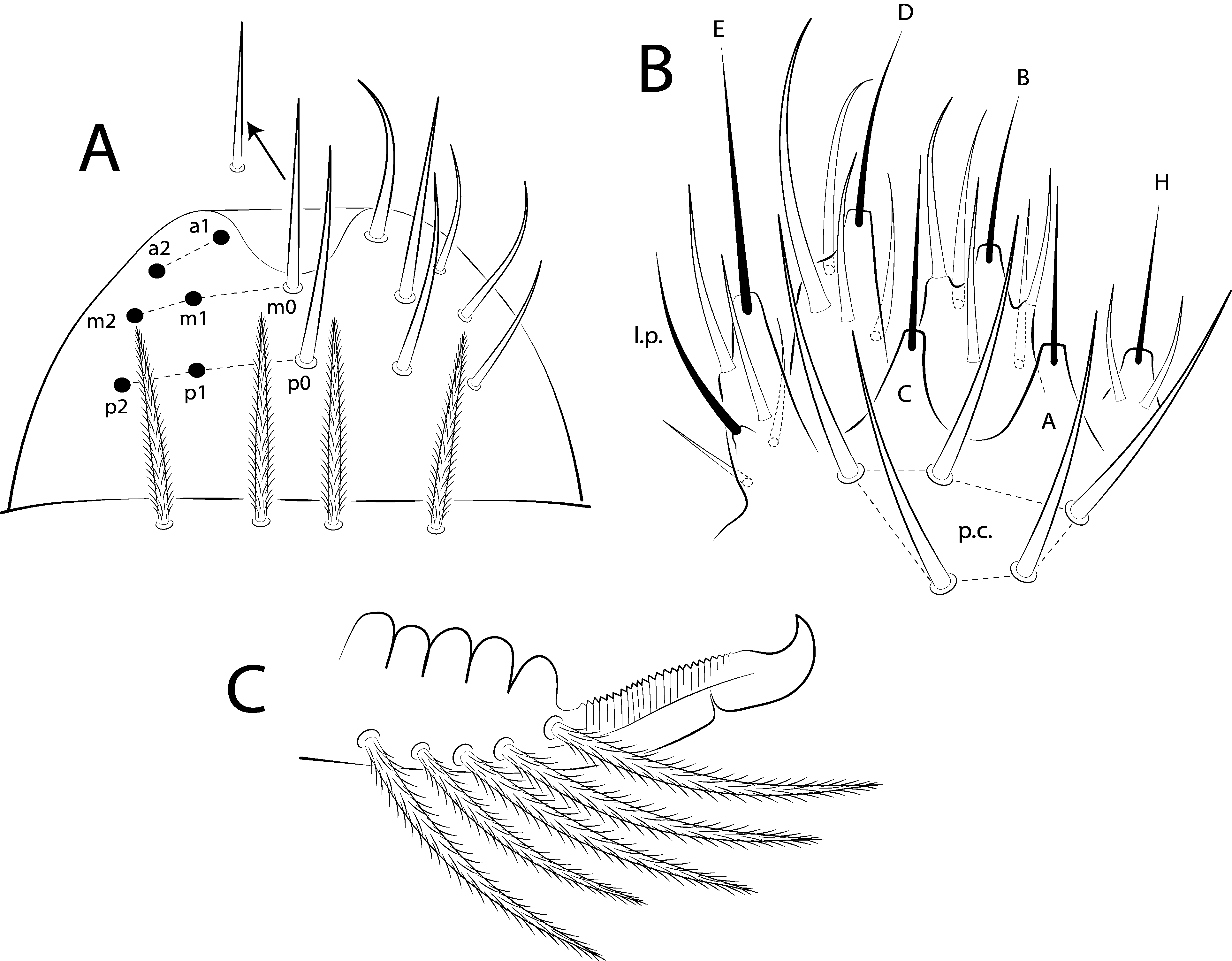
FIGURE 4A–C. Characteristics present in description of Seira species: A, prelabral and labral chaetotaxy (dorsal view), arrow indicates m0 chaeta smaller in some species; B, labial palp and proximal chaetae (ventral view), “p.c.” is proximal chaetae, “l.p.” is lateral process of papilla E; C, distal dens and mucro (lateral view).
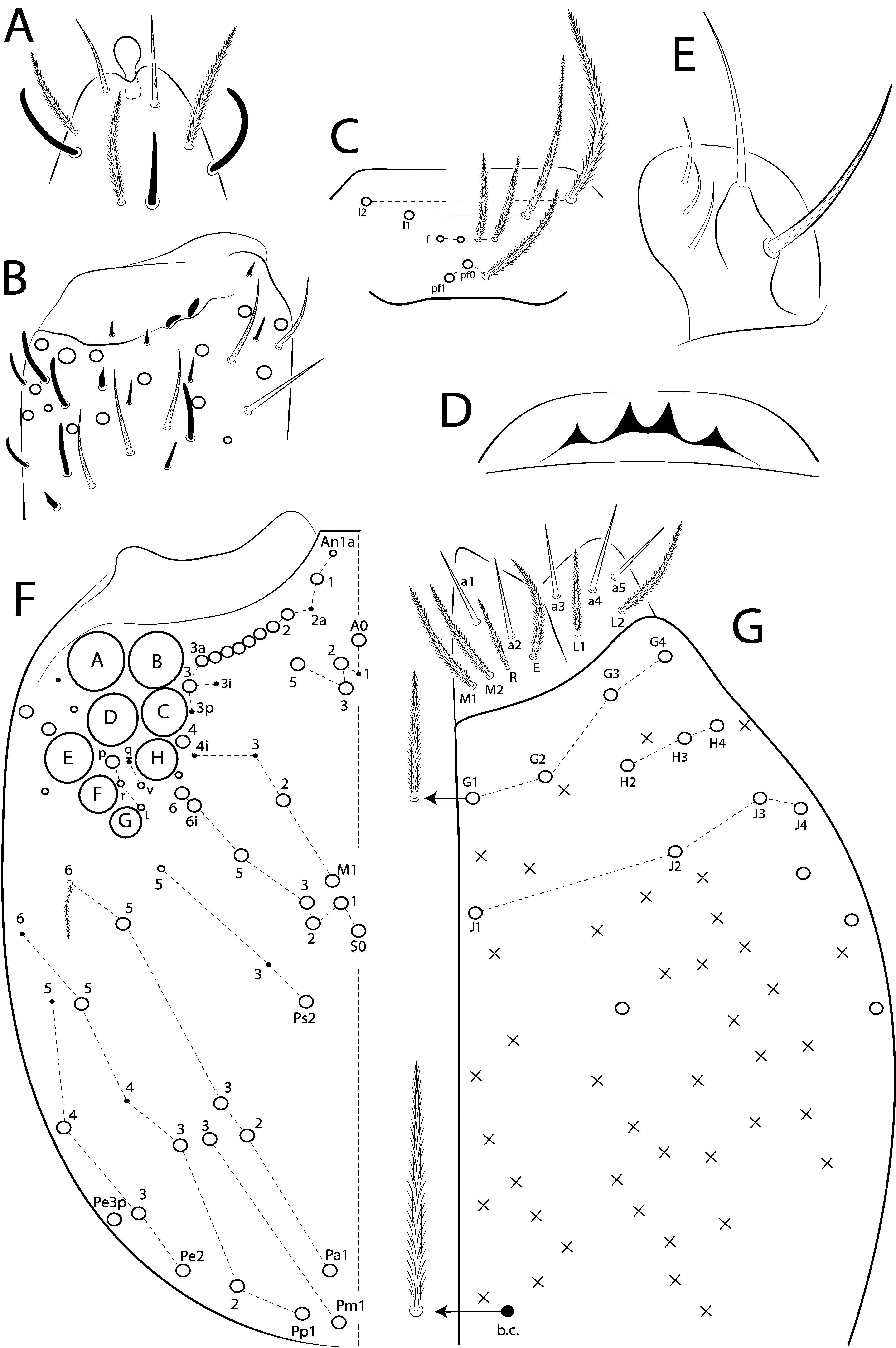
FIGURE 5A–G. Seira domestica: head; A, apex of Ant IV; B, Ant III apical organ; C, clypeal chaetotaxy; D, labral papillae; E, maxillary outer lobe; F, dorsal cephalic chaetotaxy (valid also for S. ferrari, except for interocular chaeta q); G, ventral head and basomedian and basolateral labial fields, arrows indicate normal ciliated chaetae and basal chaeta (b.c.), respectively.
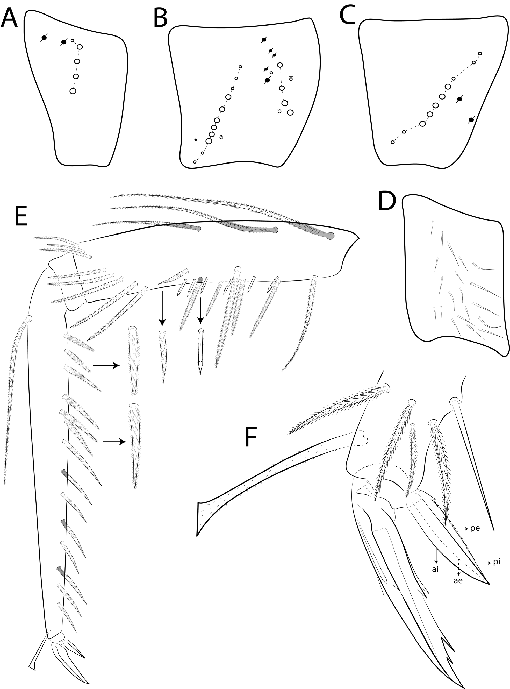
FIGURE 8A–H. Seira domestica: A, subcoxa I; B, subcoxa II; C, subcoxa III; D, trochanteral organ; E, chaetotaxy of femur and tibiotarsus I of male (anterior view), arrows indicate mac finely ciliate, small truncate chaetae, robust spine-like chaeta (in femur) and two types of tibiotarsal spine-like chaetae finely ciliate apically rounded and acuminate; F, distal tibiotarsus and empodial complex III (posterior view).
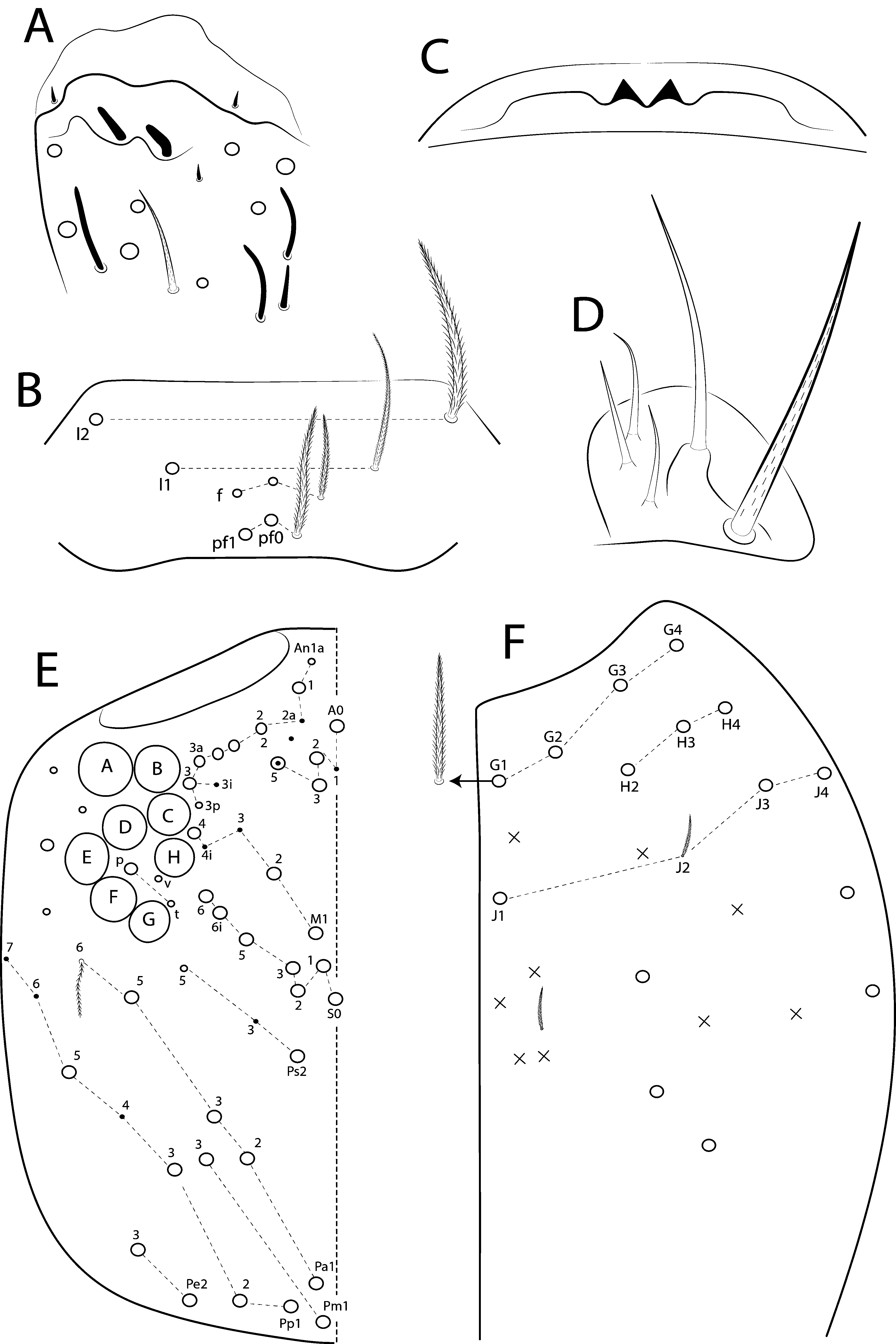
FIGURE 10A–F. Seira dinizi: head; A, Ant III apical organ; B, clypeal chaetotaxy; C, labral papillae; D, maxillary outer lobe; E, dorsal cephalic chaetotaxy; F, postlabial ventral head, arrow indicates normal ciliated chaetae.
IMAGES