AnimaliaNot EvaluatedacceptedspeciesAccepted
Pyura gangelion
(Savigny, 1816)
GBIF:148692794
0year
0
Synonyms
ABOUT
Descriptions(2)
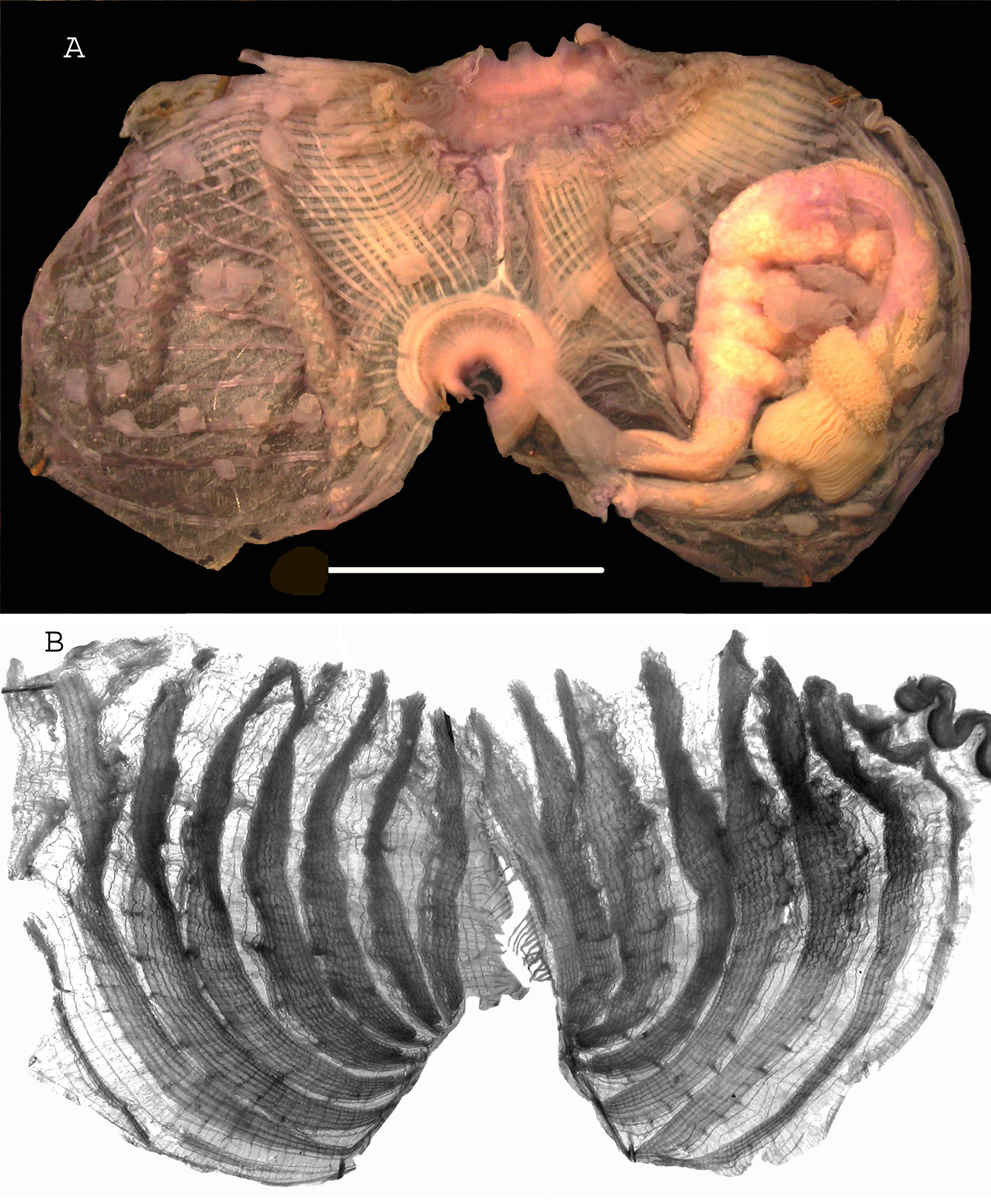
The specimens are fixed by their ventral side (Fig. 12 A, B). The tunic is wrinkled, more or less covered with epibionts. In formalin the tunic is beige on the body and brown near the siphons. The siphon apertures are surrounded by protuberances and they have dense spinules. The internal tunic lining at both siphon apertures is brown and thick with spinules of different sizes as long as 0.25 mm with a more or less inflated centre (Figs 12 E, F; 15 B). This internal tunic is prolonged more interiorly in a thinner bulbous membrane without spinules but with chalk-white round inclusions (Fig. 12 C). This deep part of the siphonal lining is tightly applied on the thin internal velum and is divided into 4 wide lobes only at the atrial siphon (Figs 12 C, 15 A). This atrial lobed membrane is also present in specimens from Eilat and Djibouti (MNHN collection) (Fig. 15 A). The body wall is thin, except on the red and much contracted siphons. The musculature issued from each siphon forms well separated bundles regularly crossed on both body sides (Fig. 13 A). There are about 18 large oral tentacles with short pinnules and smaller tentacles intercalated at the base of the velum. The prepharyngeal band, in a deep dorsal V, encloses a dorsal tubercle with horns variably rolled (Fig. 13 B). The dorsal lamina has numerous long thin papillae erect along the rim of an imperforated band. The 6 high branchial folds on each body side end by papillae at the oesophagus entrance (Fig. 14 A). The longitudinal vessels are equally spaced over and between the folds and it is difficult to know when they belong to a fold or to the interspace. There is an average of 5 stigmata in a mesh between the folds and 4 stigmata on the sides of the folds. There are parastigmatic vessels and the main transverse vessels are particularly wide. A branchial formula on the right side in a specimen 5 cm large is: E- 8 (30) 7 (34) 6 (34) 6 (35) 6 (32) 6 (22) 5 - DL. The gut loop widely opened extends far anteriorly occupying a large part of the left body side (Figs 13 A; 14 B). It is totally attached to the body wall. The stomach is slightly enlarged at the cardia and wears few digitate hepatic lobes; on the pyloric part there is a hepatic massive pedunculate gland in shape of cauliflower with dark green papillae in a convoluted design (Figs 13 A; 14 B). The intestine is narrow and isodiametric with the wall of the descending limb wrinkled in transverse ridges. Foliated endocarps are numerous on the gut along the exterior side of the loop from the oesophagus to the anus. The anus in a funnel has a slim rim in few low lobes (Fig. 13 C). There is one curved gonad on each side with 15 to 20 lobes on the left gonad and up to 25 lobes on the right one (Figs 13 A; 14 B). The left gonad is totally included inside the gut loop and all lobes wear endocarps. The right gonad is parallel to the endostyle and curves posteriorly. The short gonoducts open near to the atrial siphon (Fig. 13 A). In the space between the right gonad and the endostyle is a long and inflated vessel wearing aligned endocarps (Fig. 13 A; 14 C). There are no endocarps on the body wall.
Monniot, Françoise (2018): Ascidians collected during the Madibenthos expedition in Martinique 3. Stolidobranchia, Pyuridae and Molgulidae. Zootaxa 4459 (3): 401-430, DOI: 10.11646/zootaxa.4459.3.1
Remarks: Specimens described under the name Pyura torpida by Monniot (1983) from Martinique have been re-examined and have the same anatomy as the Madibenthos samples. The characters described above also well correspond to re-examined specimens of P. gangelion from Eilat redescribed by Monniot C. (1973) and specimens from Djibouti (Fig. 15). In the same publication Monniot (1973) suggested a possible synonymy between P. gangelion and P. sansibarica Michaelsen 1908; this is emphasised by precisions given by Millar (1956) for a specimen identified as P. sansibarica from Mozambique (he noted “ white spherical bodies ” different from spicules). The Indo-Pacific material named P. albanyensis (Monniot F. & C. 1996 Fig. 58 and 2001 Fig. 106) and P. gangelion: Kott (2004) have the same organisation but the tunic is thicker and the 4 - lobed atrial membrane of the siphon is not mentioned or not present. P. gangelion by its external aspect, the spinules, the branchial sac, the gonads and the absence of endocarps on the body wall, can be confused with P. vittata. Both species are variable but they differ by the diameter of the rectum, the internal tunic lining of the siphons, dark brown in P. vittata and with a 4 - lobed atrial membrane and chalky inclusions in P. gangelion. The spinules have a similar shape in both species but are more iridescent in P. vittata.
Monniot, Françoise (2018): Ascidians collected during the Madibenthos expedition in Martinique 3. Stolidobranchia, Pyuridae and Molgulidae. Zootaxa 4459 (3): 401-430, DOI: 10.11646/zootaxa.4459.3.1
Export occurrence data
Darwin Core Archive (ZIP)
CLASSIFICATION
Taxonomic Classification Tree

MULTIMEDIA
Media Files(5)
FIGURE 3. Halocynthia microspinosa. A, dissection, scale bar = 1cm; B, branchial sac.
Imageimage/png© Monniot, FrançoiseMonniot, Françoise

FIGURE 12. Pyura gangelion. A, B, two specimens, scala bar =1cm; C, atrial membrane, scale bar = 5mm; D, rim of the atrial membrane aperture with spinules; E, F, spinules scale bar = 0.1mm.
Imageimage/png© Monniot, FrançoiseMonniot, Françoise

FIGURE 13. Pyura gangelion (speciment st. 100). A, dissection, scale bar = 1cm; B, oral siphon; C, anus.
Imageimage/png© Monniot, FrançoiseMonniot, Françoise


IMAGES