AnimaliaNot EvaluatedacceptedspeciesAccepted
Ascidia columbiana
(Huntsman, 1912)
GBIF:159168002
0year
ABOUT
Descriptions(1)
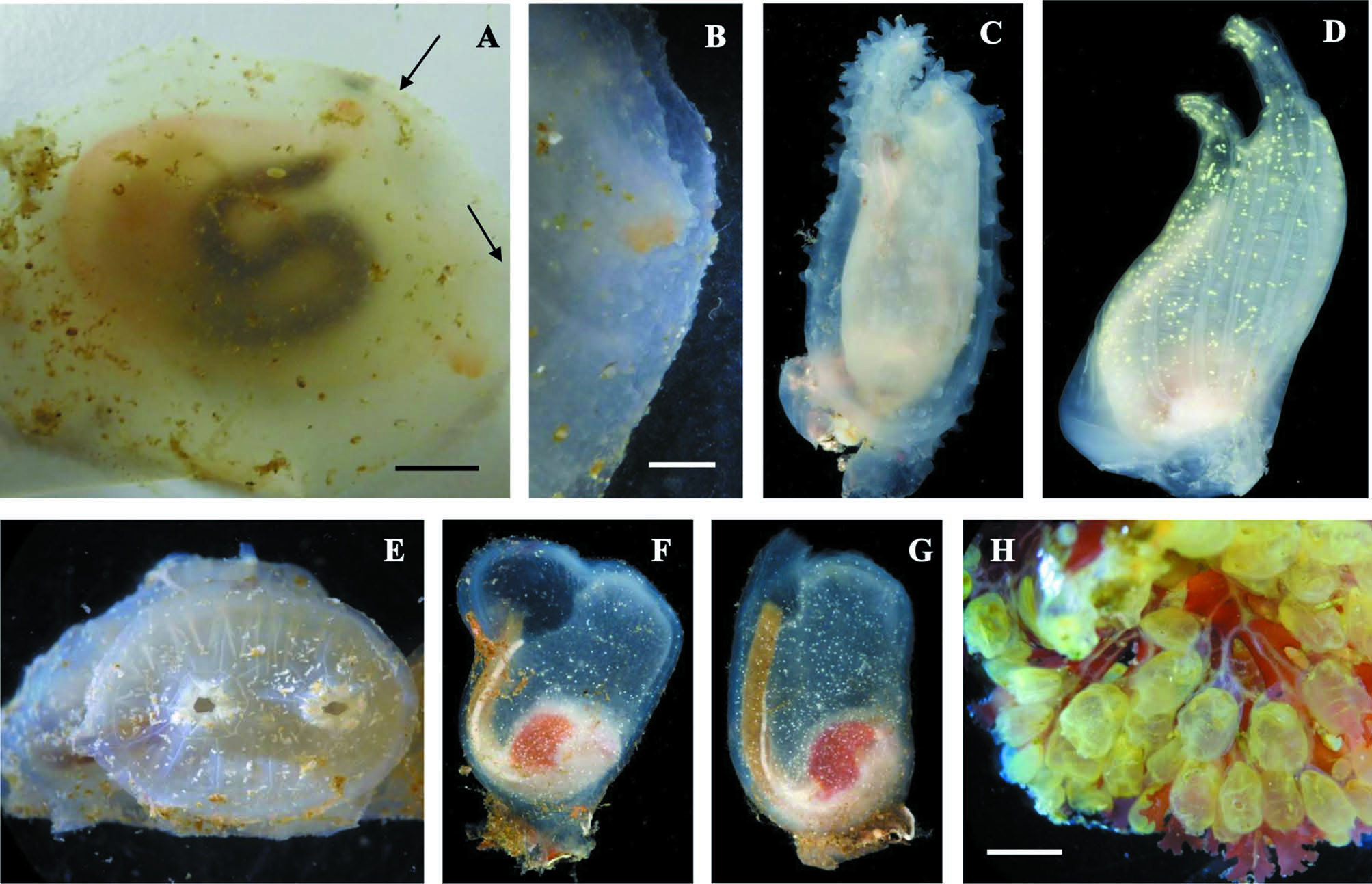
Figure 10 A, B IHAK 12 BHAK 0609 UF 2466. Under rocks low intertidal across small bay from Hakai dock. Three parasitic copepods in pharyngeal sac vouchered as BHAK 0618. XHAK 1 BHAK 1281 UF 2511. Maey Channel ARMS, 7.3 m. One specimen on plate, 2.6 cm long in papillated tunic. XHAK 9 Kelpie Point ARMS, 5 m. One on plate. Specimen from IHAK 12 with typical morphology for this species; 6.5 cm long in tunic, 3.5 cm long out of tunic. Body very flattened, attached on the left side. Tunic colorless, opaque, papillated. Oral siphon at anterior end, eight lobes and red spot between each lobe; atrial siphon slightly posterior with seven lobes and red spot between each lobe. Both siphons very short. Although a good description was given by Huntsman (1912 a, b as Ascidiopsis columbiana), Van Name (1945) mistakenly synonymized A. columbiana under A. callosa, thus combining the morphological characters. A. callosa Stimpson, 1852 (not found during the present survey) is a more northern circumpolar species (Van Name 1945); it has a smooth tunic and broods its embryos, while A. columbiana is a free-spawner. Another difference is that in A. columbiana the sperm duct crosses the intestine; in A. callosa it does not. For a detailed description see Huntsman (1912 a, b) and Lambert & Sanamyan (2001). Distribution: Alaska to Washington (Huntsman 1912 a, b; Lambert CC et al. 1996 as A. callosa; Lambert & Sanamyan 2001).
Lambert, Gretchen (2019): The Ascidiacea collected during the 2017 British Columbia Hakai MarineGEO BioBlitz. Zootaxa 4657 (3): 401-436, DOI: 10.11646/zootaxa.4657.3.1
Export occurrence data
Darwin Core Archive (ZIP)
CLASSIFICATION
Taxonomic Classification Tree

MULTIMEDIA
Media Files(1)
FIGURE 10. Figure 10. Phlebobranchia. A, B: Ascidia columbiana. A: whole animal right side, anterior on the right. Arrows indicate oral siphon opening (on right) and atrial opening above. B: anterior end around oral opening showing tunic papillations. C: Ascidia paratropa 9 cm in length; D: Ciona savignyi 6.2 cm in length; E: Chelyosoma productum 1.5 cm in diameter; F: Corella inflata about 3 cm in length; G: Corella willmeriana about 3 cm in length; H: Perophora annectens. Scale bars: A, 1.5 cm; B, 2 mm; H, 4 mm. C, D, F, G photos by G. Paulay.
Imageimage/png© Lambert, GretchenLambert, Gretchen
IMAGES