AnimaliaNot EvaluatedacceptedspeciesAccepted
Chelyosoma productum
Stimpson, 1864
GBIF:159168009
0year
ABOUT
Descriptions(1)
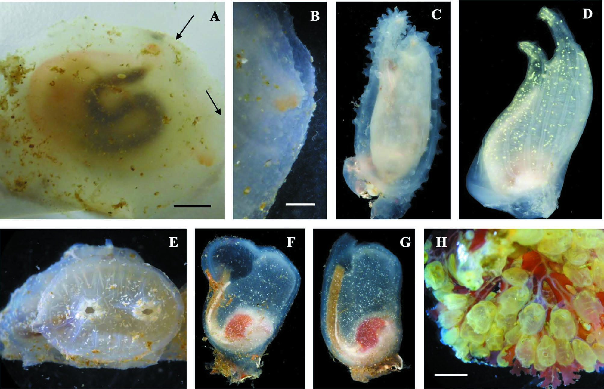
Figure 10 E IHAK 12 BHAK 0617. Low rocky intertidal across small bay from Hakai dock. One very small (tissue sample only). IHAK 18 BHAK 0642, 0643 UF 2493. Two small specimens, from under lab dock. IHAK 36 C in bulk sample. MHAK 14 BHAK 0621. Tippy Rock Bay low intertidal on red alga Neorhodomela Masuda, 1982. Tissue sample only. XHAK 1 Maey Channel ARMS 7.3 m. With Corella inflata (Huntsman, 1912) on plate. XHAK 4 Spider Island ARMS, 9 m. Four on cross pieces, nine on plates. XHAK 9 BHAK 2832, 2835 UF 2555, 2556. Kelpie Point ARMS, 5 m. Two small specimens on plates. Like all members of the Corellidae, Chelysoma productum has spiral stigmata. Its most distinguishing characteristic, however, is the flat oval disk at the anterior end that bears the oral and atrial openings. This disk is composed of a species-specific pattern of translucent plates, a tunic adaptation peculiar to the genus. Huntsman (1912 b) and Van Name (1945) give a detailed description of the plate pattern and other aspects of the morphology. There are six small triangular plates around each siphonal opening, two larger ones between the two openings, and a single row of plates of various sizes around the perimeter of the disk. The plates are annulated, suggesting possible growth rings, and are slightly separated from one another by tunic. An unusual pattern of muscles controls opening and closing of the siphons. The body is oriented obliquely anteroposteriorly, as shown in Fig. 10 B. The digestive tract and gonad are on the right side, another trait of this family discussed at length by Huntsman (1912 b). Distribution: Alaska to southern California (Huntsman 1912 b; Van Name 1945; Abbott & Newberry 1980; O’Clair & O’Clair 1998; Lamb & Hanby 2005).
Lambert, Gretchen (2019): The Ascidiacea collected during the 2017 British Columbia Hakai MarineGEO BioBlitz. Zootaxa 4657 (3): 401-436, DOI: 10.11646/zootaxa.4657.3.1
Export occurrence data
Darwin Core Archive (ZIP)
CLASSIFICATION
Taxonomic Classification Tree

MULTIMEDIA
Media Files(1)
FIGURE 10. Figure 10. Phlebobranchia. A, B: Ascidia columbiana. A: whole animal right side, anterior on the right. Arrows indicate oral siphon opening (on right) and atrial opening above. B: anterior end around oral opening showing tunic papillations. C: Ascidia paratropa 9 cm in length; D: Ciona savignyi 6.2 cm in length; E: Chelyosoma productum 1.5 cm in diameter; F: Corella inflata about 3 cm in length; G: Corella willmeriana about 3 cm in length; H: Perophora annectens. Scale bars: A, 1.5 cm; B, 2 mm; H, 4 mm. C, D, F, G photos by G. Paulay.
Imageimage/png© Lambert, GretchenLambert, Gretchen
IMAGES