AnimaliaacceptedfamilyAccepted
Discoglossidae

Discoglossidae

GBIF:159397247

ABOUT

Descriptions(1)

Figure 7 A-I, L-Q MAXILLAE: The maxillary elements have been grouped informally into three divisions: (1) Large frog with sculptured maxilla (discoglossid A). (2) Large frog with smooth maxilla (discoglossid B). (3) Small frog with sculptured maxilla (discoglossid C). Discoglossid A The external sculpturing in AMNH 8461 (fig. 7 P, Q) is basically of the pattern found in the smaller group of frogs represented by AMNH 8460, and can be distinguished from them by the greater depth of the pits and the greater prominence of the surrounding ridges, which occasionally become isolated into tubercle-like projections. As in the smaller specimens, the external sculpturing is not present at the ventral border of the maxilla. On the maxilla about 20 teeth are present in a distance of 8 mm. Discoglossid B These are much larger than the small frogs with sculptured maxillae and only slightly smaller than the larger forms with sculptured maxillae. The maxilla in AMNH 8462 is essentially smooth. A few faint anteroposteriorly elongate grooves are present in some specimens. The height of the teeth is the same as in AMNH 8461 and 20 teeth occupy a distance of 5.6 mm. on the maxilla (fig. 7 N, 0). Discoglossid C A number of maxillary fragments have been recovered which are dimensionally and morphologically distinct from other anuran maxillae. AMNH 8460 is a slender maxilla containing a number of teeth, the crowns of which have been broken (fig. 7 L, M). The teeth are about 0.25 mm. in height and 20 teeth are present in a distance of 3.6 mm. The external sculpturing is quite distinctive and consists of shallow pitted depressions bounded by ridges. The ventral edge of the maxilla is free of sculpturing. HUMERI: The criteria for differentiating distal ends of frog humeri were discussed by Hecht and Estes (1960) in their study of the amphibians from the Jurassic Como Bluff localities in Wyoming. Some of these features are: the distance from the lateral to the medial epicondyle; the diameter of the distal ball; the relative development of the fossa cubitus ventralis; the development of the crests originating from the condyles, and the shape and size of the olecranon scar including the position of its dorsal termination. Discoglossid A A distinct group of humeri is represented by AMNH 8446 (fig. 7 A, B). These humeral fragments are easily differentiated from other humeri on the basis of size and other criteria. These robust humeri measure 6.4 mm. from the lateral epicondyle to the medial epicondyle, and the diameter of the head ball is 4.4 mm. Apart from their large size, these humeri can be distinguished by better development of the lateral epicondyle than in either AMNH 8450 or 8449. The medial crest does not originate from the medial epicondyle but usually from a point between the medial epicondyle and the ball. The crest is distinct and a small foramen lies medial to it. The lateral crest is much smaller and less distinct, and originates slightly lateral to the ball. The two crests do not meet although the medial crest continues dorsally along with humeral shaft. The olecranon scar is well developed and the dorsal apex is not so pointed as in AMNH 8450 and 8449 nor is it situated so far laterally. Discoglossid B A fragment of a humerus, AMNH 8449, is distinct from the other humeri in the collection (fig. 7 C, D). It is dissimilar from AMNH 8450 in the development of the epicondyles, particularly in the position and degree of separation of the lateral epicondyle from the ball, and also in size. The diameter of the ball is 3.2 mm. and the distance between the epicondyles is 4.5 mm. The crests are developed as in AMNH 8450, and the medial crest is distinctly more prominent than the lateral crest. The crests do not join dorsal to the fossa cubitus ventralis and gradually become indistinct. The fossa cubitus ventralis is a fairly deep crescentic depression. Most of the olecranon scar is broken away but its dorsal termination appears to be also close to the lateral border. Just dorsal to the fossa cubitus ventralis, the shaft is dorsoventrally compressed. Discoglossid C The distal part of a humerus, AMNH 8450 (fig. 7 E, F), is referable to this category. Its humeral ball is spherical with a diameter of 2 mm. The lateral epicondyle is closely approximated to the ball and not well formed, but the medial condyle is strongly developed and well separated from the ball. A prominent crest originates at the ventral edge of the medial epicondyle and extends dorsally. The medial crest is much better developed than the lateral crest, the latter originating at the dorsal limit of the lateral epicondyle. The fossa cubitus ventralis is shallow. Neither of the crests is very distinct. The area dorsal to the lateral epicondyle is laterally expanded and flattened. The olecranon scar is relatively large and subtriangular; the dorsal apex of the scar is situated close to the lateral border. The shaft narrows considerably at the point of dorsal termination of the olecranon scar and the cross section of the shaft becomes rounded. ILIA: Six specimens are referred to the Discoglossidae on the basis of characters listed by Estes (1964, p. 56). AMNH 8456 is a partially complete right ilium (fig. 7 G). The acetabulum is relatively larger and more rounded than in the pelobatid specimens. The pubic process is short and the shaft relatively straight, as can be seen in AMNH 8457 (fig. 7 H). The dorsal crest is not well developed but an elongate prominent groove is present ventral to it. A single specimen, AMNH 8459, is different from ilia represented by AMNH 8452 and 8453, and has been referred to the Discoglossidae (fig. 71). The most obvious difference is in the expansion of the acetabulum anteriorly. The acetabulum is shallowly depressed and well separated from the rest of the bone by a prominent raised rim. The anterior edge of the acetabulum is pointed and projects anteriorly. The pubic process of the ilium is greatly reduced and the ischial process is shorter than in the other anuran ilia recovered.
Sahni, Ashok (1972): The vertebrate Fauna of the Judith River formation, Montana. Bulletin of the American Museum of Natural History 147 (6): 319-416, DOI: 10.5281/zenodo.3382461, Hdl: http://hdl.handle.net/2246/1099

Export occurrence data

Darwin Core Archive (ZIP)

CLASSIFICATION

Taxonomic Classification Tree

MULTIMEDIA

Media Files(1)

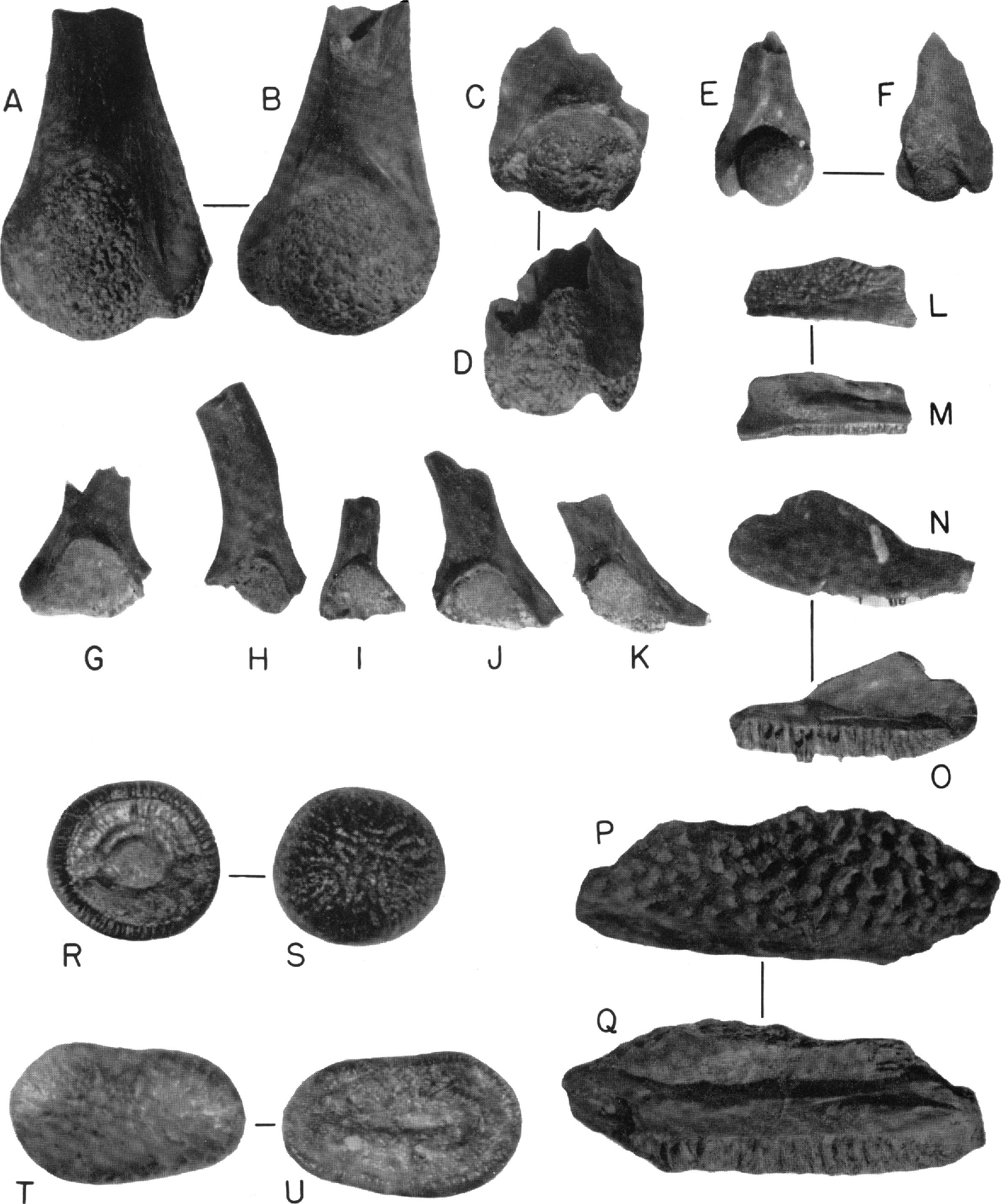
FIG. 7. A. AMNH 8446, ventral view of left discoglossid (A) humerus. B. Dorsal view. Both x 5. C. AMNH 8449, ventral view of left discoglossid (B) humerus. D. Dorsal view. Both x 5. E. AMNH 8450, ventral view of left discoglossid (C) humerus. F. Dorsal view. Both x 5. G. AMNH 8456, lateral view of distal portion of right discoglossid ilium, x 5. H. AMNH 8457, lateral view of distal portion of left discoglossid ilium, x 5. I. AMNH 8459, lateral view of distal portion of left discoglossid ilium, x 5. J. AMNH 8452, lateral view of distal portion of left pelobatid ilium, x 5. K. AMNH 8453, lateral view of distal portion of pelobatid ilium, x 5. L. AMNH 8460, external view of left discoglossid (C) maxilla. M. Internal view. Both x 5. N. AMNH 8462, external view of right discoglossid (B) maxilla. 0. Internal view. Both x 5. P. AMNH 8461, external view of left discoglossid (A) maxilla. Q. Internal view. Both x 5. R. AMNH 10100, occlusal view of?Paralbula sp. S. Basal view. Both x 10. T. AMNH 10101, occlusal view of?Paralbula sp. U. Basal view. Both x 1.0.

Imageimage/png© Sahni, AshokSahni, Ashok

IMAGES

Gallery(1)

See Gallery

Occurrences with images

Source Information

The vertebrate Fauna of the Judith River formation, Montana

checklist

This dataset contains the digitized treatments in Plazi based on the original journal article Sahni, Ashok (1972): The vertebrate Fauna of the Judith River formation, Montana. Bulletin of the American Museum of Natural History 147 (6): 319-416, DOI: 10.5281/zenodo.3382461, Hdl: http://hdl.handle.net/2246/1099

GEOLOGIC INVESTIGATION of the Late Cretaceous Judith River Formation in the area just north of the Judith River on the Missouri River in north-central Montana has resulted in the discovery of varied vertebrate forms. The beds are mainly freshwater continental deposits consisting of crossbedded channel sandstones, gray siltstones, and carbonaceous shales with occasional seams of lignitic coal. The stratigraphic sequence consists of the Marias River Shale overlain by the Eagle, Claggett, Judith River, and Bearpaw formations. The last four constitute the Montana Group. The Judith River Formation is Campanian in age as determined by its position between the fossiliferous marine Claggett and Bearpaw shales.

The fauna was obtained from the upper 50 feet of the formation. The bone concentration in the productive sandstone is the result of size sorting leading to underrepresentation of the larger dinosaurs. Three orders of mammals are represented, the Eutheria by a single genus, the Allotheria by five, and the Metatheria by at least three genera. Teiid and parasaniwid lizards are frequent. Only a fraction of the large number of described dinosaur genera, however, is represented in the collection by isolated teeth. Fish and amphibians form a sizable portion of the fauna.

Vertebrates from the Judith River Formation are more primitive than, but generally similar to, later Maestrichtian species. The mammals differ from their descendants in the Lance Formation at the species level. The community structure and the paleoecology of the fauna of the Judith River Formation resemble those of the Lance Formation. The greatest difference between the two communities is the greater variety of dinosaurs in the earlier formation.

Sahni A, plazi (1972). The vertebrate Fauna of the Judith River formation, Montana. Plazi.org taxonomic treatments database. Checklist dataset https://doi.org/10.5281/zenodo.3382461 accessed via GBIF.org on 2026-06-15.

CC0Published 12/31/1972View dataset
GBIF Usage Key
159397247
Dataset Key
fd03f669-c046-46cd-861a-6bf56b7fc989
Origin
source
Backbone Key
3241294
Taxon ID
1A7187CFFFF41742FEE5FCB8E0A553A8.taxon
Last Crawled
6/10/2026
Last Interpreted
6/10/2026