
Adeonella lichenoides
(Lamarck, 1816)
GBIF:163752450
ABOUT
Descriptions(4)
Export occurrence data
Darwin Core Archive (ZIP)
CLASSIFICATION
Taxonomic Classification Tree

MULTIMEDIA
Media Files(4)
FIGURES 35–40. Adeonella cf. lichenoides (Lamarck, 1816), RGM.1350554, early Pleistocene, Java. 35. View of a bifurcated branch fragment. 36. Group of autozooids with frontal adventitious avicularia and rounded vicarious avicularium. 37. Group of zooids, some with sealed orifices. 38. Group of autozooids with suboral avicularium variable in size, shape and orientation, and a fertile zooid. 39. Close-up of an autozooid with suboral and frontal avicularia. 40. Zooids with frontal avicularia variable in shape. Scale bars: Fig. 35 = 500 µm; Figs 36, 38–40 = 100 µm; Fig. 37 = 200 µm.
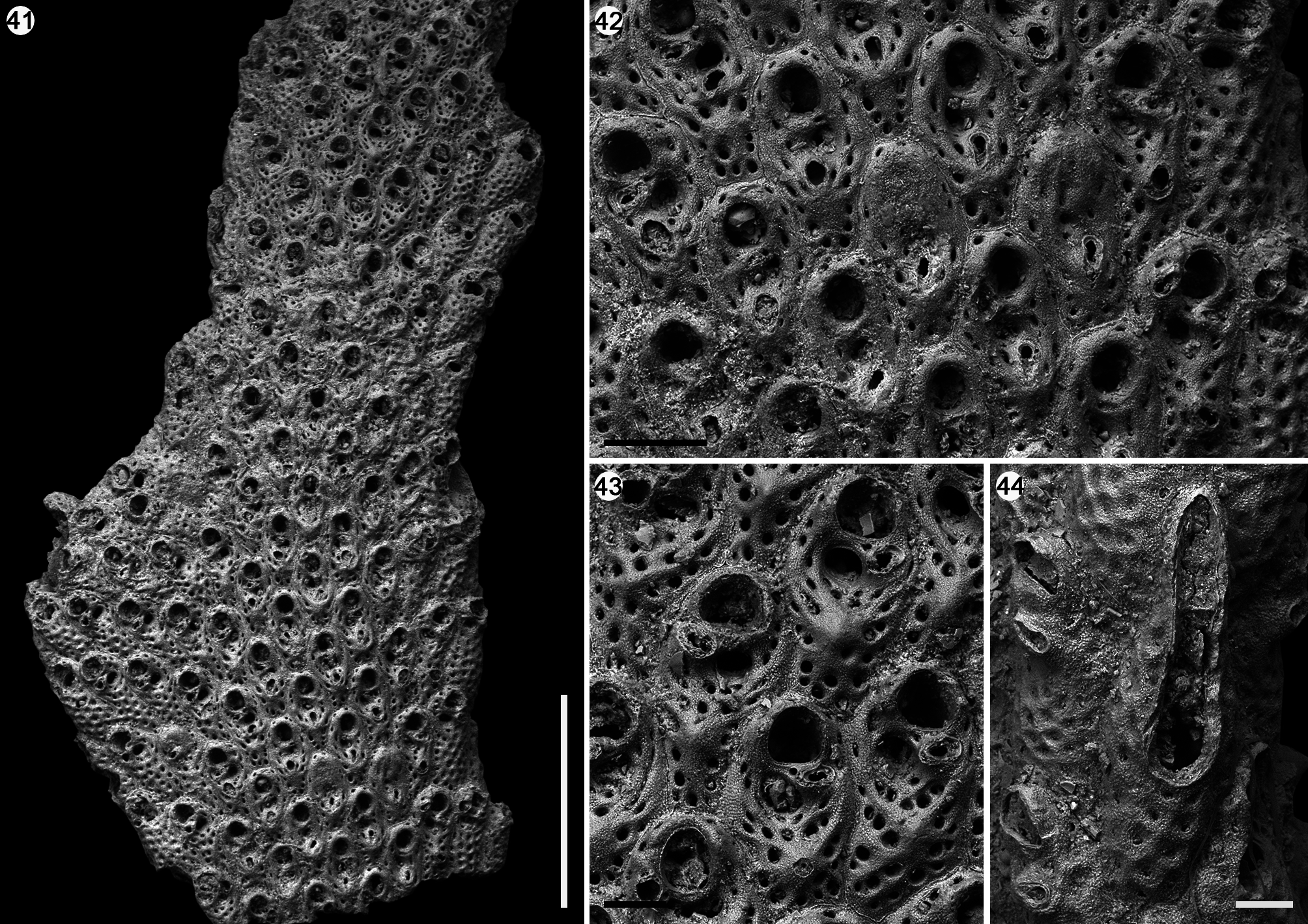
FIGURES 41–44. Adeonella cf. lichenoides (Lamarck, 1816), RGM.1350555, Holocene, UPGG041, off South Sulawesi. 41. View of a branch fragment. 42. Group of autozooids with frontal avicularia and sealed orifice. 43. Group of zooids with suboral avicularium and tubercle on the frontal shield. 44. Close-up of a lateral subtriangular vicarious avicularium. Scale bars: Fig. 41 = 1 mm; Fig. 42 = 200 µm; Figs 43, 44 = 100 µm.
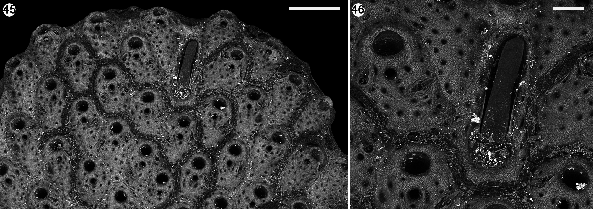
FIGURES 45, 46. Adeonella lichenoides (Lamarck, 1816), syntype of Adeonella platalea (Busk, 1854), Recent, off Cape Capricorn (Queensland), NHMUK 1854.11.15.185. Group of autozooids and rounded vicarious avicularium. 46. Close-up of an autozooid and a vicarious avicularium. Scale bars: Fig. 45 = 400 µm; Fig. 46 = 100 µm.
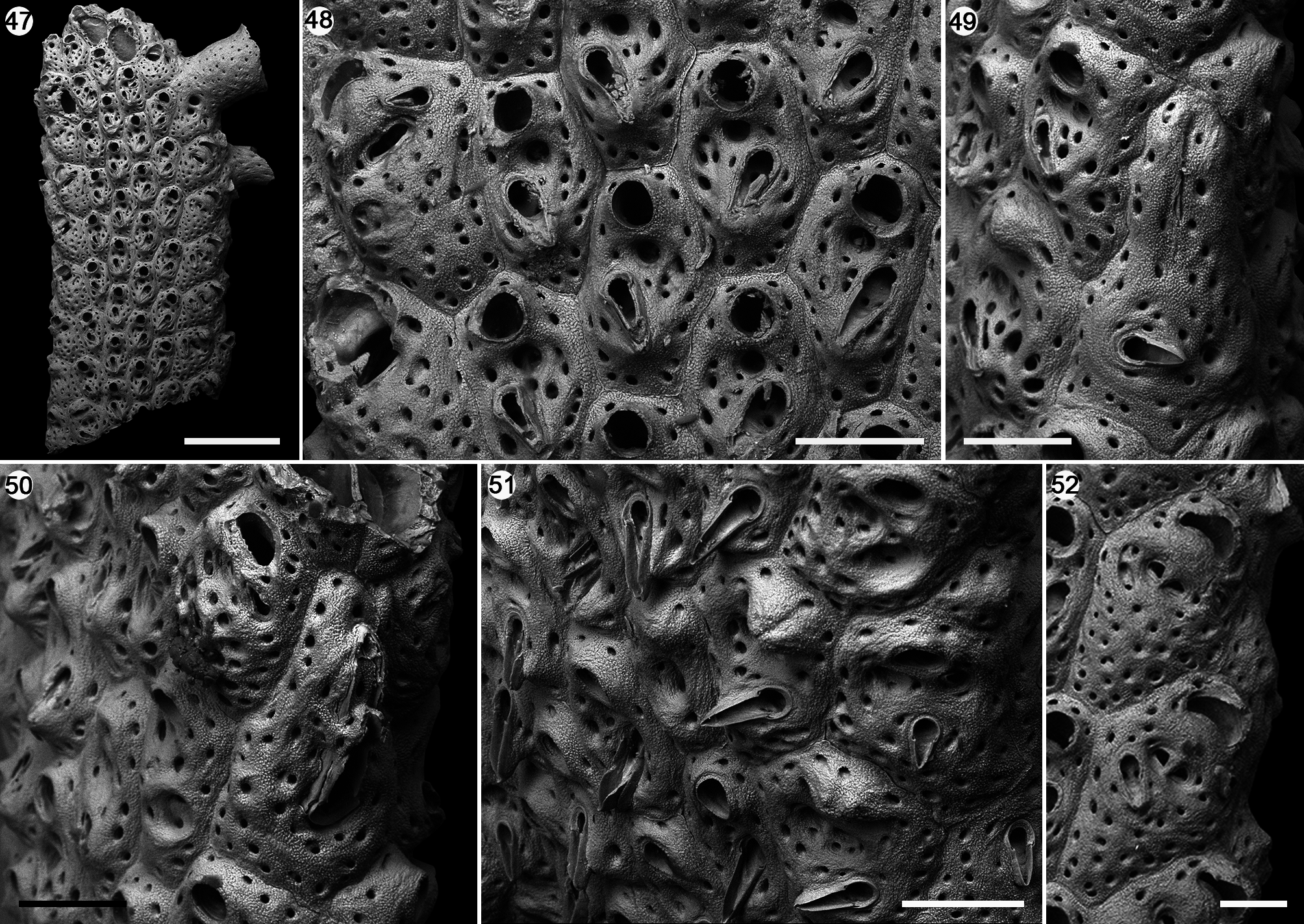
FIGURES 47–52. Adeonella intricaria (Busk, 1884), syntype, Recent, Challenger Expedition, Station 190 (8˚56’S; 136˚5’E), NHMUK 1899.7.1.2519. 47. View of a branch fragment. 48. Group of autozooids with frontal avicularia and fertile zooids. 49. Close-up of a lateral triangular vicarious avicularium with sealed opening. 50. Close-up of a lateral triangular vicarious avicularium with intramural budding. 51. Autozooids with long and acute frontal avicularia. 52. Close-up of two fertile zooids at the peripheral margin of the branch. Scale bars: Fig. 47 = 500 µm; Figs 48–51 = 200 µm; Fig. 52 = 100 µm.
IMAGES