Corynosoma australe
Johnston, 1937
GBIF:163829644
ABOUT
Descriptions(8)
Export occurrence data
Darwin Core Archive (ZIP)
CLASSIFICATION
Taxonomic Classification Tree

MULTIMEDIA
Media Files(2)
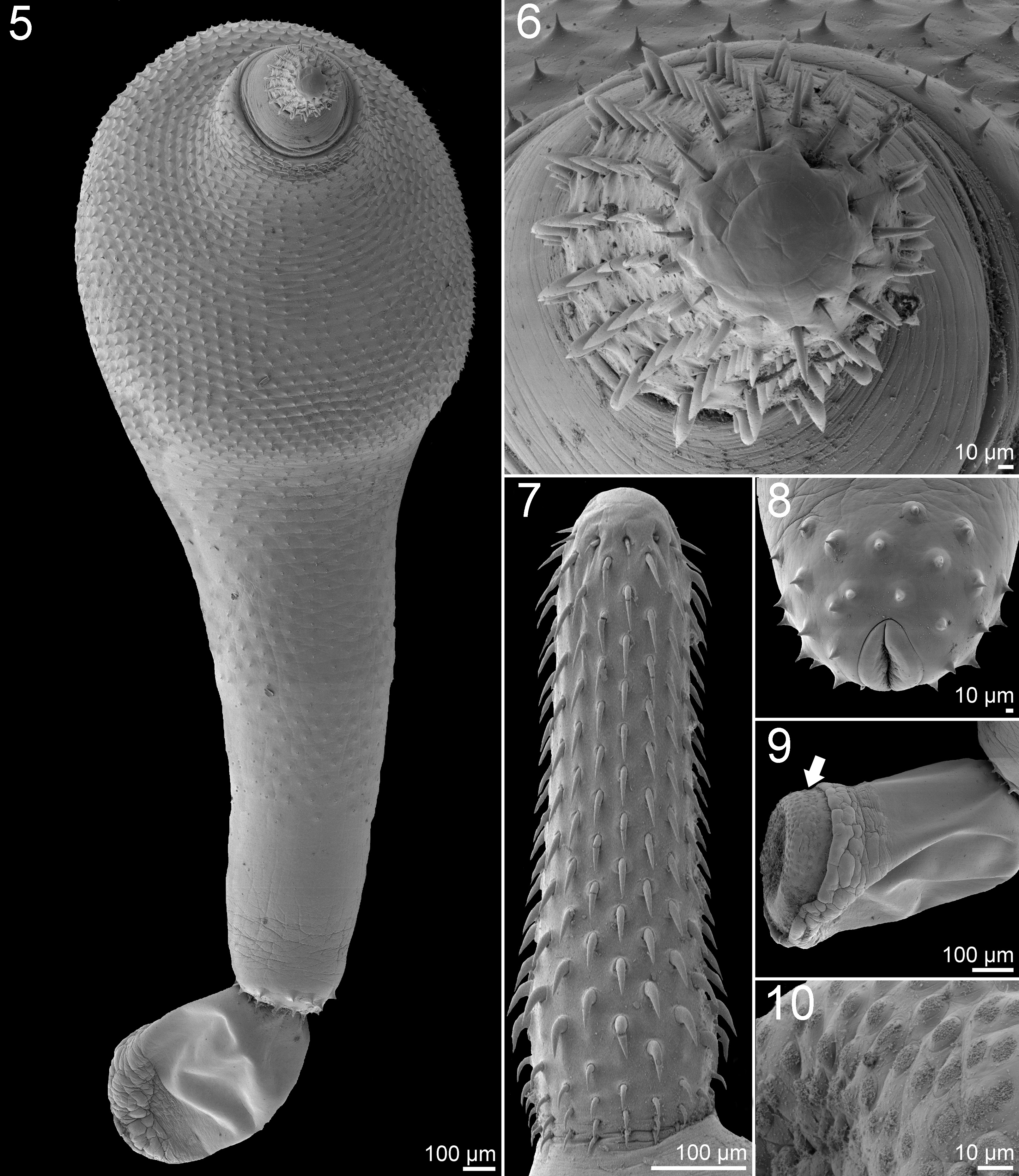
FIGURES 5–10. Scanning electron micrographs of adult males of Corynosoma australe Johnston, 1937 from South American sea lion Otaria flavescens. 5. Whole worm, ventral view. 6. Proboscis, apical view. 7. Proboscis, lateral view. 8. Genital spines and genital pore, basal view. 9. Copulatory bursa, lateral view. The white arrows point sensory receptors. 10. Detail of sensory receptors in the copulatory bursa.
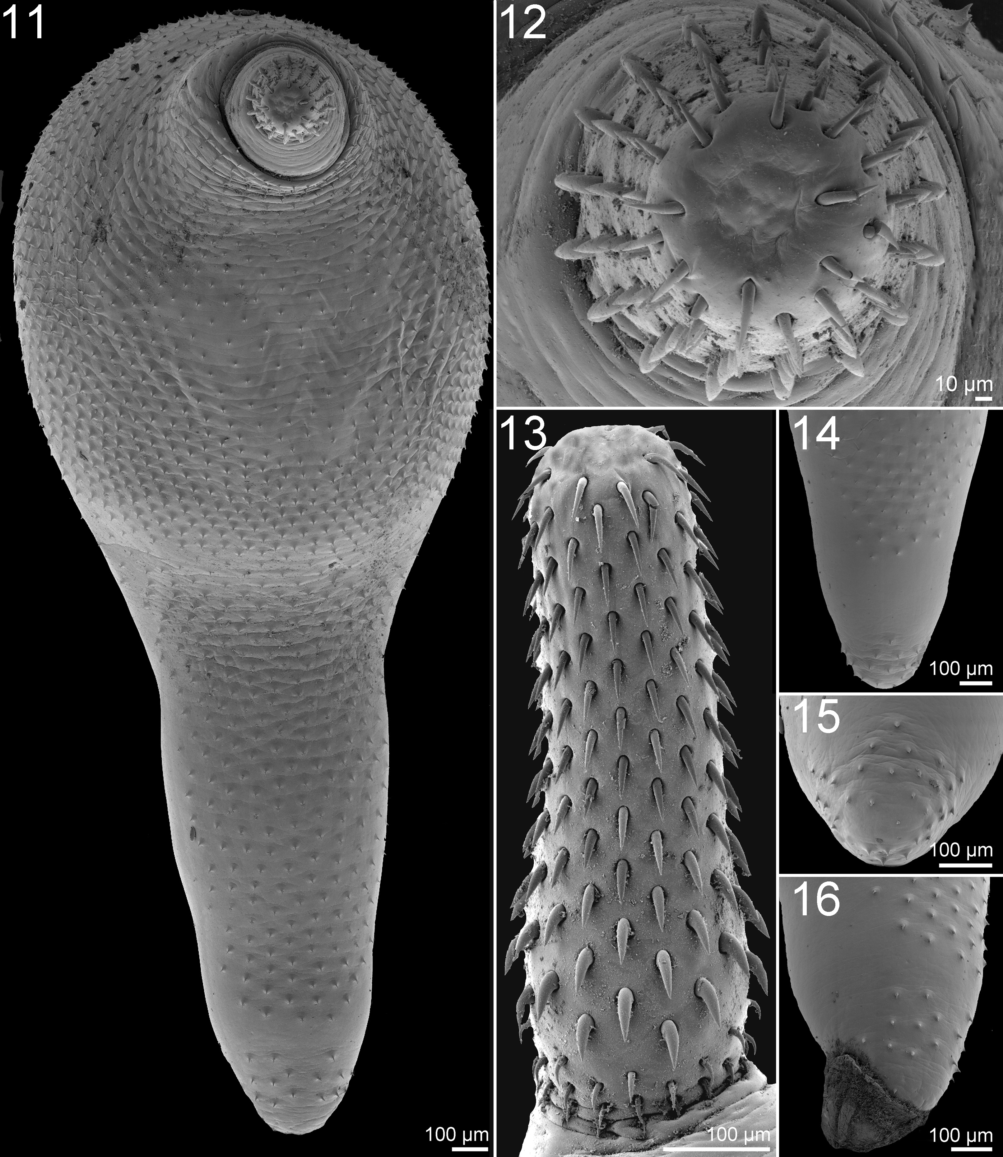
FIGURES 11–16. Scanning electron micrographs of adult females of Corynosoma australe Johnston, 1937 from South American sea lion Otaria flavescens. 11.Whole worm, ventral view. 12. Proboscis, apical view. 13. Proboscis, lateral view. 14. Posterior end of trunk, ventral view. 15. Spines near genital pore, basal view. 16. Posterior end of trunk with copulatory caps, lateral view.
IMAGES