AnimaliaNot EvaluatedacceptedspeciesAccepted
Adeonellopsis japonica

Adeonellopsis japonica

(Ortmann, 1890)

GBIF:164249544

0year

0

Synonyms

ABOUT

Descriptions(5)

Figs 7 – 9, 10 A
Hirose, Masato (2016): Diversity and distribution of adeonid bryozoans (Cheilostomata: Adeonidae) in Japanese waters. European Journal of Taxonomy 203 (203): 1-41, DOI: 10.5852/ejt.2016.203
Measurements Autozooids. ZL: 404 ̅ 750 (553 ± 71); ZW: 184 ̅ 419 (308 ± 52); n = 115. SOrL: 69 ̅ 109 (87 ± 11); SOrW: 56 ̅ 127 (102 ± 14); n = 67. SAvL: 134 ̅ 232 (176 ± 20); SAvW: 62 ̅ 133 (90 ± 14); n = 81. FAvL: 75 – 138 (110 ± 15); FAvW: 40 – 72 (59 ± 8); n = 47. SpL: 16 ̅ 92 (43 ± 16); SpW: 20 ̅ 75 (44 ± 14); n = 70. Gonozooids. ZL: 453 ̅ 722 (612 ± 60); ZW: 329 ̅ 524 (415 ± 53); n = 35. SOrL: 57 ̅ 125 (86 ± 13); SOrW: 100 ̅ 166 (134 ± 14); n = 34. SAvL: 109 ̅ 176 (135 ± 17); SAvW: 52 ̅ 104 (73 ± 12); n = 25. SpL: 102 ̅ 205 (146 ± 24); SpW: 98 ̅ 174 (134 ± 21); n = 33. Vicarious avicularia at branch margins. L: 358 ̅ 399 (385 ± 23); W: 141 ̅ 172 (158 ± 16); n = 3. Description Colony yellowish brown, erect, dichotomously branching, irregularly spreading; up to 10 cm high (Fig. 7 A). Branches flattened (Figs 7 B, 9 A), multiserial, with zooids opening both sides, rounded at end; 1.6 – 4.6 mm wide (average 2.8 mm; n = 27). Autozooids oval or hexagonal, outlined by a distinct deep marginal groove (Figs 7 C, 8 C, 9 B), with 6 – 9 small pores along each lateral margin and two or three small frontal pores just proximal to orifice. Frontal shield umbonuloid (Fig. 10 A). Young autozooids at growing margin of branch have concave frontal shields surrounded by broad, swollen rim with rows of granulation arranged perpendicular to margin (Fig. 7 E – F); rim width 41 – 76 μm (average 57 μm) (n = 31). Mature frontal shield convex, entirely covered with minute granules. Spiramen abuts proximal end of suboral avicularium; subdivided into two or three openings separated by narrow septa, or lobate due to incomplete septa (Fig. 7 C); with increased calcification, sometimes appearing as single opening (Fig. 7 D); reduced in size during ontogeny, associated with depression of avicularian rostrum, sometimes occluded (Figs 7 D, 8 D). Secondary orifice nearly circular (Fig. 7 C) or broader than long (Figs 7 D, 8 C); peristomial rim lacking; primary orifice submerged deep in peristome. Large, suboral avicularium in center of frontal shield, extending from spiramen to proximolateral corner of orifice, or nearly so, usually pointing distolaterally (Figs 7 C, F, 9 B); rostrum slightly raised distally, not hooked; mandible elongate-triangular; no crossbar. Additional smaller frontal avicularium often present near proximal end of zooid; identical in form to suboral avicularium; pointing in any direction (Fig. 7 C). Gonozooids occur at branch bifurcations (Fig. 8 B, E); larger than autozooids, with broader, slightly curving orifice; bearing suboral avicularium; smaller frontal avicularia either lacking (Fig. 8 B, E) or up to three per zooid (Fig. 7 G – H). Spiramen large, roughly circular in outline, divided into 6 – 10 pores (Figs 7 G – H, 8 B). Spiramen lacking marginal denticles in both autozooids and gonozooids (Figs 7 C, F, 8 E), although incomplete septa may appear as denticles. Vicarious avicularia at branch margins (Figs 7 D, 8 A, 9 C – D) smaller than autozooids; triangular, with mandible directed distally. Basal part of branch consists of kenozooids with secondary orifice occluded by secondary calcification (Fig. 8 D); about same size as autozooids, 486 – 631 μm long (average 541 μm) by 199 – 351 μm wide (average 314 μm) (n = 11). Kenozooids also bearing several small avicularia budded from areolae, near proximal margin of zooid, associated with other marginal pores (Fig. 8 D), 101 – 152 μm long (average 121 μm) by 47 – 75 μm wide (average 59 μm) (n = 15).
Hirose, Masato (2016): Diversity and distribution of adeonid bryozoans (Cheilostomata: Adeonidae) in Japanese waters. European Journal of Taxonomy 203 (203): 1-41, DOI: 10.5852/ejt.2016.203
Remarks Ortmann (1890) first described Adeonellopsis japonica as Adeonella japonica, based on Döderlein’s specimens from Sagami Bay. Hayward (1988) discussed this species in his revision of Adeonella, though he did not examine Ortmann’s material; he argued that Adeonella japonica almost certainly belongs in Adeonella because of the elongate avicularia along the edge of the branches, as described by Ortmann. However, the frontal shield of A. japonica is umbonuloid, rather than the lepralioid frontal shield characteristic of Adeonella; therefore, A. japonica clearly belongs in Adeonellopsis. Harmer (1957) reported a Japanese adeonid species resembling Adeonellopsis parvipuncta MacGillivray (1886), which has two kinds of spiramina: a small, undivided, slightly denticulate single pore, or a much larger pore region comprising many pores. These features of the spiramen completely correspond to the spiramina of autozooids and gonozooids in A. japonica. Harmer (1957) noted that a few zooids in the middle of the branches have a larger spiramen, which is the condition of gonozooids in A. japonica. Furthermore, his sketch of the larger zooid also indicates a broader, slightly curved orifice. I thus consider Harmer’s A. parvipuncta to be a synonym of A. japonica.
Hirose, Masato (2016): Diversity and distribution of adeonid bryozoans (Cheilostomata: Adeonidae) in Japanese waters. European Journal of Taxonomy 203 (203): 1-41, DOI: 10.5852/ejt.2016.203
Distribution Japan: Sagami Bay, Sagami Sea, Suruga Bay, Kii Peninsula, Kouchi, and Otsuchi; 48.7 – 493 m depth. The current species has also been collected from Hawaii (off Oahu Island), 549 – 605 m depth, by the R / V Albatross (USNM 271601, IZ cat 8450; RB 8450) (Fig. 9). Okada & Mawatari (1938) reported this species from Kata, Seto, and off Wakayama, around the Kii Peninsula; the specimens in the SMBL include both A. japonica and Adeonella cf. lichenoides. Mawatari (1952) also reported A. japonica from Wakayama Prefecture, without a description, but with an illustration (pl. XII, fig. 1), which apparently shows proximally swollen zooids and the absence of suboral avicularia, and is more similar to A. cf. lichenoides. Adeonellopsis japonica is one of the common erect species in the Sagami Sea (Hirose et al. 2012).
Hirose, Masato (2016): Diversity and distribution of adeonid bryozoans (Cheilostomata: Adeonidae) in Japanese waters. European Journal of Taxonomy 203 (203): 1-41, DOI: 10.5852/ejt.2016.203
Material examined Lectotype [designated herein] JAPAN: branched colony, Sagami Bay, 1882, 111 ̅ 277 m depth, collected by L. Döderlein, MZS 3 - 3 (SEM specimen NSMT-Te 741). Paralectotypes [designated herein] JAPAN: branched colonies, Jogashima, 1882, 185 ̅ 370 m depth, collected by L. Döderlein, MZS 3 - 4 (SEM specimen NSMT-Te 742); branched colony, Sagami Bay, 1882, collected by L. Döderlein, MZS 3 - 1; branched colonies, Sagami Bay, MZS 3 - 2 (SEM specimen NSMT-Te 739), “ 15 August 1889 / 29 July 1890, 111 m depth, southeast of Jogashima, Sagami Bay ” is written on a small label, but the collector and the date are unclear). Other material examined JAPAN: eastern side of Sagami Bay (NSMT-BryR 36, Bry R 71, Bry R 193, BryR 206, BryR 225, BryR 254, BryR 255, BryR 259, BryR 260, BryR 261, BryR 277, all in Emperor Showa collection); eastern side of Sagami Bay, collected by NSMT (NSMT-TeS 15 to TeS 18); NE off Ohakozaki, Otsuchi, collected by M. Hirose (NSMT-Te 758, Te 759, Te 760); west-southwest of Jogashima, Sagami Bay, collected by H. Koutsuka and M. Hirose (NSMT-Te 795, Te 798); south of Kanae-zaki, Tosashimizu, Kochi (NSMT- Te 1051); Seto, Kii Peninsula, Aug. 1936, probably specimen studied by Okada & Mawatari (1938) (SMBL-Brz. 17, some fragments NSMT-Te 1052); Pacific Ocean, off Honshu Island, 7 May 1900, Albatross Station D. 3704, 107 ̅ 275 m depth (USNM PAL 271600 A ̅ D). HAWAI’I: off Oahu Island, Albatross Station D. 3916, 549 ̅ 605 m depth, collected 6 May 1902 (USNM PAL 271601); south coast of Oahu Island, Albatross Station D. 3916 (USNM RB 8450).
Hirose, Masato (2016): Diversity and distribution of adeonid bryozoans (Cheilostomata: Adeonidae) in Japanese waters. European Journal of Taxonomy 203 (203): 1-41, DOI: 10.5852/ejt.2016.203

Export occurrence data

Darwin Core Archive (ZIP)

CLASSIFICATION

Taxonomic Classification Tree

MULTIMEDIA

Media Files(4)

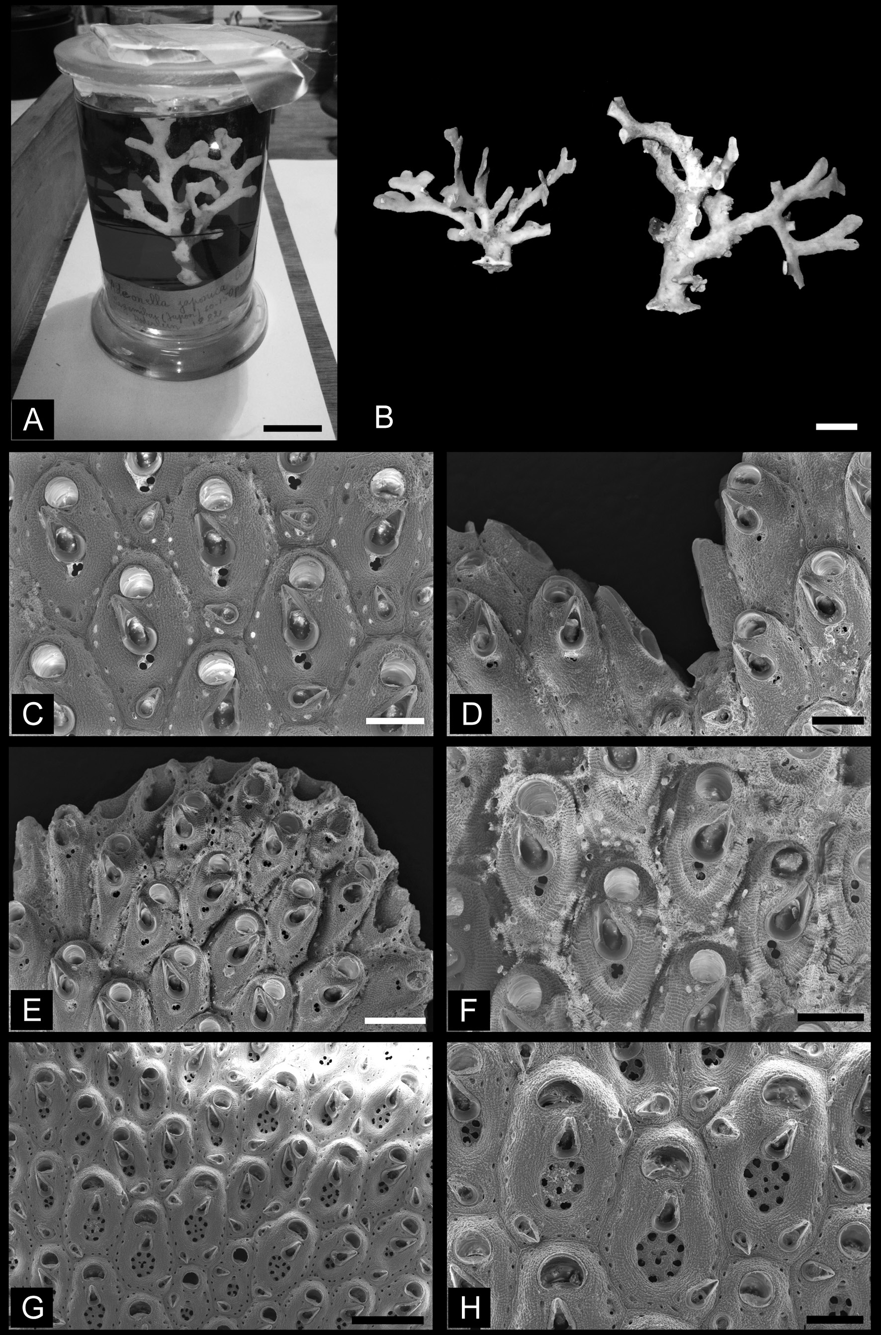
Fig. 7. Adeonellopsis japonica (Ortmann, 1890). A. Lectotype from among Ortmann’s specimens in the Döderlein Collection (MZS 3-3). B. Colonies collected at Otsuchi (NSMT-Te760). C. Autozooids with large suboral avicularium and biporous spiramen (NSMT-Te741, from lectotype, MZS 3-3). D. Lateral vicarious avicularia at branch bifurcation (NSMT-Te741, from lectotype, MZS 3-3). E. Young autozooids at distal end of branch (NSMT-Te741, from lectotype, MZS 3-3). F. Young autozooids, with biporous spiramen surrounded by thick, swollen rim bearing rows of granulation (NSMT-Te741, from lectotype, MZS 3-3). G. Autozooids and gonozooids at branch bifurcation (NSMT-Te742, from paralectotype MZS 3-4). H. Gonozooids showing large multiporous spiramen (NSMT-Te742, from paralectotype MZS 3-4). A–B = optical photographs; C–H = SEM images. Scale bars: A = 2 cm; B = 1 cm; C–D, F, H = 200 μm; E = 300 μm; G = 500 μm.

Imageimage/png© Hirose, MasatoHirose, Masato

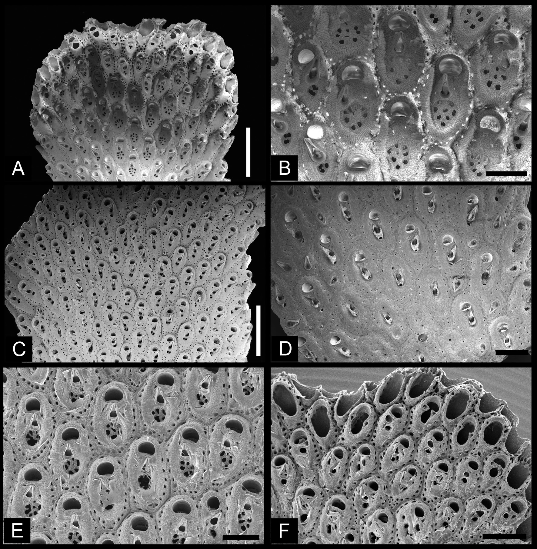
Fig. 8. Adeonellopsis japonica (Ortmann, 1890). A. Distal end of branch just starting to bifurcate, showing young autozooids and gonozooids (NSMT-BryR277). B. Enlargement of young gonozooids, showing peripheral rim with rows of granulation perpendicular to margin, and large multiporous spiramen (NSMT-BryR277). C. Autozooids in middle of branch, showing immersed spiramen (NSMT- Bry R 193). D. Basal part of branch, showing autozooids, and some kenozooids with closed orifice (NSMT-BryR36). E. Young gonozooids, showing distinct marginal pores and multiporous spiramen (NSMT-Te758). F. Distal end of branch, showing autozooid formation (NSMT-Te758). SEM images. Scale bars: A, C = 1 mm, B, D–E = 300 μm; F = 500 μm.

Imageimage/png© Hirose, MasatoHirose, Masato

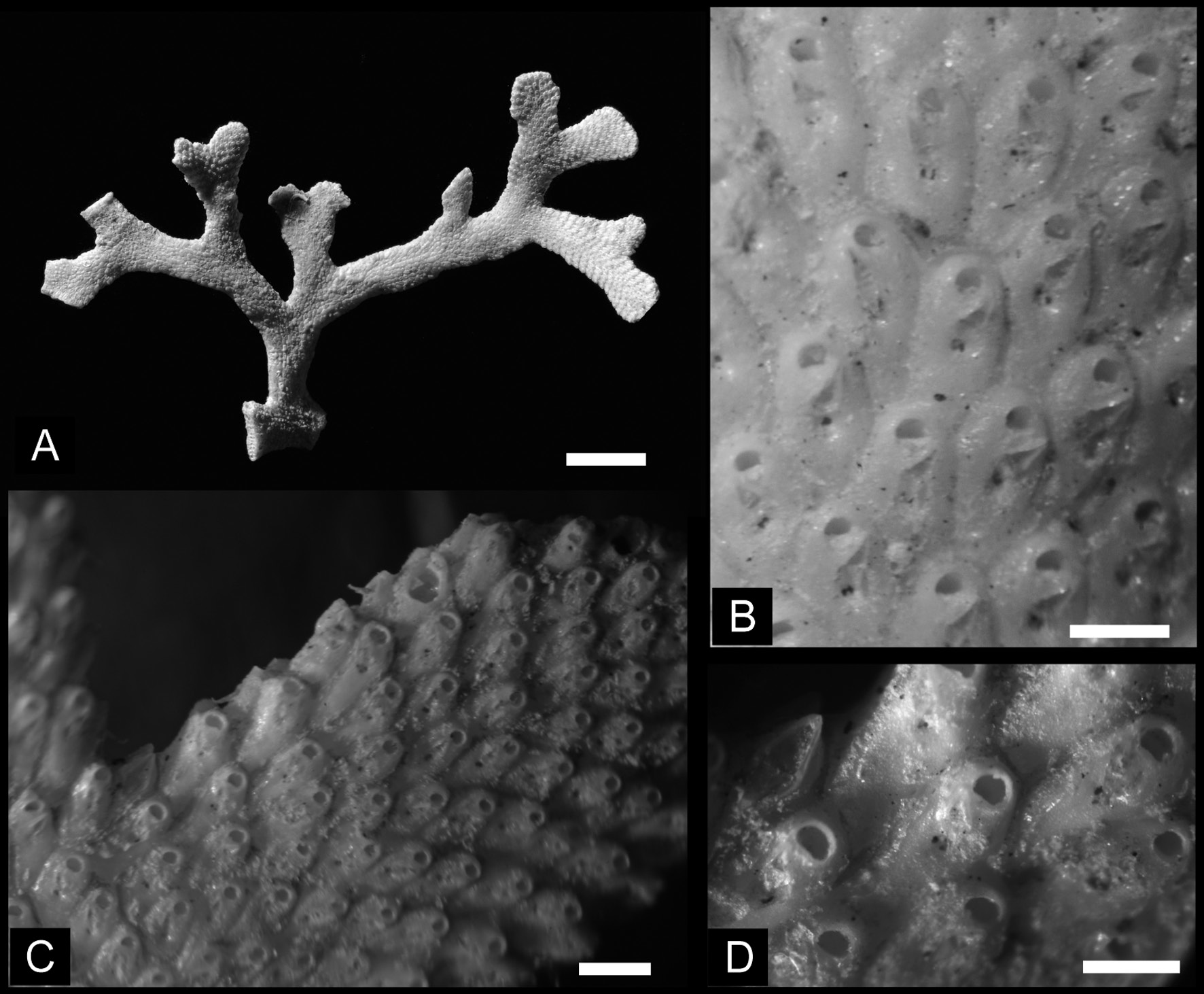
Fig. 9. Adeonellopsis japonica (Ortmann, 1890). A. Colony collected at Albatross Station D, off Oahu Island, Hawaii (USNM 271601). B. Autozooids of Hawaii specimen, showing suboral avicularia and biporous spiramen (USNM 271601, Hawaii). C. Autozooids and lateral vicarious avicularia at a branch bifurcation (USNM 271601 paper box, Hawaii). D. Enlargement of lateral vicarious avicularia at branch bifurcation (USNM 271601 paper box, Hawaii). A = optical photograph; B–D = photomicrographs. Scale bars: A = 5 mm; B, D = 300 μm; C = 500 μm.

Imageimage/png© Hirose, MasatoHirose, Masato

Fig. 10. Interior frontal shield. A. Adeonellopsis japonica (Ortmann, 1890) (NSMT-Te1052). B. A. parvirostrum sp. nov. (NSMT-Te807). Arrowheads indicate ring scar. SEM images. Scale bars: A = 50 μm; B = 25 μm.

Imageimage/png© Hirose, MasatoHirose, Masato

IMAGES

Gallery(4)

See Gallery

Occurrences with images

Source Information

Diversity and distribution of adeonid bryozoans (Cheilostomata: Adeonidae) in Japanese waters

checklist

This dataset contains the digitized treatments in Plazi based on the original journal article Hirose, Masato (2016): Diversity and distribution of adeonid bryozoans (Cheilostomata: Adeonidae) in Japanese waters. European Journal of Taxonomy 203 (203): 1-41, DOI: 10.5852/ejt.2016.203

Abstract. Adeonid bryozoans construct antler-like erect colonies and are common in bryozoan assemblages along the Japanese Pacific coast. The taxonomy of Japanese adeonid species, however, has not been studied since their original descriptions more than 100 years ago. In the present study, adeonid specimens from historical collections and material recently collected along the Japanese coast are examined. Eight adeonid species in two genera were detected, of which Adeonella jahanai sp. nov., Adeonellopsis parvirostrum sp. nov., and Adeonellopsis toyoshioae sp. nov. are described as new species based on the branch width, size and morphology of frontal or suboral avicularia, shape and size of areolar pores, and size of the spiramen. Adeonellopsis arculifera (Canu & Bassler, 1929) is a new record for Japan. Lectotypes for Adeonellopsis japonica (Ortmann, 1890) and Adeonella sparassis (Ortmann, 1890) were selected among Ortmann’s syntypes. Most species of Adeonellopsis around Japan have a southern distribution from Sagami Bay to Okinawa, while A. japonica shows a more northern distribution from Kouchi to Otsuchi. In contrast, Adeonellopsis arculifera was collected only from southwestern Japan. A key to Japanese adeonid species is provided.

Hirose M, valdenar (2016). Diversity and distribution of adeonid bryozoans (Cheilostomata: Adeonidae) in Japanese waters. European Journal of Taxonomy. Checklist dataset https://doi.org/10.5852/ejt.2016.203 accessed via GBIF.org on 2026-06-18.

CC0Published 6/8/2016View dataset
GBIF Usage Key
164249544
Dataset Key
af31502e-3df0-4f1a-bea6-e24ed3f38a88
Origin
source
Backbone Key
8963773
Taxon ID
C55487F1FFD0FFB2FDAAFB84FC53FAB7.taxon
Last Crawled
6/10/2026
Last Interpreted
6/10/2026