Palaeovaranus cayluxi
Zittel, 1887
GBIF:181601118
ABOUT
Descriptions(5)
Export occurrence data
Darwin Core Archive (ZIP)
CLASSIFICATION
Taxonomic Classification Tree

MULTIMEDIA
Media Files(5)
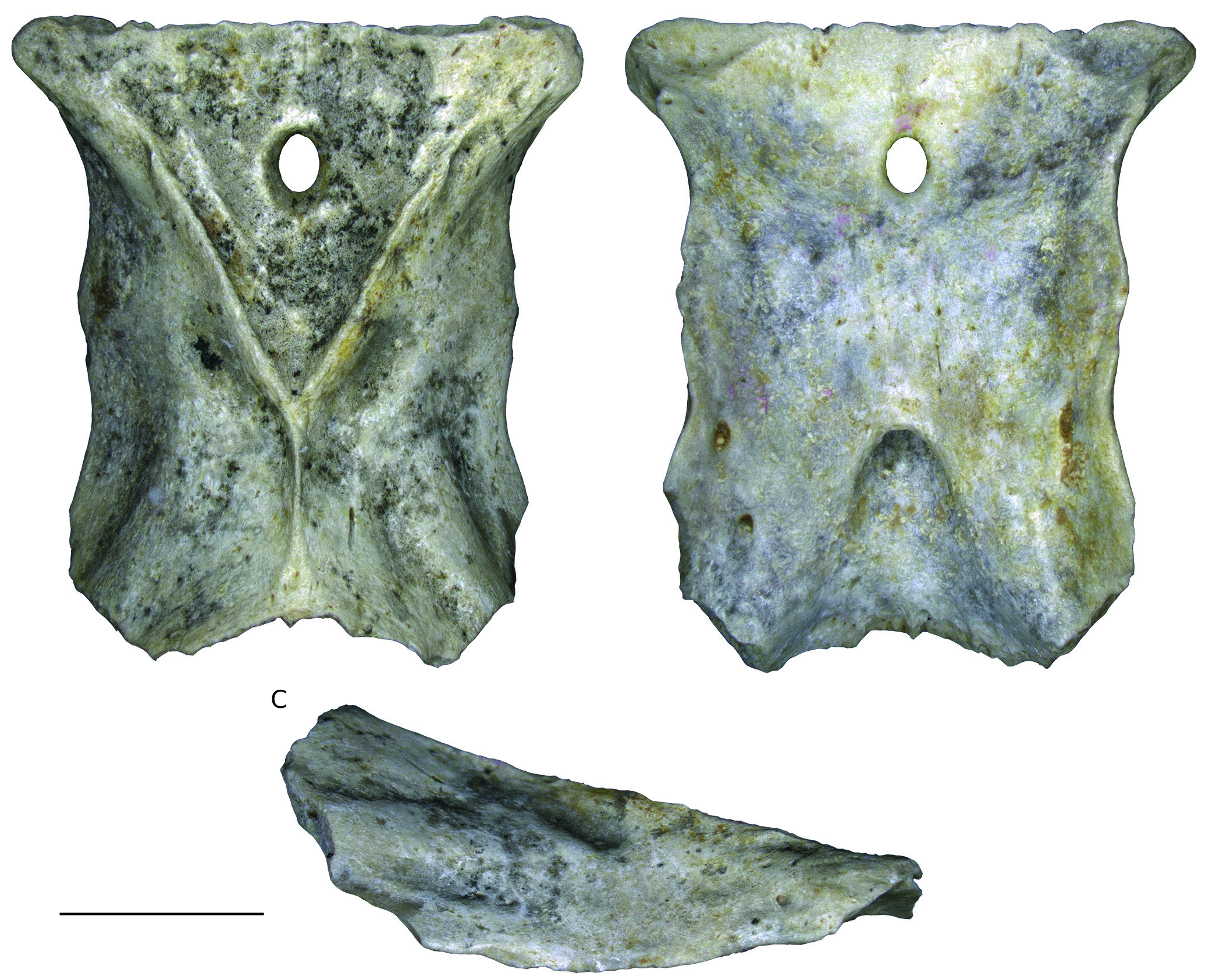
FIG. 34. — Palaeovaranus cayluxiZittel,1887-1890.Photographs of parietal NHMW 2019/0048/0001 in dorsal (A), ventral (B), and right lateral (C) views.Scale bar:5 mm.
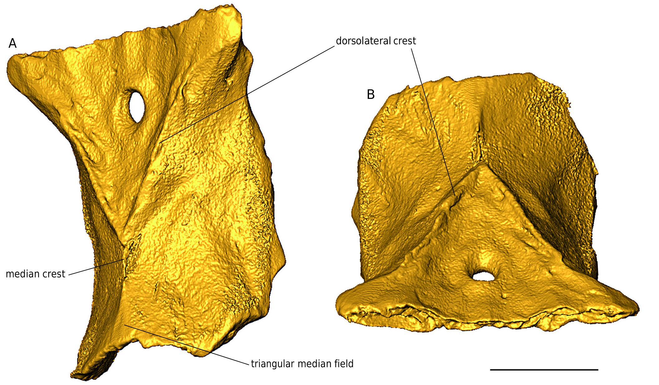
FIG. 35. — Palaeovaranus cayluxi Zittel,1887-1890.Virtual 3D models of parietal NHMW 2019/0048/0001 in dorsolateral (A) and anterior (B) views.Scale bar:5 mm.
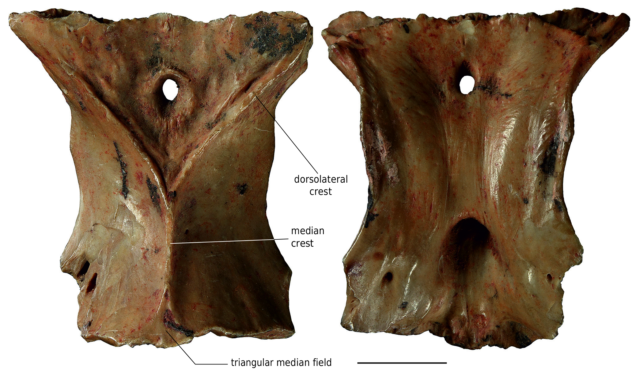
FIG. 36. — Palaeovaranus cayluxi Zittel, 1887-1890. Photographs of parietal MNHN.F.QU17176 in dorsal (A) and ventral (B) views. This specimen originally appeared as drawings in Augé (2005: fig. 188). Scale bar: 5 mm.
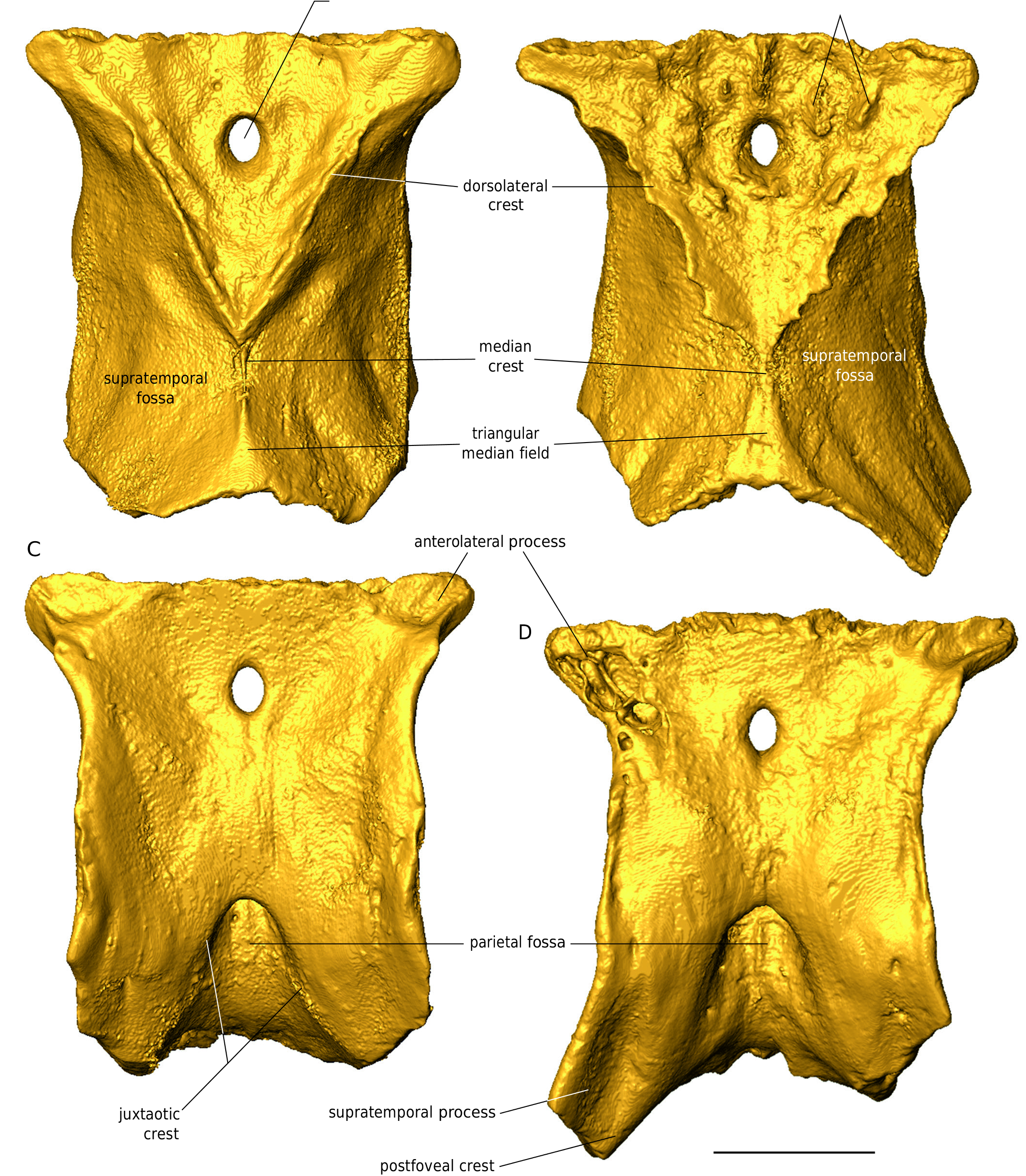
FIG. 37. — Palaeovaranus cayluxi Zittel, 1887-1890 and Palaeovaranus lismonimenos n. sp. A, C, virtual 3D models of parietal NHMW 2019/0048/0001 of Palaeovaranus cayluxi in dorsal (A) and ventral (C) views; B, D, virtual 3D models of the holotype parietal NHMW 2019/0047/0001 of Palaeovaranus lismonimenos n. sp. in dorsal (B) and ventral (D) views. Scale bar: 5 mm.
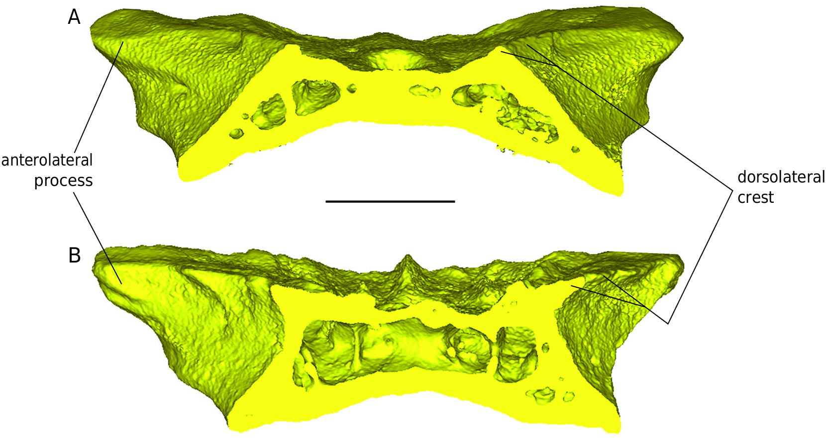
FIG. 41. — Palaeovaranus cayluxi Zittel, 1887-1890 and Palaeovaranus lismonimenos n. sp. Transverse sections of 3D models of parietals. A, parietal NHMW 2019/0048/0001 of Palaeovaranus cayluxi; B, holotype parietal NHMW 2019/0047/0001 of Palaeovaranus lismonimenos n. sp. Scale bar: 3 mm.
IMAGES