
Charinus alagoanus
Miranda, Giupponi, Prendini & Scharff, 2021
GBIF:188640119
ABOUT
Descriptions(7)
Export occurrence data
Darwin Core Archive (ZIP)
CLASSIFICATION
Taxonomic Classification Tree

MULTIMEDIA
Media Files(5)
Fig. 10. Charinus Simon, 1892, chelicerae, prolateral and retrolateral views. A–B. Charinus loko sp. nov. (SMNS). C–D. Charinus miskito sp. nov. (SMNS). E–F. Charinus mocoa sp. nov. (SMF 68). G–H. Charinus alagoanus sp. nov. (MNRJ 9295). I–J. Charinus renneri sp. nov. (MNRJ 9198). K–L. Charinus madagascariensis Fage, 1954 (MNHN). Scale bars: 0.5 mm.
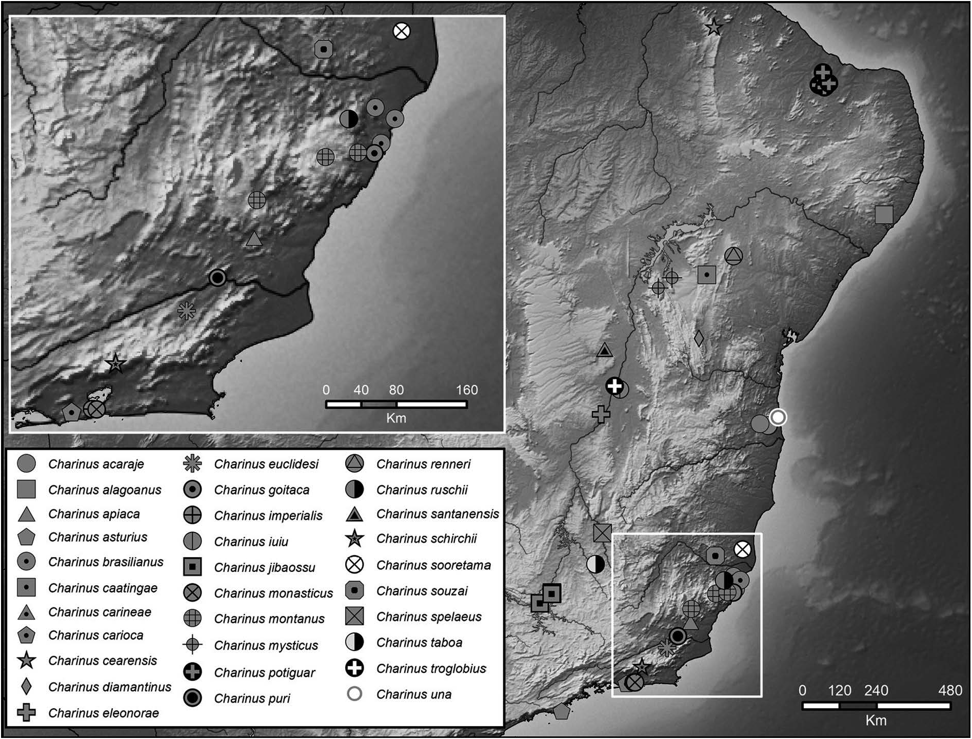
Fig. 43. Map plotting known distributions of species of Charinus Simon, 1892 in eastern South America, with inset for southeastern Brazil.

Fig. 44. Charinus alagoanus sp. nov. (MNRJ 9294), general morphology, ♀. A. Habitus, dorsal view. B. Sternum, ventral view. C. Frontal process. D. Pedipalp tarsus, frontal view. E. Pedipalp, dorsal view. F. Pedipalp, ventral view. Scale bars: A, E–F = 1 mm; B, D = 0.5 mm; C = 0.25 mm.
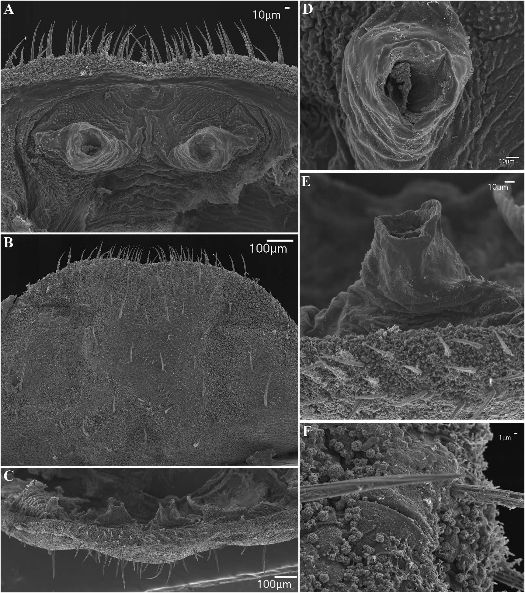
Fig. 45. Charinus alagoanus sp. nov. (MNRJ 9295), female gonopod and genital operculum. A. Suckerlike gonopod, dorsal view.B. Genital operculum, ventral view. C. Gonopods, posterior view.D. Aperture on sinistral side of sucker-like gonopod, dorsal view. E. Projection on sinistral side of sucker-like gonopod, posterior view. F. Gland opening near lateral margin of genital operculum.

Fig. 46. Charinus alagoanus sp. nov. (MNRJ 9295), antenniform leg I, ♀. A. Apex of distal article. B. Distal segment of tarsus, lateral view. C. Apex of distal article of tarsus showing claw and tarsal organ. D. Detail of claw and tarsal organ. E. Rod sensilla and olfactory setae.
IMAGES