
Sarax bispinosus
(Nair, 1934) Miranda, Giupponi, Prendini & Scharff, 2021
GBIF:188640180
0
Synonyms
ABOUT
Descriptions(8)
Export occurrence data
Darwin Core Archive (ZIP)
CLASSIFICATION
Taxonomic Classification Tree

MULTIMEDIA
Media Files(7)
Fig. 3. Charinidae Quintero, 1986, pedipalp femur, prolateral and dorsal views. A–B. Charinus carinae sp. nov. (MNRJ 9293). C–D. Charinus gertschi Goodnight & Goodnight, 1946 (AMCC [LP 10076]). E–F. Sarax bispinosus (Nair, 1934) (AMCC [LP 12298]). G–H. Sarax willeyi Gravely, 1915 (SMF). Scale bars: A–D, G–H = 1 mm; E–F = 0.5 mm.

Fig. 4. Charinidae Quintero, 1986, pedipalp patellar articles, dorsal and prolateral views. A–B. Charinus carinae sp. nov. (MNRJ 9293). C–D. Charinus gertschi Goodnight & Goodnight, 1946 (AMCC [LP 10076]). E–F. Sarax bispinosus (Nair, 1934) (AMCC [LP 12298]). G–H. Sarax willeyi Gravely, 1915 (SMF). Scale bars: A–D, G–H = 1 mm; E–F = 0.5 mm.
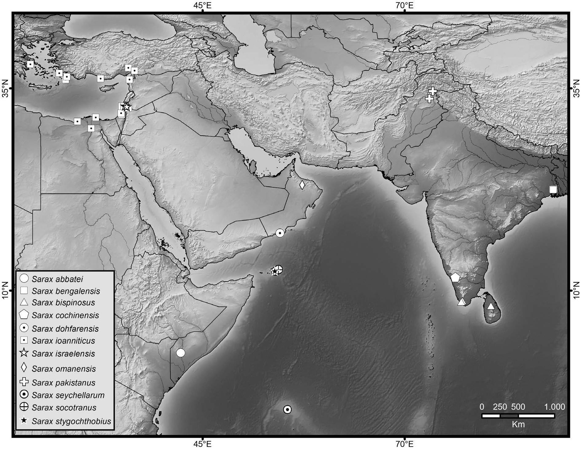
Fig. 135. Map plotting known distributions of species of Sarax Simon, 1892 in Africa, the Middle East and South Asia.

Fig. 137. Sarax bispinosus (Nair, 1934) (AMCC [LP 12298]), general morphology, ♂. A. Habitus, dorsal view. B. Sternum, ventral view. C. Frontal process. D. Pedipalp tarsus, frontal view. E. Pedipalp, dorsal view. F. Pedipalp, ventral view. Scale bars: A–B, E–F = 1 mm; C = 0.1 mm; D = 0.5 mm.
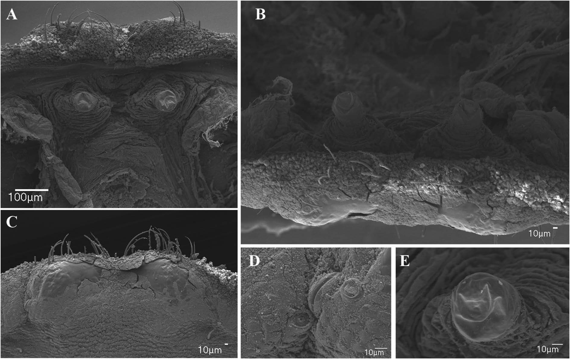
Fig. 138. Sarax bispinosus (Nair, 1934) (AMNH), female gonopod and genital operculum. A. Fingerlike gonopod, dorsal view. B. Gonopods, posterior view. C. Posterior margin of genital operculum, ventral view. D. Glandular opening on margin of genital operculum. E. Dextral gonopod.
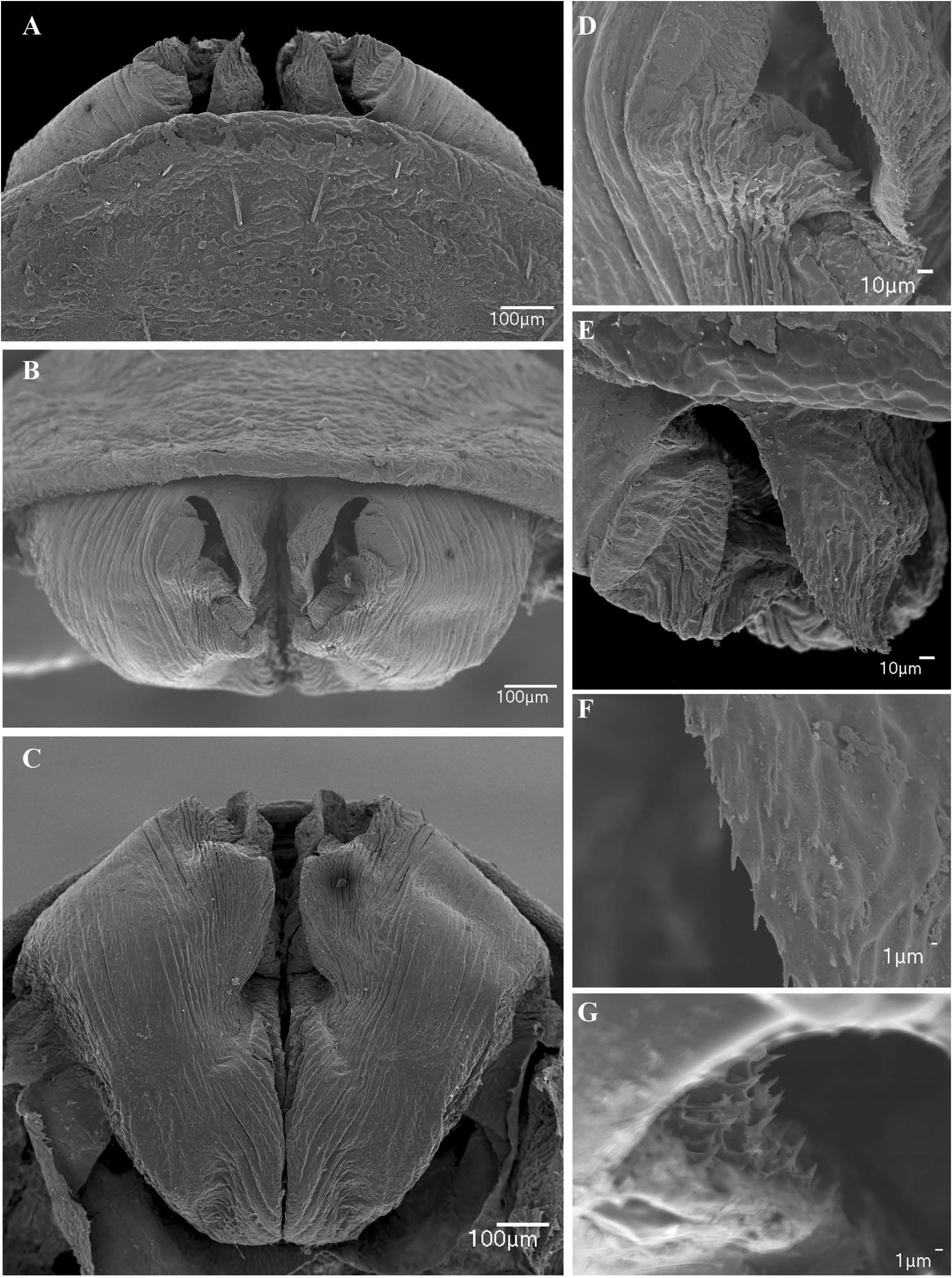
Fig. 139. Sarax bispinosus (Nair, 1934) (AMNH), male gonopod. A. Ventral view. B. Posterior view. C. Dorsal view. D. Sinistral side of gonopod. E. Lamina medialis. F. Margin of lamina medialis. G. Internal surface of fistula with spiny projections.
IMAGES