
Charinus
Simon, 1892
GBIF:190223677
ABOUT
Descriptions(1)
Key to the identification of the species of Charinus in eastern South America
1. Leg IV distitibia with four trichobothria in frontal and caudal series; leg IV basitibia with three pseudo-articles ............................................................................. C. alagoanus sp. nov. (Figs 44–46)
– Leg IV distitibia with five or six trichobothria frontal and caudal series; leg IV basitibia with two or four pseudo-articles ........................................................................................................................... 2
2. Leg IV distitibia with five trichobothria in frontal and caudal series ............................................... 3
– Leg IV distitibia with six trichobothria in frontal and caudal series .............................................. 12
3. Median eyes absent............................................................................................................................ 4
– Median eyes present .......................................................................................................................... 5
4. Tibia of leg I with 21 articles; leg I tarsus with 37 articles; leg IV basitibia with two pseudoarticles........................................................................................ C. monasticus sp. nov. (Figs 69–70)
– Tibia of leg I with 23 articles; leg I tarsus with 41 articles; leg IV basitibia with four pseudoarticles............................................................................... C. troglobious Baptista & Giupponi, 2002
5. Pedipalp femur with three dorsal spines; pedipalp patella with two ventral spines ............................ ............................................................................................................... C. una sp. nov. (Figs 78–79)
– Pedipalp femur with more than three dorsal spines; pedipalp patella with more than two ventral spines ................................................................................................................................................. 6
6. Median and lateral eyes reduced........................................................................................................ 7
– Median and lateral eyes unmodified.................................................................................................. 8
7. Median ocular tubercle present but reduced; reduced median eyes with dark pigmentation; pedipalp patella dorsal spine IV two-thirds length of dorsal spine III ............................................................... .............................................................................. C. taboa Vasconcelos, Giupponi & Ferreira, 2016
– Median ocular tubercle absent, reduced eyes situated directly on tegument; reduced median eyes unpigmented; pedipalp patella dorsal spine IV one-fifth length of dorsal spine III ............................ .......................................................................................... C. spelaeus Vasconcelos & Ferreira, 2017
8. Cheliceral basal segment with long (distinct) tooth adjacent to bifid tooth ........................................ ..................................................................................... C. sooretama sp. nov. (Figs 7E–F, 9C–D, 75)
– Cheliceral basal segment with short (indistinct) tooth adjacent to bifid tooth .................................. 9
9. Base of female gonopod unsclerotized ................... C. euclidesi sp. nov. (Figs 6C–D, 9A–B, 61–63)
– Base of female gonopod sclerotized................................................................................................ 10
10. Pedipalp tibia with two ventral spines ..................................... C. diamantinus sp. nov. (Figs 57–60)
– Pedipalp tibia with one ventral spine................................................................................................11
11. Curved carina present between ocular triads and lateral margin of carapace; cheliceral claw with nine teeth............................................................................................ C. souzai sp. nov. (Figs 76–77)
– Curved carina absent between ocular triads and lateral margin of carapace (Fig. 48A–B); cheliceral claw with six or seven teeth..................................... C. brasilianus Weygoldt, 1972 (Figs 8C–D, 48)
12. Leg I tarsus with 28 articles.................................................................. C. montanus Weygoldt, 1972
– Leg I tarsus with 41 articles............................................................................................................. 13
13. Carapace anterior margin with eight or ten setae ............................................................................ 14
– Carapace anterior margin with six setae.......................................................................................... 16
14. Carapace anterior margin with ten setae; median ocular tubercle absent; pedipalp patella with spine between ventral spine I and distal margin .......................... C. eleonorae Baptista & Giupponi, 2003
– Carapace anterior margin with eight setae; median ocular tubercle present; pedipalp patella with setiferous tubercles between ventral spine I and distal margin ....................................................... 15
15. Pedipalp tarsus with two dorsal spines (Fig. 47D) ...................... C. apiaca sp. nov. (Figs 5A–B, 47)
– Pedipalp tarsus with three dorsal spines ......................... C. caatingae Vasconcelos & Ferreira, 2016
16. Carapace anterior margin projected anteriorly ............ C. santanensis Vasconcelos & Ferreira, 2017
– Carapace anterior margin rounded................................................................................................... 17
17. Leg I tarsus, first article equal in length to subsequent three articles.................................................. ......................................................................... C. jibaossu Vasconcelos, Giupponi & Ferreira, 2014
– Leg I tarsus, first article equal in length to all subsequent articles ................................................. 18
18. Pedipalp tarsus with three dorsal spines .......................................................................................... 19
– Pedipalp tarsus with two dorsal spines ............................................................................................ 23
19. Cheliceral claw with four teeth.................................................. C. mysticus Giupponi & Kury, 2002
– Cheliceral claw with 9–13 teeth ...................................................................................................... 20
20. Pedipalp coxa, rounded dorsal carina containing seven setae ......................................................... 21
– Pedipalp coxa, rounded dorsal carina containing two or five setae................................................. 22
21. Pedipalp patella with six ventral spines (Fig. 49F); cheliceral claw with 13 teeth ............................. .................................................................................. C. carinae sp. nov. (Figs 3A–B, 4A–B, 49–51)
– Pedipalp patella with three or four ventral spines; cheliceral claw with ten teeth............................... .......................................................................................................... C. goitaca sp. nov. (Figs 64–65)
22. Pedipalp coxa, rounded dorsal carina containing five setae; pedipalp patella with six dorsal spines .................... C. ruschii Miranda, Milleri-Pinto, Gonçalves-Souza, Giupponi & Scharff, 2016
– Pedipalp coxa, rounded dorsal carina containing two setae; pedipalp patella with four dorsal spines ..................................................................................... C. carioca sp. nov. (Figs 6E–F, 52–53)
23. Pedipalp coxa, rounded dorsal carina containing no setae .............................................................. 24
– Pedipalp coxa, rounded dorsal carina containing setae ................................................................... 26
24. Pedipalp patella with three dorsal spines............................................................................................. .................................. C. acaraje Pinto-da-Rocha, Machado & Weygoldt, 2002 (Figs 7A–B, 8A–B)
– Pedipalp patella with five dorsal spines........................................................................................... 25
25. Pedipalp femur with four dorsal spines ..................... C. renneri sp. nov. (Figs 6A–B, 10I–J, 73–74)
– Pedipalp femur with five dorsal spines..... C. asturius Pinto-da-Rocha, Machado & Weygoldt, 2002
26. Pedipalp femur with five dorsal spines......................................... C. cearensis sp. nov. (Figs 54–56)
– Pedipalp femur with four dorsal spines ........................................................................................... 27
27. Pedipalp femur with five ventral spines........................................................................................... 28
– Pedipalp femur with four ventral spines.......................................................................................... 29
28. Pedipalp femur with secondary row of dorsal spines; pedipalp patella, distance from ventral spine I to distal margin half length of spine ...................................................... C. puri sp. nov. (Figs 71–72)
– Pedipalp femur without secondary row of dorsal spines; pedipalp patella, distance from ventral spine I to distal margin greater than length of spine ............... C. iuiu Vasconcelos & Ferreira, 2016
29. Cheliceral claw with up to 13 teeth; median and lateral eyes reduced................................................ ......................................................................... C. potiguar Vasconcelos, Giupponi & Ferreira, 2013
– Cheliceral claw with 11 teeth; median and lateral eyes unmodified.................................................... ..................................................................................................... C. imperialis sp. nov. (Figs 66–68)
Export occurrence data
Darwin Core Archive (ZIP)
CLASSIFICATION
Taxonomic Classification Tree

MULTIMEDIA
Media Files(44)
Fig. 3. Charinidae Quintero, 1986, pedipalp femur, prolateral and dorsal views. A–B. Charinus carinae sp. nov. (MNRJ 9293). C–D. Charinus gertschi Goodnight & Goodnight, 1946 (AMCC [LP 10076]). E–F. Sarax bispinosus (Nair, 1934) (AMCC [LP 12298]). G–H. Sarax willeyi Gravely, 1915 (SMF). Scale bars: A–D, G–H = 1 mm; E–F = 0.5 mm.

Fig. 4. Charinidae Quintero, 1986, pedipalp patellar articles, dorsal and prolateral views. A–B. Charinus carinae sp. nov. (MNRJ 9293). C–D. Charinus gertschi Goodnight & Goodnight, 1946 (AMCC [LP 10076]). E–F. Sarax bispinosus (Nair, 1934) (AMCC [LP 12298]). G–H. Sarax willeyi Gravely, 1915 (SMF). Scale bars: A–D, G–H = 1 mm; E–F = 0.5 mm.
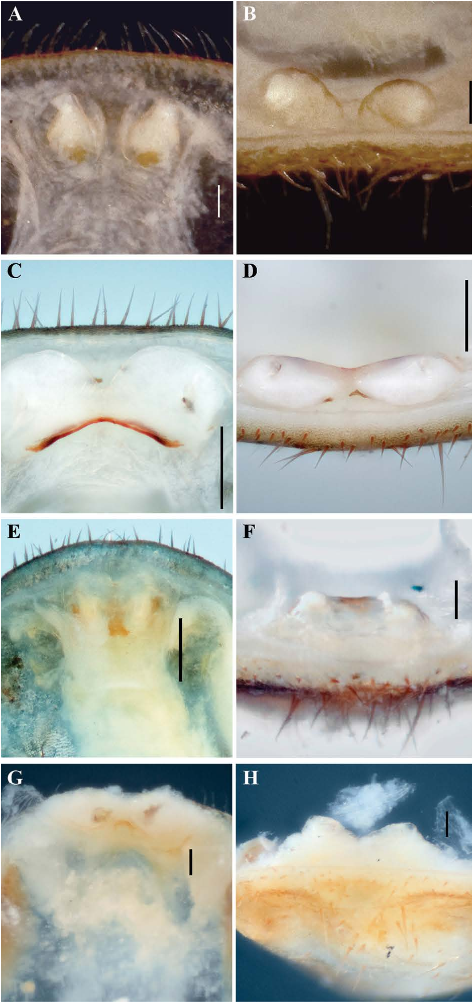
Fig. 5. Charinus Simon, 1892, female gonopods, dorsal (left column) and posterior (right column) views. A–B. Charinus apiaca sp. nov. (MNRJ 9286). C–D. Charinus gertschi Goodnight & Goodnight, 1946 (AMCC [LP 10076]). E–F. Charinus miskito sp. nov. (SMNS). G–H. Charinus mocoa sp. nov. (SMF 68). Scale bars: A–B, F–H = 0.1 mm; C–E, I–L = 0.25 mm.

Fig. 6. Charinus Simon, 1892, female gonopods, dorsal (left column) and posterior (right column) views. A–B. Charinus renneri sp. nov. (MNRJ 9198). C–D. Charinus euclidesi sp. nov. (MNRJ 9099). E–F. Charinus carioca sp. nov. (MNRJ 9201). Scale bars: A–D = 0.25 mm; E–F = 0.1 mm.
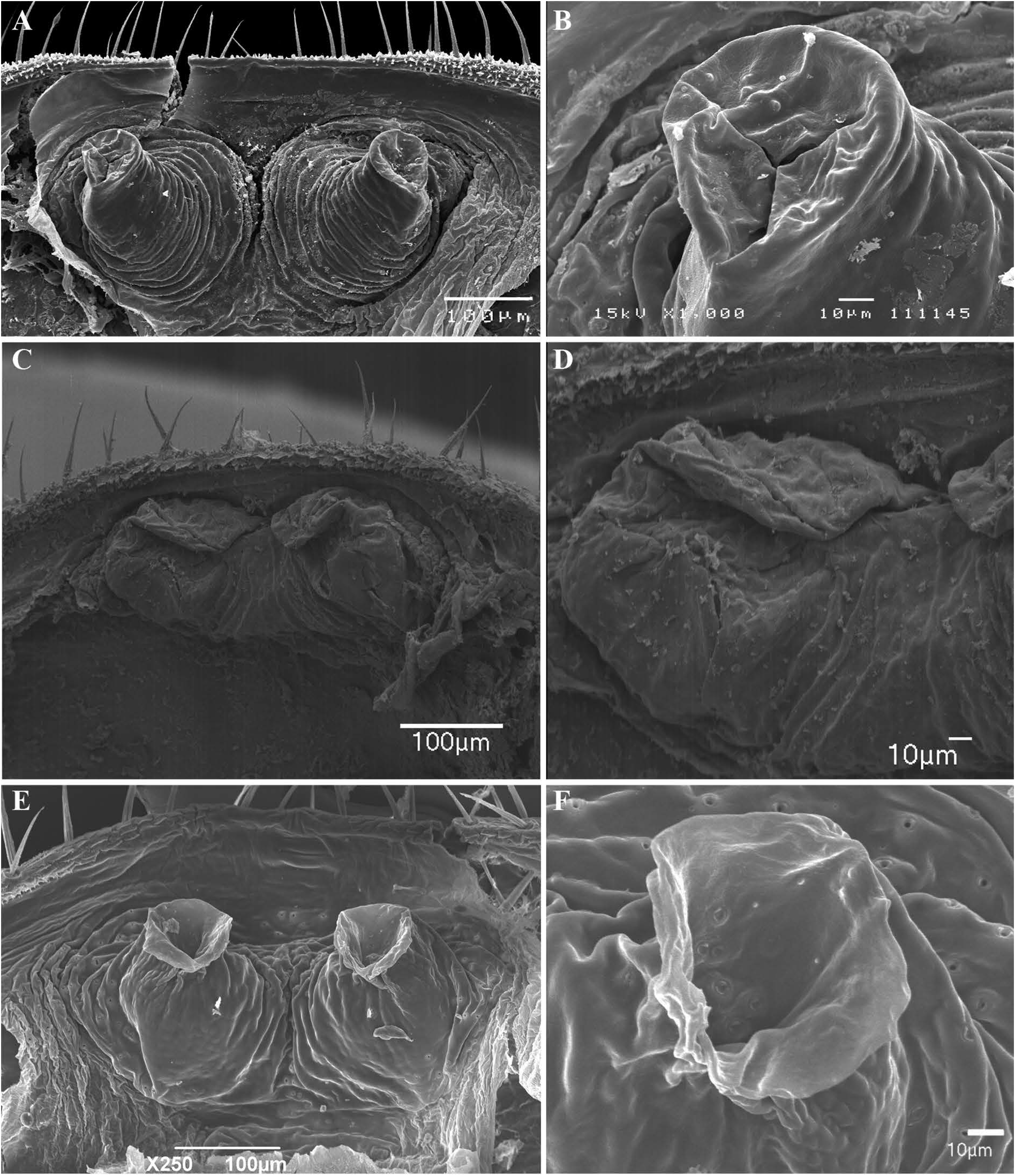
Fig. 7. Charinus Simon, 1892, female gonopods, dorsal view (left column) and detail (right column). A–B. Charinus acaraje Pinto-da-Rocha, Machado & Weygoldt, 2002 (MNRJ 9297). C–D. Charinus palikur sp. nov. (AMCC [LP 3831]) E–F. Charinus sooretama sp. nov. (MNRJ 9245).

Fig. 8. Charinus Simon, 1892, male gonopods. A–B. Charinus acaraje Pinto-da-Rocha, Machado & Weygoldt, 2002 (MNRJ 9297), ventral view (A) and detail of dorsal lobe (LoD) and lateral lobe 1 (LoL1). C–D. Charinus brasilianus Weygoldt, 1972 (MNRJ 9226), posterior view (C) and detail of lateral lobes 1 and 2 (LoL1, 2), dorsal lobe (LoD) and lamina medialis (LaM) (D). E–H. Charinus carajas Giupponi & Miranda, 2016 (MZSP 29126), ventral view of gonopod (E), detail of sinistral side of gonopod (F), detail of LoL1 and LoD (G), and detail of LoL2 (H).
IMAGES