Acanthocyrtus
Handschin, 1925
GBIF:194444123
ABOUT
Descriptions(1)
Key to species of Acanthocyrtus
1 Tenent hair chavate (Fig. 8E); Australian species............................................................ 2
- Tenent hair pointed; African species................................................ A. marginalis Salmon, 1956
2 Prelabral chaetae ciliate................................................................................ 3
- Prelabral chaetae smooth............................................................................... 4
3 Head with 7 sutural mac; Th II–III respectively with 9 and 7 mac in p1–3 complex; Abd I with 4 mac; Abd IV with 19 central mac.......................................................................... A. lineatus Womersley, 1934
- Head with 5 sutural mac; Th II–III respectively with 4 and 5 mac in p1–3 complex; Abd I with 2 mac; Abd IV with 15 central mac............................................................................ A. spinosus (Schött, 1917)
4 Labral papillae present (Figs 3E, 9C)..................................................................... 5
- Labral papillae absent.................................................................................. 6
5 Head and Abd III–IV with lateral spots (Fig. 2); labral inner papillae conical, outer papillae reduced to a small rounded projection (Fig. 3E); labial papilla E l.p. surpass the base of a.a. (Fig. 4A); manubrium ventro-distally with 3 inner chaetae (Fig. 7B) A. necropolitanus sp. nov.
- Head and trunk depigmented (Fig. 8); labral inner papillae with 3 projections and outer papillae with 4 projections (Fig. 9C); labial papilla E l.p. not reach the base of a.a. (Fig. 9E); manubrium ventro-distal with 2 inner chaetae (Fig. 11F)................................................................................................ A. pallidus sp. nov.
6 Ant IV apical bulb bilobed; Th II with 24 mac in p1–3 complex; Abd III with 5 lateral mac; Abd IV with 20 central mac; mucronal proximal tooth clearly smaller than distal, mucronal basal spine surpassing the proximal tooth........................................................................... A. barrowensis Zhang, 2009 (in: Zhang et al. 2009)
- Ant IV apical bulb absent; Th II with 17 or less mac in p1–3 complex; Abd III with 4 lateral mac; Abd IV with 15 or less central mac; mucronal teeth subequal, mucronal spine reaching the apex of proximal tooth................................. 7
7 Head S1 and S4 mac present and Pe5 absent; labial basomedian field with 3 smaller chaetae (M2, R and RS); Th II with 6 and 17 mac respectively in m2 and p3 complex; unguis a.t. absent............ A. loftyensis Zhang, 2009 (in: Zhang et al. 2009)
- Head S1 and S4 mac absent and Pe5 present; labial basomedian field with 1 smaller chaetae (M2, RS absent); Th II with 5 and 15 mac respectively in m2 and p3 complex; unguis a.t. present............. A. yolngui Zhang, 2009 (in: Zhang et al. 2009)
Export occurrence data
Darwin Core Archive (ZIP)
CLASSIFICATION
Taxonomic Classification Tree

MULTIMEDIA
Media Files(7)
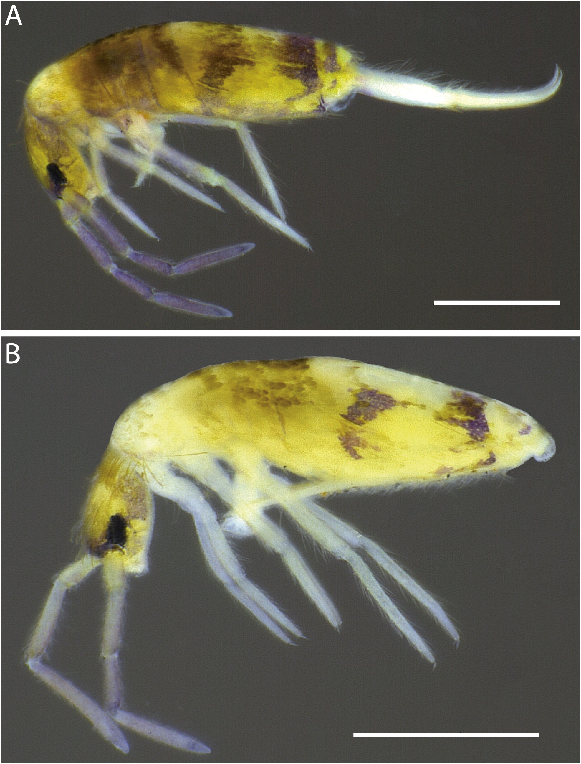
FIGURE 2A–B. Acanthocyrtus necropolitanus sp. nov., habitus of specimens fixed in ethanol (lateral view): A, specimen with weak pigments on trunk lateral; B, pigments absent on trunk lateral. Scale bars: 0.5mm.

FIGURE 4A–D. Acanthocyrtus necropolitanus sp. nov., ventral head (right side); A, labial papillae E (right side); B, maxillary palp and sublobal plate (right side); C, basomedian and basolateral labial fields and proximal chaetae; D, complete postlabial chaetotaxy.
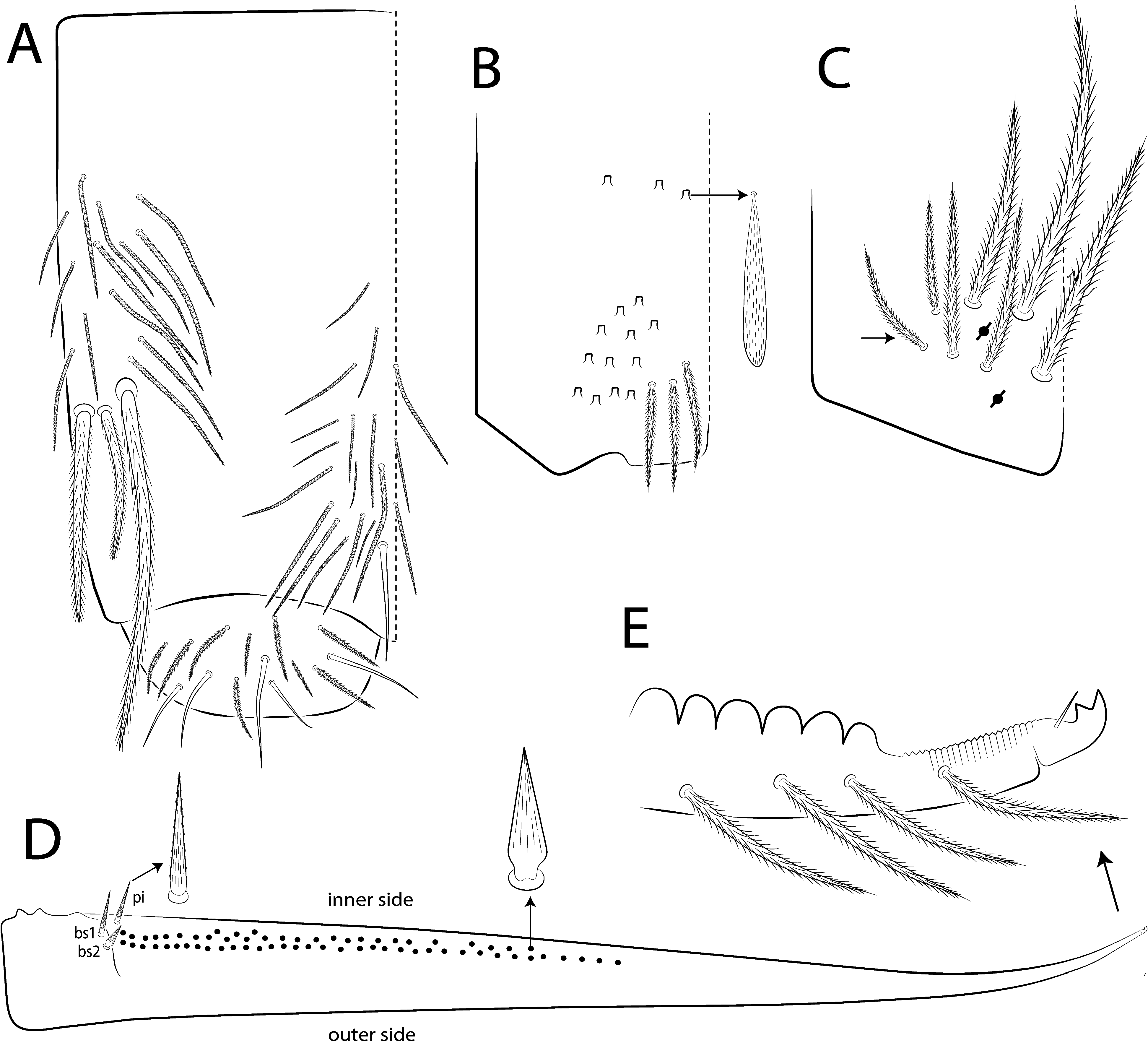
FIGURE 7A–E. Acanthocyrtus necropolitanus sp. nov., abdominal appendages; A, collophore (lateral view); B, distal manubrium (ventral view); C, manubrial plate (dorsal view), arrow indicates chaeta present or absent; D, dens spines chaetotaxy (dorsal view), E, distal dens and mucro (dorsal view).

FIGURE 8. Acanthocyrtus pallidus sp. nov., habitus of specimens fixed in ethanol (lateral view). Scale bars: 0.5mm.
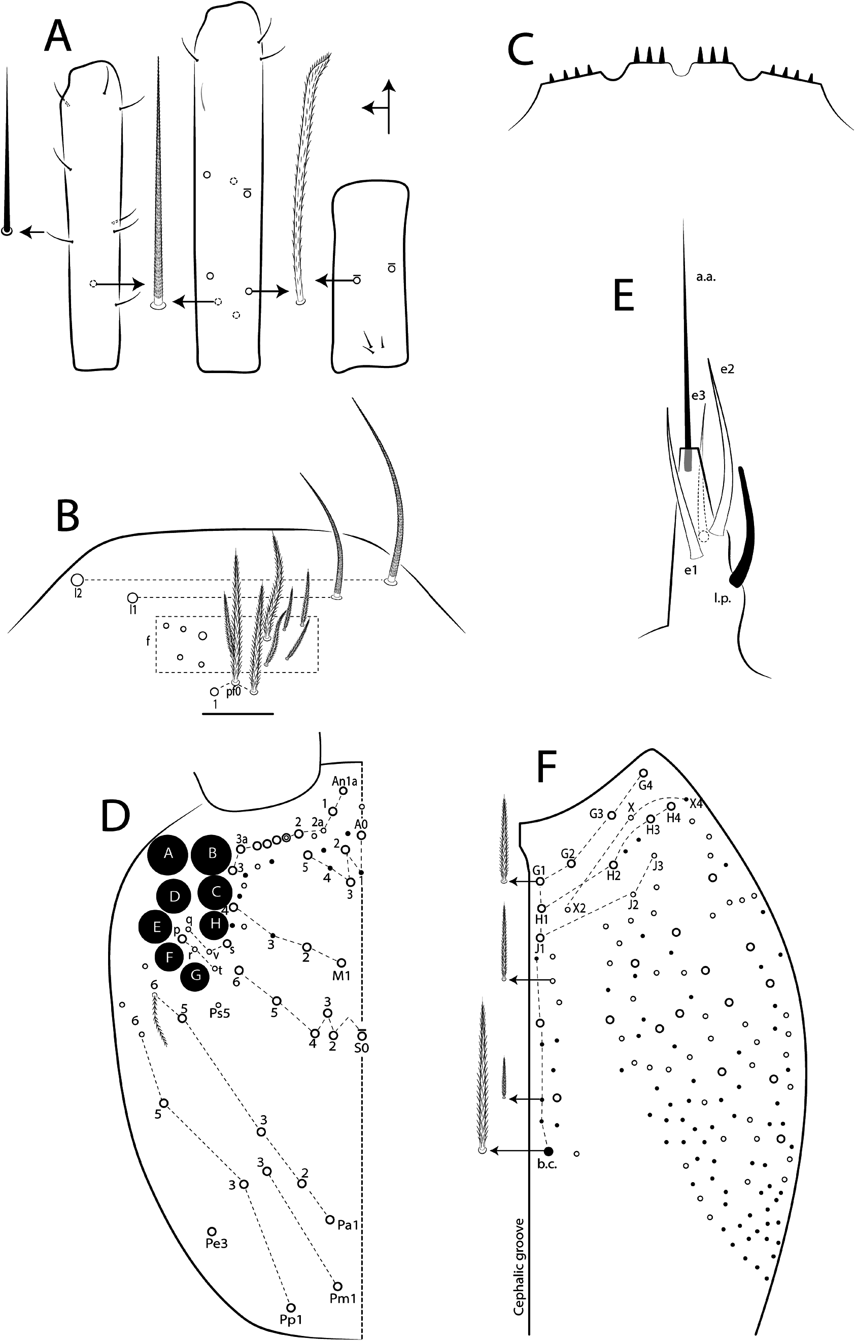
FIGURE 9A–F. Acanthocyrtus pallidus sp. nov., antennae and head; A, chaetotaxy (mac and elongated sens) of left Ant III–I (dorsal view, from left to right), respectively; B, chaetotaxy of clypeus (dorsal view); C, labral papillae (ventral view); D, head dorsal chaetotaxy and eyes (dorsal view, left side); E, labial papillae E (ventral view, right side); F, complete postlabial chaetotaxy (ventral view, right side).

FIGURE 11A–H. Acanthocyrtus pallidus sp. nov., legs and abdominal appendages; A–C, chaetotaxy of left subcoxa I–III respectively (outer side); D, trochanteral organ (posterior view); E, collophore (lateral view), arrow on lateral flap indicate smooth chaeta present or absent; F, distal manubrium (ventral view); G, manubrial plate (dorsal view); H, dens spines chaetotaxy (dorsal view),
IMAGES