AnimaliaNot EvaluatedacceptedspeciesAccepted
Pharyngodictyon magnifili
Monniot C. & Monniot F., 1991
GBIF:202595046
0year
ABOUT
Descriptions(1)
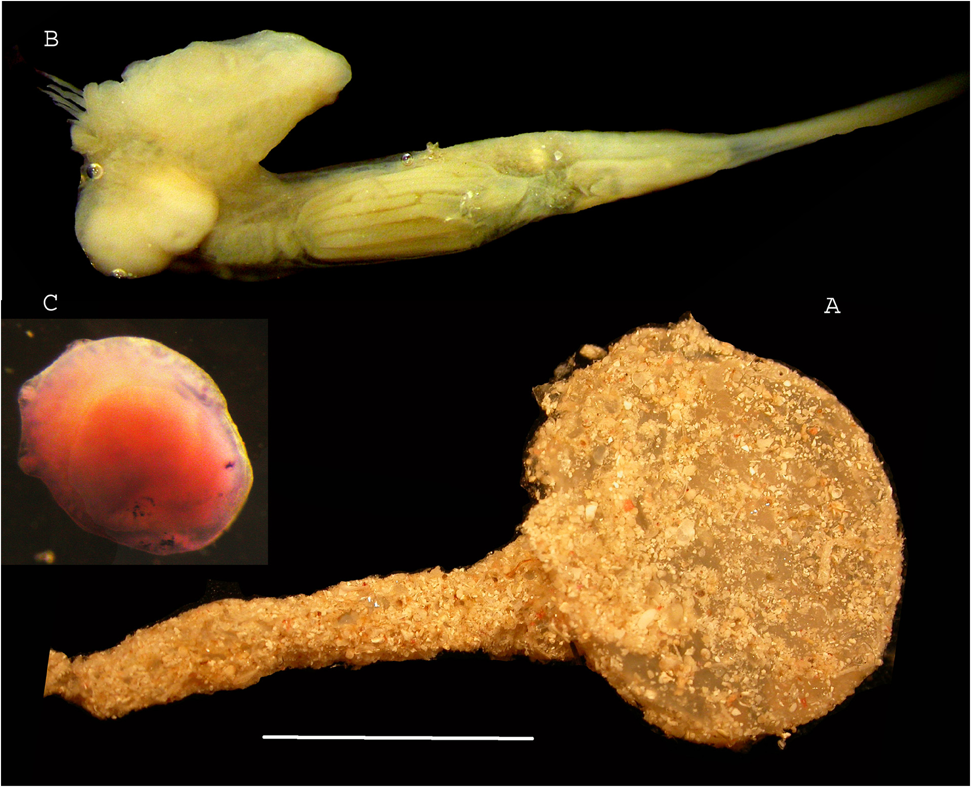
Figure 1 Stations: SPANBIOS: CP 5217: 4 specimens. CP 5237: 1 specimen. The largest specimen has a spherical head 15 mm in diameter on a narrow peduncle 20 mm long (Fig. 1 A). The tunic is vitreous with some encrusted sand. The apertures cannot be seen, and all zooids are contracted. Thoraces and abdomens are located in the head and the long post-abdomens extend inside the peduncle. The oral tentacles are thin, long, protruding out of the siphon (Fig. 1 B). The branchial tissue has at least seven transverse bars with triangular dorsal languets. The stomach is elongated with longitudinal folds (Fig. 1 B). The ovary is anterior to a clump of testis lobes. In one zooid two immature embryos are incubated in the thoracic cavity (Fig. 1 B). One tadpole (Fig. 1 C) was found free inside the colony head. It has three adhesive papillae in a line circled by very numerous ramified vesicles (Fig. 1 C). Sensitive organs cannot be clearly seen. All characters of the SPANBIOS specimens well correspond to those already described from New Caledonia and figured in Monniot C. & Monniot F. 1991 (Figs 7 F; 10).
Exbodi, Françoise Monniot (2022): Additional records of bathyal ascidians (Tunicata) from the New Caledonia region. Zootaxa 5195 (3): 201-223, DOI: 10.11646/zootaxa.5195.3.1
Export occurrence data
Darwin Core Archive (ZIP)
CLASSIFICATION
Taxonomic Classification Tree

MULTIMEDIA
Media Files(3)
FIGURE 1. Pharyngodictyon magnifili. A, colony, scale bar = 1cm. B, zooid. C. larva stained with hemalum.
Imageimage/png© Exbodi, Françoise MonniotExbodi, Françoise Monniot
IMAGES