AnimaliaNot EvaluatedacceptedspeciesAccepted
Bathyoncus lanatus
Monniot C. & Monniot F., 1991
GBIF:202595067
0year
0
Synonyms
ABOUT
Descriptions(1)
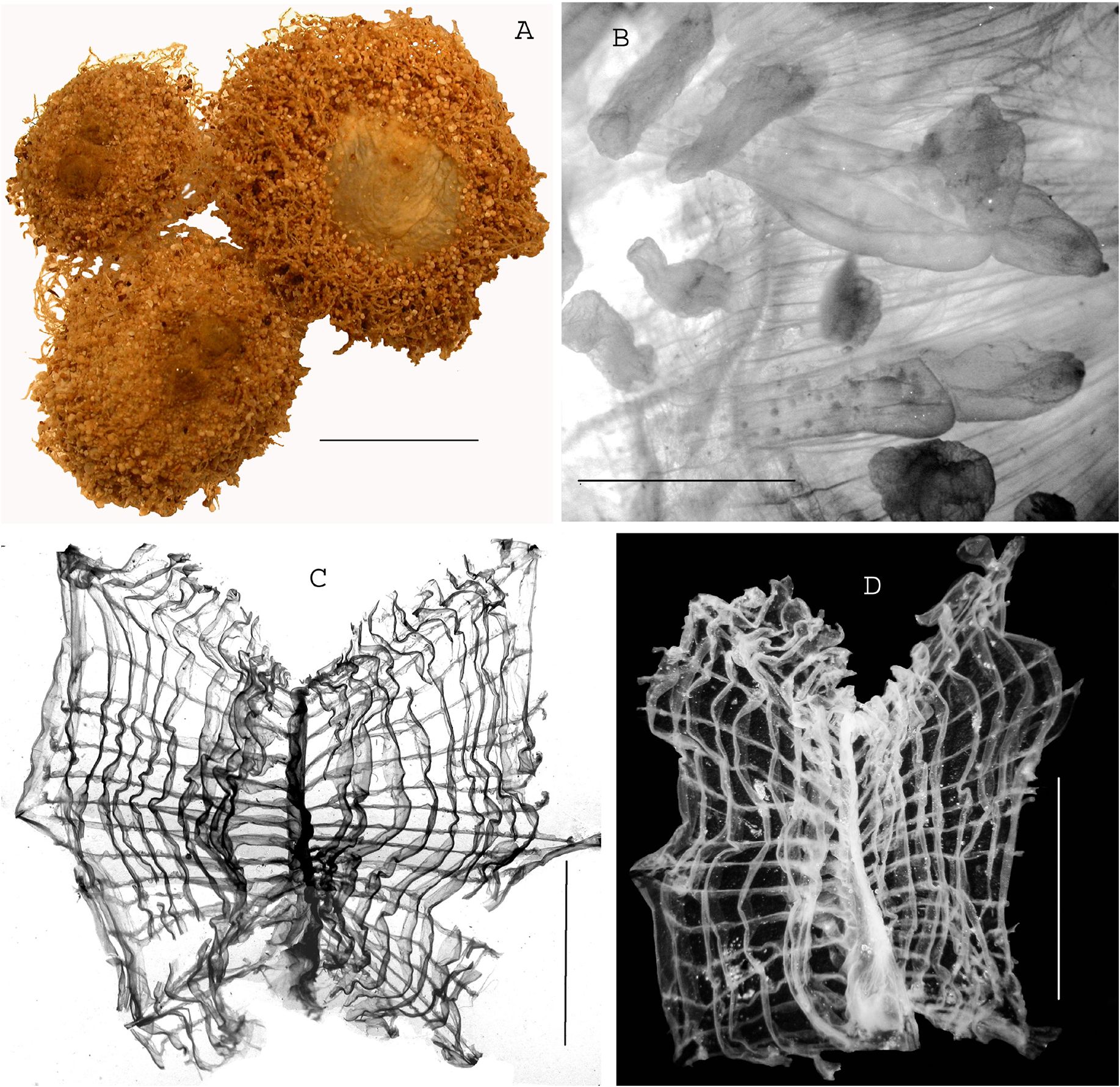
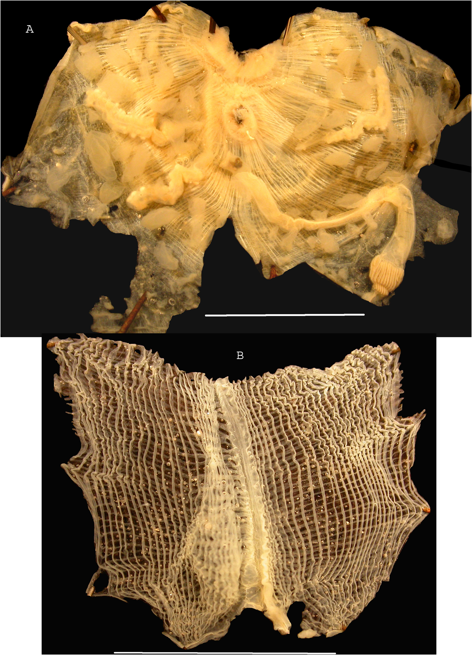
Figures 13 – 15. Stations: SPANBIOS: CP 5220; DW 5257, 11 specimens. EXBODI: CP 3892, 1 specimen. B. lanatus was previously colllected several times in different cruises around New Caledonia from 400 to 1100 m depth and in Fiji (Monniot C. & Monniot F. 1991). Some of them have been re-examined. The different specimens have variable anatomical characters even at the same station concerning the number of branchial vessels and number of gonads but the external aspect is always the same (Fig. 13 A). They certainly represent a single species. The body is round, enclosed in a tunic encrusted with sand with dense rhizoids except on a ventral disc which is naked. Both siphons are placed close to each other at the upper side of the body (Fig. 13 A). The body wall is thin, with a network of crossed muscles thinner on the ventral body side (Fig. 14 A). The siphon sphincters are strong. The oral tentacles are long and thin. The prepharyngeal band does not curve at the level of the button-like dorsal tubercle. The branchial tissue (Fig. 13 C, D) without true stigmata is made of crossed longitudinal and transverse vessels variable in number and not related to the number of gonads and their development. Most of the specimens of the SPANBIOS stations have no more than seven longitudinal vessels on each side but one specimen has up to 12 vessels on each side. On the right side three longitudinal vessels are grouped in a kind of fold (Fig. 13 C, D). Occasionally on the left side one or two dorsal vessels do not reach the base of the branchial sac (Fig. 13 D). The dorsal lamina is smooth. A single specimen from the EXBODI expedition may belong to the same species. It has many more longitudinal vessels with some grouped in a fold (Fig. 15 B). In all specimens the gonads (Fig. 14 B) are long hermaphrodite tubes ending in short, closed male and female papillae. The sperm-duct runs along the ovary. There are either two gonads on each side (Figs 14 A) or one gonad on each side (Fig. 14 C) or two gonads on the right and one gonad on the left (Fig. 15 A). Ampular endocarps are scattered on the body wall on both sides (Fig. 14 A, B, C; 15 A). The digestive loop is widely open (Figs 14 A, B, C; 15 A )). The olive-shaped stomach has 15 to 20 longitudinal folds and a curved caecum. The anus rim is thick and undulated. The morphological characters of B. lanatus seem highly variable among the numerous specimens present in New Caledonia. There remains some doubt about the specific position of the specimen from the EXBODI expedition collected at the same geographic area, having a much more developed branchial tissue but with the same absence of true stigmata; all other organs match well. Two other species of the genus Bathyoncus are present in New Caledonia: Bathyoncus tantulus Monniot & Monniot, 1991 has a body in two parts: a round head and a thick peduncle anchored by basal filaments. There are five longitudinal branchial vessels, one gonad in a polycarp-shape on each side and a large endocarp on the right side.
Exbodi, Françoise Monniot (2022): Additional records of bathyal ascidians (Tunicata) from the New Caledonia region. Zootaxa 5195 (3): 201-223, DOI: 10.11646/zootaxa.5195.3.1
Export occurrence data
Darwin Core Archive (ZIP)
CLASSIFICATION
Taxonomic Classification Tree

NOMENCLATURE
Synonyms(1)
MULTIMEDIA
Media Files(3)
FIGURE 13. Bathyoncus lanatus. A, three specimens from SPANBIOS expedition, scale bar = 1cm. B, detail of two gonads of the right side, scale bar = 2mm. C, D, branchial tissue of different specimens, scale bars = 5mm.
Imageimage/png© Exbodi, Françoise MonniotExbodi, Françoise Monniot

IMAGES