Rhadinorhynchus hiansi
Soota & Bhattacharya, 1981
GBIF:209457594
ABOUT
Descriptions(4)
Export occurrence data
Darwin Core Archive (ZIP)
CLASSIFICATION
Taxonomic Classification Tree

MULTIMEDIA
Media Files(9)
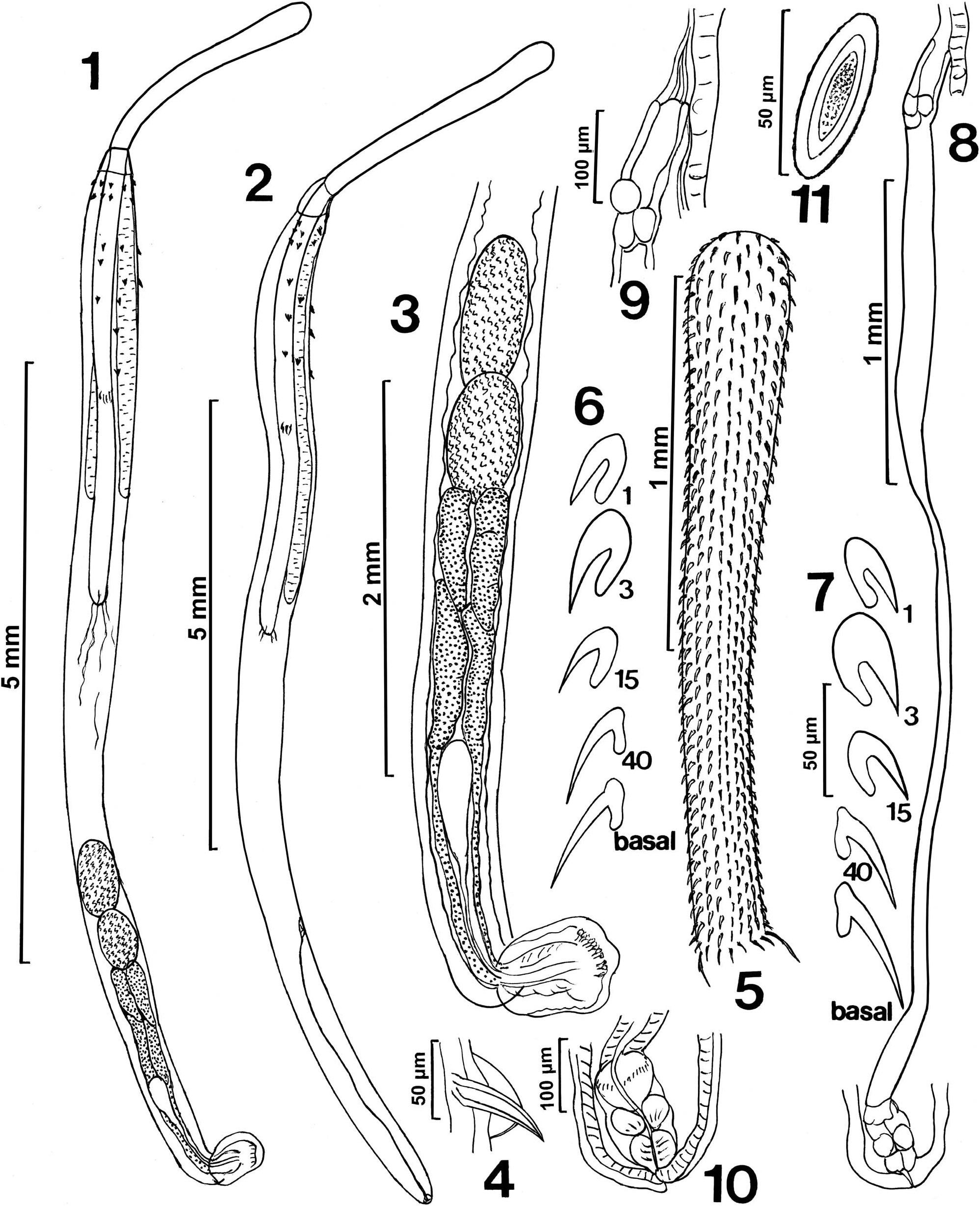
Figures 1–11. Line drawings of whole mounted specimens of Rhadinorhynchus hiansi from Sarda orientalis collected off the southern Pacific coast of Vietnam at Nha Trang. (1) A young male specimen. (2) A young female specimen. (3) Detail of the reproductive system of the male specimen in Figure 1. (4) A posterior trunk spine. (5) The proboscis of a male specimen. (6, 7) Profiles of selected dorsal (6) and ventral (7) proboscis hooks from Figure 5 showing 5 types of hooks. Hook no. 1 (apical), no. 3 (largest), no. 15 (smaller mid-proboscis hook), no. 40 (larger hooks near posterior end), basal crown (longest). (8) Reproductive system of an 11 mm long female showing long uterus and the attachment of the simple uterine bell to the ventral wall of the trunk. (9) A larger magnification of the uterine bell. (10) Larger magnification of the vagina. (11) An egg.
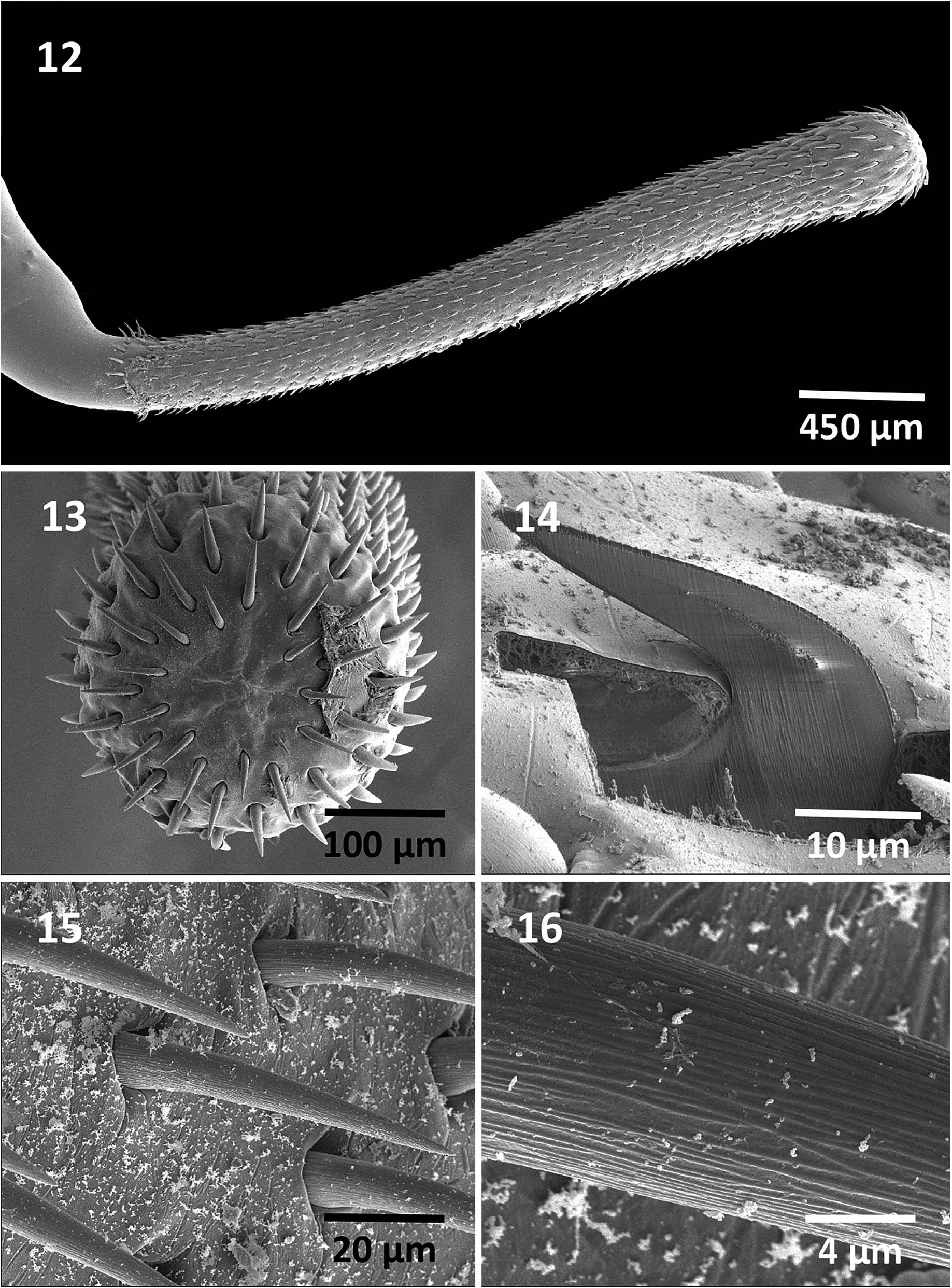
Figures 12–16. SEM of specimens from Sarda orientalis collected off the southern Pacific coast of Vietnam at Nha Trang. (12) A fully extended proboscis of a male specimen. (13) An apical view of the proboscis of specimen in Figure 12 showing the smaller apical hooks and the arrangement of hook rows. (14) A gallium cut hook showing the thin cortical layer and the dense prominent core. (15) A view of hooks at mid-proboscis showing their shape, external texture, and orientation. (16) A higher magnification of a hook showing detail of its external striations. All hooks are striated.
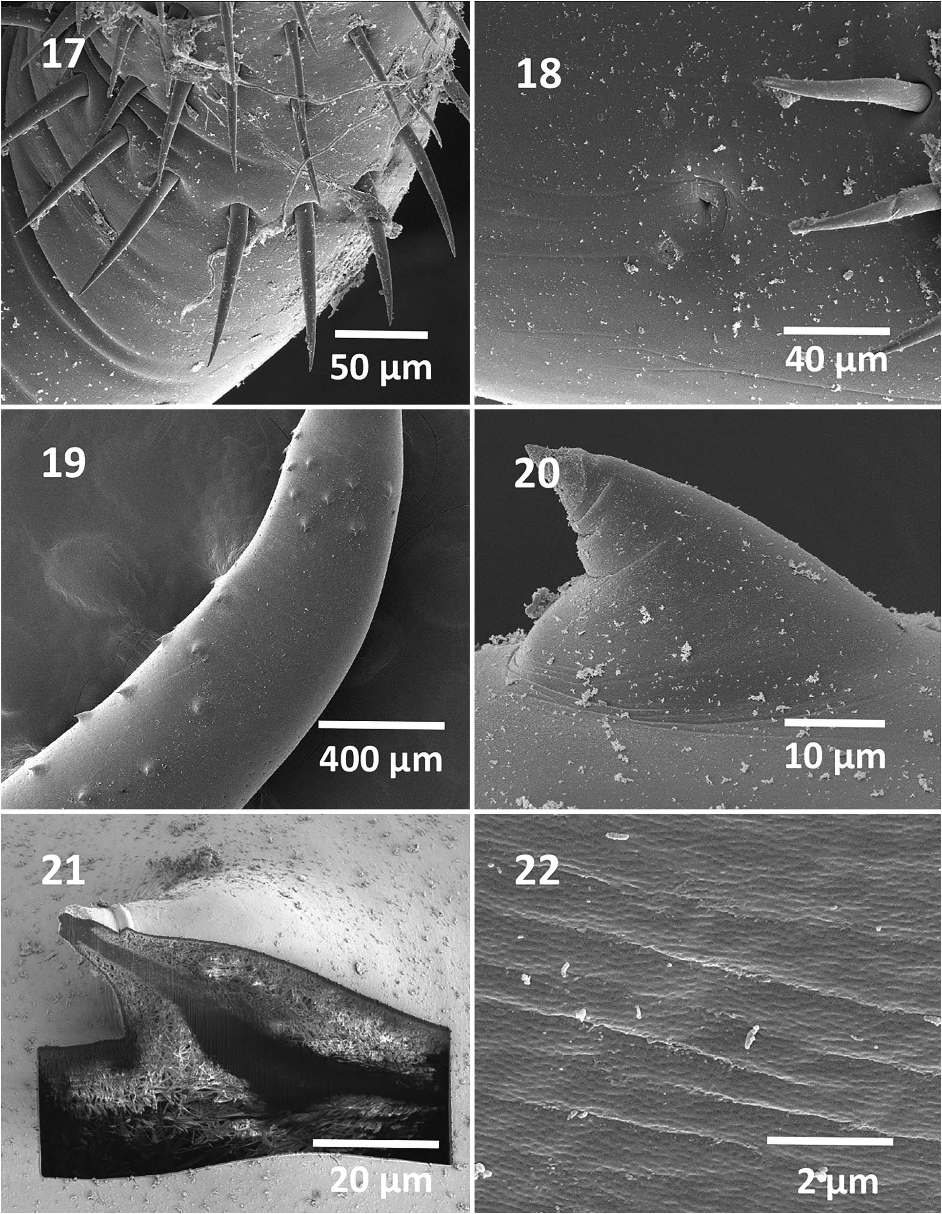
Figures 17–22. SEM of specimens from Sarda orientalis collected off the southern Pacific coast of Vietnam at Nha Trang. (17) Posterior proboscis hooks are longest and in a perfect ring. (18) Sensory pore just posterior to basal hooks. (19) Anterior trunk of a male specimen showing anterior rings of spines and posterior ventral and lateral spines characteristic of this species. (20) A higher magnification of a trunk spine. (21) A gallium cut spine showing a middle dense core and a think spongy outer layer under the cuticle. (22) An example of micropores in the middle of trunk.

Figures 23–28. SEM of specimens from Sarda orientalis collected off the southern Pacific coast of Vietnam at Nha Trang. (23) Rounded posterior end of a female specimen showing the sub-ventral position of gonopore appearing as a lateral slit. (24) Eggs with rounded poles and prominent coarse surface. (25) The posterior end of a male specimen showing the terminal position of an invaginated bursa. (26) A lateral view of terminal bursa. (27) A face view of the thick bursa showing the distribution of sensory structures in an outer ring of clusters and inner rings of single units. (28) Higher magnification of a sensory cluster from the outer ring showing its organization and the elevated dome-shaped center of each unit. The organization of sensory structures of the bursa is species specific and is a useful diagnostic tool for species recognition.

Figure 29. Energy dispersive X-ray spectrum of the base center of a gallium cut large anterior hook of a Rhadinorhynchus hiansi specimen showing high levels of calcium and phosphorus. The x-ray data are the elemental analysis of the hook base (see boldfaced figures in Table III). Insert: SEM of a cross and lateral longitudinal gallium cut hook.

Figure 30. Energy dispersive X-ray spectrum of the tip of a gallium cut small base hook of a Rhadinorhynchus hiansi specimen showing high levels of sulfur. The x-ray data are the elemental analysis of the hook tip (see boldfaced figures in Table IV). Insert: SEM of a cross and lateral longitudinal gallium cut hook.
IMAGES