AnimaliaNot EvaluatedacceptedspeciesAccepted
Cirrodrilus japonicus

Cirrodrilus japonicus

(Pierantoni, 1912) A.Lateral

GBIF:210361552

0year

0

Synonyms

ABOUT

Descriptions(7)

Host. Cambaroides japonicus (De Haan, 1841), the “ Japanese crayfish ” or “ Nihon-zarigani ” in Japanese. Habitat. Specimens have been observed alive on all parts of the exposed host and in the gill chambers. Additional information. Yamaguchi (1934: 198) observed that some individuals lacked a spermatheca but had eggs in segment 7, while in other specimens, spermatozoa were seen in the glandular atrium. Further studies of the life cycle of this species are needed to fully explain these observations.
Ohtaka, Akifumi, Gelder, Stuart R. (2023): A taxonomic reassessment of Cirrodrilus japonicus (Pierantoni, 1912) (Annelida, Clitellata, Branchiobdellida). Zootaxa 5263 (4): 557-565, DOI: 10.11646/zootaxa.5263.4.7, URL: http://dx.doi.org/10.11646/zootaxa.5263.4.7
(Figures 2, 3)
Ohtaka, Akifumi, Gelder, Stuart R. (2023): A taxonomic reassessment of Cirrodrilus japonicus (Pierantoni, 1912) (Annelida, Clitellata, Branchiobdellida). Zootaxa 5263 (4): 557-565, DOI: 10.11646/zootaxa.5263.4.7, URL: http://dx.doi.org/10.11646/zootaxa.5263.4.7
Description. Total body length 2.0 – 3.0 mm, the head width is usually subequal to segment 1, and the club-shaped body has distinct segments (Fig. 2 A). Dorsal ridges and projections, supernumerary muscles and lateral paired lobes on segments 8 and 9 are all absent. Peristomium has four lobes on the dorsal lip, two pairs of lateral lobes, a median emargination in the ventral lip (Fig. 2 B), and the mouth is surrounded by 16 oral papillae. The jaws differ in size and shape; dorsal being 1.5 x the ventral. Dorsal jaw has a crescent-shaped base about 45 μm wide, with a large median tooth and small lateral teeth across the anterior surface along with ridges parallel to the median axes (Fig. 2 C). The base of the ventral jaw is ovoid, smaller, 35 μm wide, also with a large median tooth and small lateral teeth across the anterior surface along with ridges parallel to the median axes. The dental formula is about 9 / 9 (4 - 1 - 4 / 4 - 1 - 4) and there is one pharyngeal sulcus. A pair of vasa deferentia or sperm tubes open separately into the glandular atrium; their exact entry has not been resolved. The tubular glandular atrium is about 0.7 x the segment diameter in length; deferent lobes and a prostate gland are absent. A terete muscular atrium, about 0.2 x segment diameter in length, and a sub-spherical muscular bursa length is 0.3 x the segment diameter (Fig. 2 D), and its atrium opens externally through the genital pore. The penis is eversible and when retracted, occupies the penial sheath that extends from the ental bursa back into the muscular atrium for about half the latter’s length. The spermatheca is about 0.7 x segment diameter in length and consists of a spermathecal bulb (tubular when empty) about 0.2 x the organ’s length and a terete muscular duct about 0.8 x the organ’s length which opens externally through a spermathecal pore (Fig. 2 D). The anterior excretory ducts open separately close to the median line on the dorsal surface of segment 3. Variations. Body length of adults vary from 2.0 mm to 3.0 mm depending on the response to the preservative before death. The dorsal jaw base varies from crescent-shaped to elongate oval, while the ventral jaw base is oval to banana-shaped. Measuring the width of the jaws was found to be less influenced by aspect than other jaw dimensions, e. g., height of the median tooth. The dorsal jaw widths ranged from 41.0 μm to 52.0 μm (N = 22, mean value 44.6 μm) and for the ventral jaw, 28.9 μm to 46.0 μm (N = 18, mean value 34.9 μm). Yamaguchi (1934: 197) drew the jaws and gave their magnification, and from his figures (7 a & b) in a reprint, it was calculated that the dorsal jaw was 68 μm and the ventral 38 μm wide, respectively. Ridges parallel to the median axes on both jaws vary from distinct to absent (Fig. 2 E, F); note the ventral jaw in figure 2 F. The dental formula also varies due to the number of small teeth on each jaw, which range from 7 / 7 (3 - 1 - 3 / 3 - 1 - 3) to 11 / 13 (5 - 1 - 5 / 6 - 1 - 6). The glandular atrium is tubular and varies in appearance from straight to having two or more folds. As the atrium floats free in the coelom, other organs can push it into available spaces thus causing the folds. When the spermathecal bulb is filled with spermatozoa it can double in size, and the duct may temporarily contain spermatozoa causing it to dilate medially. Syntypes. Body lengths of the two syntypes, ZMH V- 2912 a and 2912 b are 2.3 mm and 1.8 mm long, respectively. Both specimens are slim terete, widest in segment 5 or 6, with their posterior discs being slightly narrower, and showing indistinct, partially autolyzed internal organs (Fig. 3 A). Specimen 2912 a has an everted pharynx with clearly visible jaws (Fig. 3 B), and indistinguishable peristomial lobes. The constricted peristomium in specimen 2912 b appears to have only 9, but actually 10 inwardly curved lobes (Fig. 3 C); four dorsal lobes (d), two pairs are lateral lobes (l) and a ventral lip with a median emargination (v). The jaws are well preserved with distinct ridges parallel to their median axes, and the dorsal jaws are larger than the ventral ones (Fig. 3 D, E). The base width of the dorsal jaws in 2912 a and 2912 b are 41 and 38 μm, respectively. The dental formula in 2912 a and 2912 b is 3 - 1 - 4 / 3 - 1 - 4 and 3 - 1 - 3 / 3 - 1 - 4 (Fig. 3 D, E), respectively.
Ohtaka, Akifumi, Gelder, Stuart R. (2023): A taxonomic reassessment of Cirrodrilus japonicus (Pierantoni, 1912) (Annelida, Clitellata, Branchiobdellida). Zootaxa 5263 (4): 557-565, DOI: 10.11646/zootaxa.5263.4.7, URL: http://dx.doi.org/10.11646/zootaxa.5263.4.7
Diagnosis. Length about 2.5 mm, head width usually subequal segment 1, body club-shaped, segments distinct; dorsal lip four lobes, lateral lobes two pairs, ventral lip with median emargination; 16 oral papillae; jaws size different (dorsal 1.5 x ventral), dorsal crescent-shaped, ventral ovoid, teeth single large median, small lateral, longitudinal striations, dental formula 9 / 9 (4 - 1 - 4 / 4 - 1 - 4); pharyngeal sulcus one; glandular atrium tubular, length 0.7 x segment diameter; muscular atrium terete, length 0.7 x segment diameter; bursa sub-spherical, length 0.25 x segment diameter, penial sheath, ectal 0.1 x muscular atrium, penis eversible; spermatheca shape club-like, length 0.7 x segment diameter, duct shape terete, length 0.8 x organ, bulb shape tubular (empty), length 0.2 x organ.
Ohtaka, Akifumi, Gelder, Stuart R. (2023): A taxonomic reassessment of Cirrodrilus japonicus (Pierantoni, 1912) (Annelida, Clitellata, Branchiobdellida). Zootaxa 5263 (4): 557-565, DOI: 10.11646/zootaxa.5263.4.7, URL: http://dx.doi.org/10.11646/zootaxa.5263.4.7
Distribution. Specimens have only been recorded from Hokkaido Island, Japan, under the name of Stephanodrilus japonicus by Pierantoni (1912), Stephanodrilus (St.) ezoensis by Yamaguchi (1934) and of Cirrodrilus japonicus in the present study (Fig. 1).
Ohtaka, Akifumi, Gelder, Stuart R. (2023): A taxonomic reassessment of Cirrodrilus japonicus (Pierantoni, 1912) (Annelida, Clitellata, Branchiobdellida). Zootaxa 5263 (4): 557-565, DOI: 10.11646/zootaxa.5263.4.7, URL: http://dx.doi.org/10.11646/zootaxa.5263.4.7
Type locality. Otaru, Hokkaido Island, Japan (Fig. 1, site No. 1), according to the Museum’s specimen ledger.
Ohtaka, Akifumi, Gelder, Stuart R. (2023): A taxonomic reassessment of Cirrodrilus japonicus (Pierantoni, 1912) (Annelida, Clitellata, Branchiobdellida). Zootaxa 5263 (4): 557-565, DOI: 10.11646/zootaxa.5263.4.7, URL: http://dx.doi.org/10.11646/zootaxa.5263.4.7
Type material. In 1912, Pierantoni deposited 16 specimens in a fluid-filled small jar labeled “ Stephanodrilus japonicus Syntypes ” under the catalogue number, ZMH V- 2912, in the Museum of Nature Hamburg — Zoology, Germany; two specimens were mounted whole in Canada balsam on separate slides by AO in 2023. Yamaguchi (1934) did not designate any type specimens either or a type location, but slide-mounted whole specimens of Stephanodrilus ezoensis in his collection were identified and designated syntypes according to Article 73.2. of the International Code of Zoological Nomenclature (International Commission on Zoological Nomenclature 1999) by Ohtaka et al. (2020) (ICHUM- 1666, - 1799, - 1801, - 1809, - 1802, - 1806, - 1807, - 1811, - 1815, - 1821). Yamaguchi reported collecting specimens from 15 sites on Hokkaido, Japan (Gelder & Ohtaka 2002), but only one slide (ICHUM- 1811) bore a location name, that of Soranuma (Fig. 1: site No. 6). Material investigated. Two specimens of syntype series of C. japonicus, and extant syntype series of C. ezoensis (see above). Sixty-eight non-type specimens, collected by the first author and his colleagues on Hokkaido at sites No. 2, 9, 16 – 37 (Fig. 1), together with specimens whole-mounted on slides, in the first author's collection.
Ohtaka, Akifumi, Gelder, Stuart R. (2023): A taxonomic reassessment of Cirrodrilus japonicus (Pierantoni, 1912) (Annelida, Clitellata, Branchiobdellida). Zootaxa 5263 (4): 557-565, DOI: 10.11646/zootaxa.5263.4.7, URL: http://dx.doi.org/10.11646/zootaxa.5263.4.7

Export occurrence data

Darwin Core Archive (ZIP)

CLASSIFICATION

Taxonomic Classification Tree

MULTIMEDIA

Media Files(3)

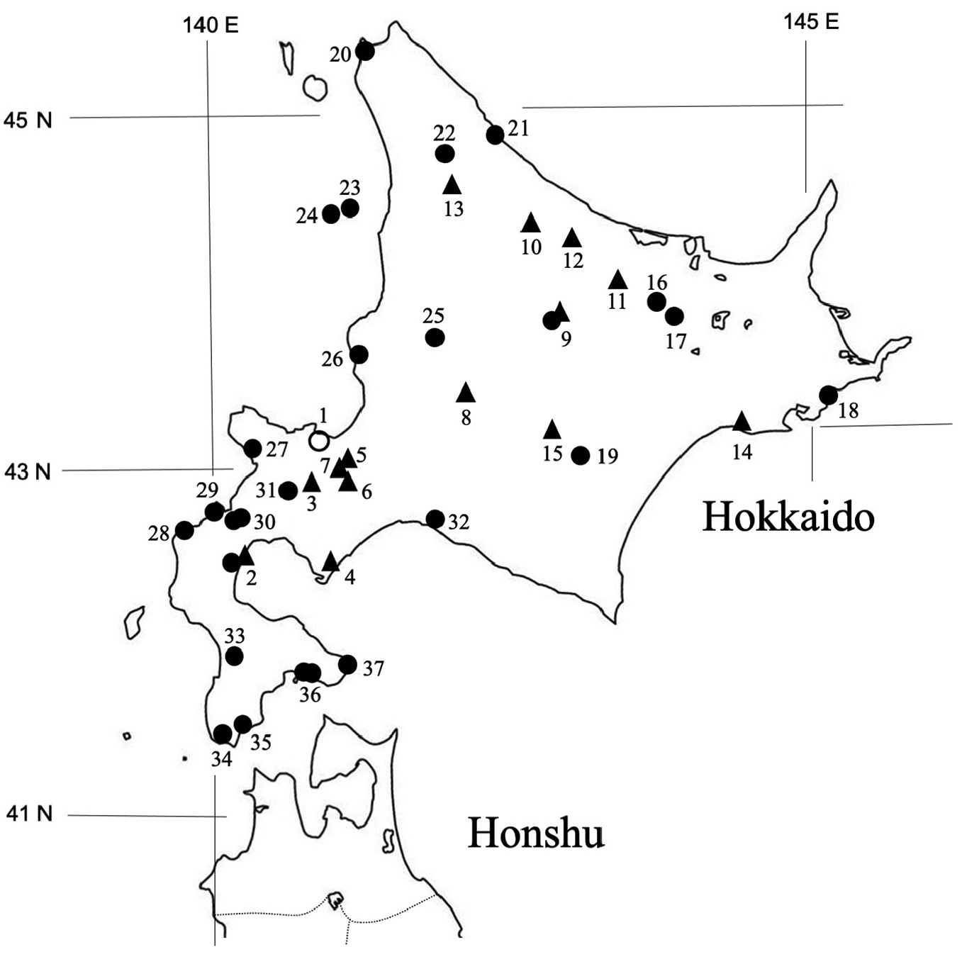
FIGURE 1. Localities of Cirrodrilus japonicus as currently known. Open circle (No. 1) indicates the type locality; triangles (Nos. 2–15) indicate the locations of Yamaguchi (1934) under the name of Stephanodrilus (St.) ezoensis; close circles (Nos. 2, 9, 16–37) indicate those from the present study. 1, Otaru; 2, Oshamanbe; 3, Kutchan; 4, Muroran; 5, Makomanai; 6, Soranuma; 7, Hattaribetsu; 8, Shimofurano; 9, Sounkei (Sounkyo); 10, Ichinohashi; 11, Rubeshibe; 12, Nokkeushi; 13, Nayoro; 14. Kushiro; 15. Shintoku; 16, Kunneppu; 17, Tsubetsu; 18, Hamanaka; 19, Obihiro; 20, Wakkanai; 21, Esashi; 22, Oumu; 23. Yagishiri Is.; 24, Teuri Is.; 25, Fukagawa; 26, Hamamasu; 27, Tomari; 28, Setana; 29, Shimamaki; 30, Suttsu; 31, Niseko; 32, Atsuma; 33, Assabu; 34, Matsumae; 35, Fukushima; 36, Hakodate; 37, Todohokke. The location of “Kitahama” in Yamaguchi (1934) could not be determined, and it was omitted from the figure.

Imageimage/png© Ohtaka, Akifumi;Gelder, Stuart R.Ohtaka, Akifumi;Gelder, Stuart R.

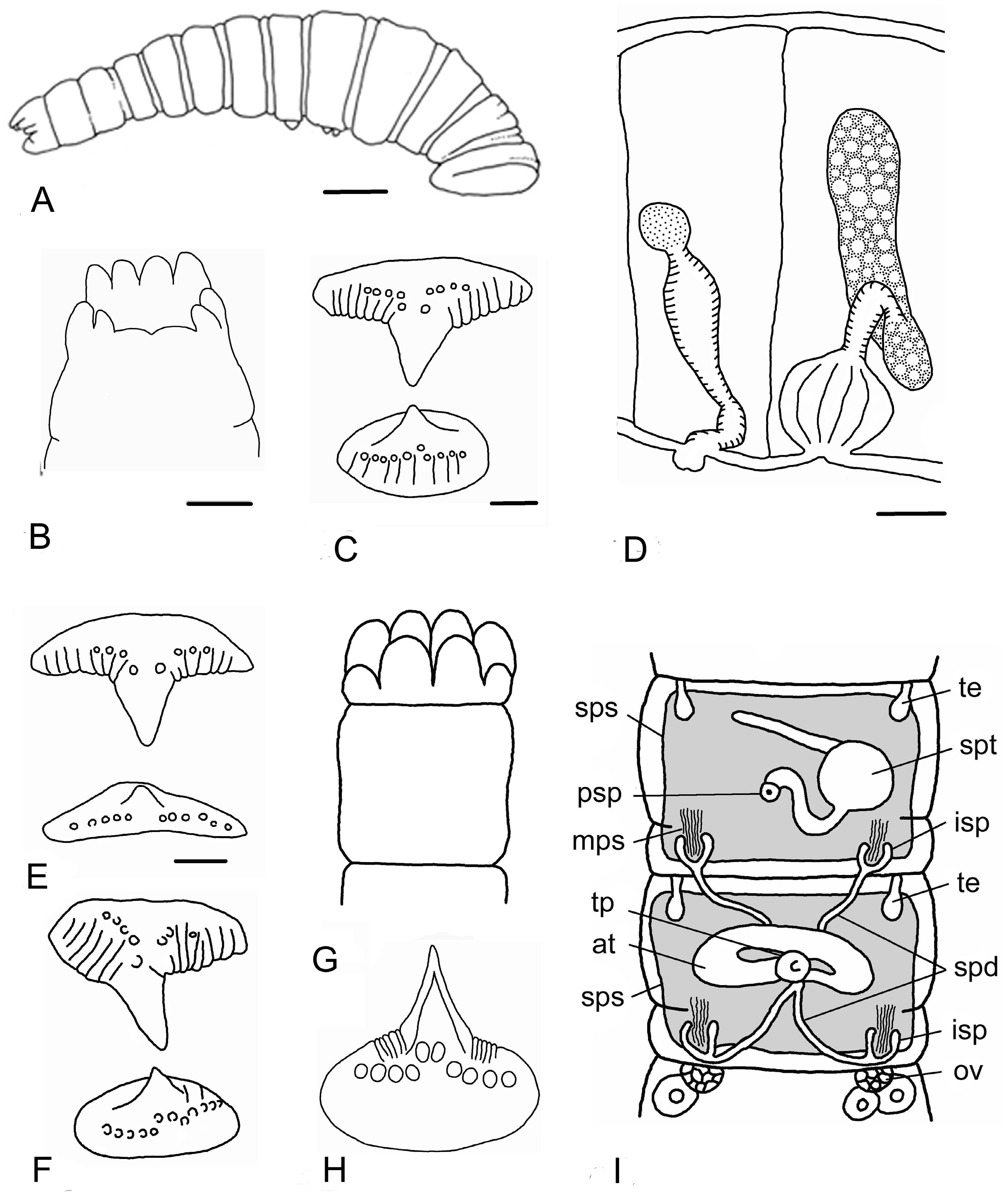
FIGURE 2. Cirrodrilus japonicus (Pierantoni, 1912) A. Lateral view of a whole specimen, redrawn from Yamaguchi (1934: Fig. 7A), scale bar = 200 μm. B. Ventral view of peristomium showing four dorsal lip lobes, two pairs of lateral lobes and the ventral lip with a median emargination, a whole-mounted specimen from Teuri Is. (see Fig. 1, site No. 24), scale bar = 50 μm. C. Anterior view of the dorsal over ventral jaws in a whole-mounted specimen from Hamanaka (see Fig. 1, site No. 18), scale bar = 10 μm. D. Lateral view of segment 5 (left) with the spermatheca and male genitalia in segment 6 (right), a whole-mounted specimen from Esashi (see Fig. 1, site No. 21), scale bar = 50 μm. Legend: Stippled, spermathecal bulb; short transverse lines, spermathecal duct and muscular atrium; stippled discs, glandular atrium; longitudinal lines, muscular bursa. Vasa deferentia not shown. E, F. Dorsal (top) and ventral (bottom) jaws of C. japonicus from an anterior aspect showing variations of the small lateral teeth and ridges. E. A specimen from Obihiro (see Fig. 1, site No. 19). F. A specimen from Hamanaka (see Fig. 1, site No. 18), scale bar = 10 μm. G–I. Original figures from Pierantoni (1912: Tab. 5, Figs. 11, 12 & 13), redrawn. G. “Fig. 11—Regione cefalica di Stephanodrilus japonicus n. sp. x120” [head region of Stephanodrilus japonicus]. H. “Fig. 13—Mascella della stessa species, x1200” [jaws of the same species]. I. “Figure 12—Regione genitale della stessa. x500.” [genital region]. Legend gives original abbreviations and, in square brackets, names in modern terminology: at, atrio [glandular atrium]; isp, imbuto spermarico [sperm funnel]; ov, ovario [ovary]; spd, spermadutto [sperm duct]; spt, spermateca [spermatheca]; te, testicolo [testis]; tp, tasco peniale [muscular bursa]. Additional features: msp, mature sperm; sps, sperm sac (grey).

Imageimage/png© Ohtaka, Akifumi;Gelder, Stuart R.Ohtaka, Akifumi;Gelder, Stuart R.

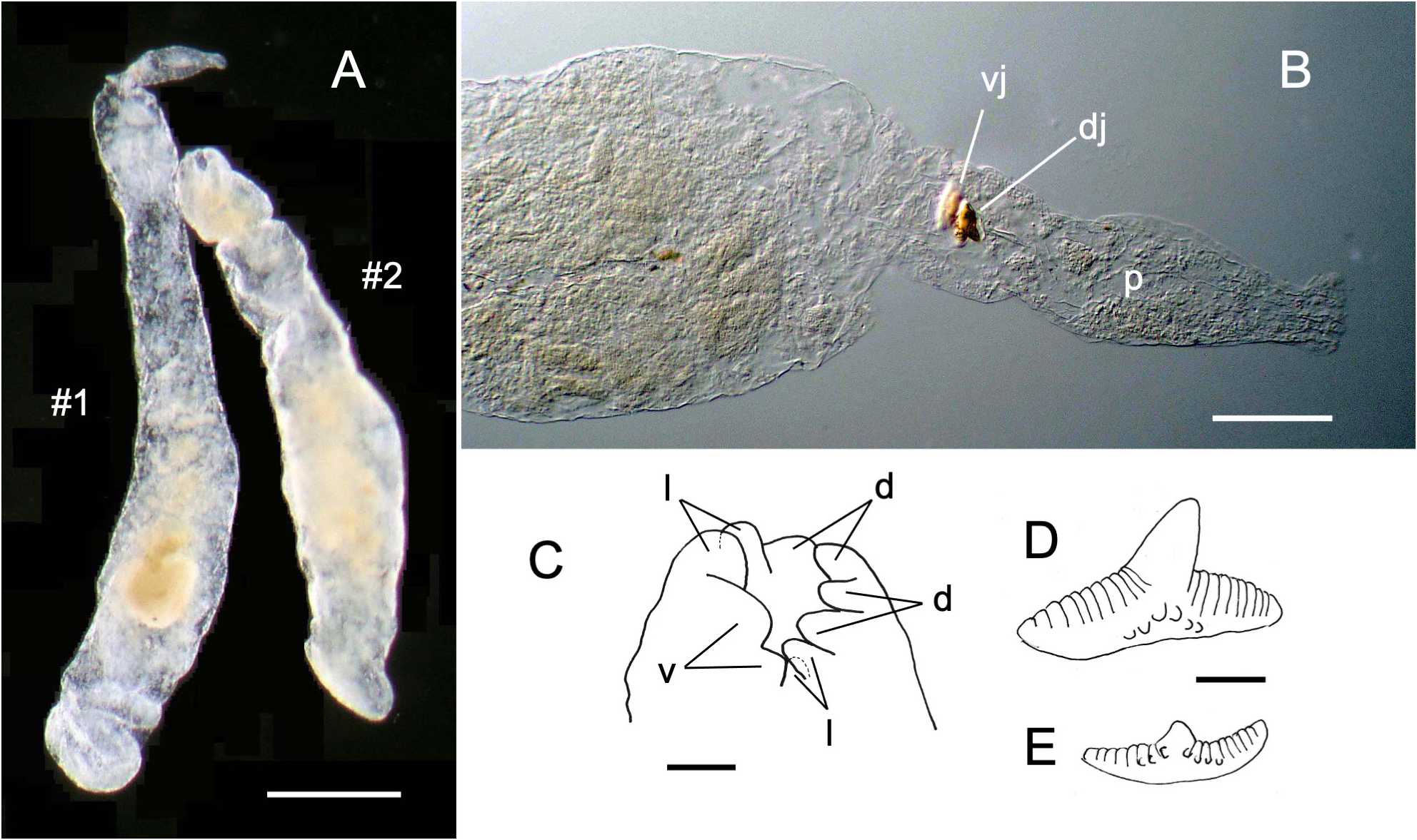
FIGURE 3. Syntypes of Cirrodrilus japonicus (Pierantoni, 1912) A. Two specimens in fluid, ZMH V-2912a (#1) and 2912b (#2). Upper indicates anterior in both worms. scale bar = 0.5 mm. B. Anterior end of body in #1 with everted pharynx, scale bar = 100 μm. C. Lateral view of peristomium in #2, scale bar = 50 μm. D, E. Dorsal (D) and ventral (E) jaw of #2 from anterior aspects, scale bar = 10 μm. Legends: d, dorsal lobe; dj, dorsal jaw; l, lateral lobe; p, pharynx; v, ventral lip; vj, ventral jaw.

Imageimage/png© Ohtaka, Akifumi;Gelder, Stuart R.Ohtaka, Akifumi;Gelder, Stuart R.

IMAGES

Gallery(3)

See Gallery

Occurrences with images

Source Information

A taxonomic reassessment of Cirrodrilus japonicus (Pierantoni, 1912) (Annelida, Clitellata, Branchiobdellida)

checklist

This dataset contains the digitized treatments in Plazi based on the original journal article Ohtaka, Akifumi, Gelder, Stuart R. (2023): A taxonomic reassessment of Cirrodrilus japonicus (Pierantoni, 1912) (Annelida, Clitellata, Branchiobdellida). Zootaxa 5263 (4): 557-565, DOI: 10.11646/zootaxa.5263.4.7, URL: http://dx.doi.org/10.11646/zootaxa.5263.4.7

Abstract

Cirrodrilus japonicus (Pierantoni, 1912) has not been identified since it was originally described from specimens removed from preserved Japanese crayfish that had been deposited in the Natural History Museum in Hamburg, Germany. A morphological comparison of the published description and recently discovered syntypes of C. japonicus from the Hamburg Museum with syntypes and newly collected material of Cirrodrilus ezoensis (Yamaguchi, 1934), supported the view that preservation artifacts in C. japonicus caused Prof. Yamaguchi to believe his well-preserved specimens were member of a separate species. Therefore, C. ezoensis becomes a junior synonym of C. japonicus.

Ohtaka A, Gelder S R, plazi (2023). A taxonomic reassessment of Cirrodrilus japonicus (Pierantoni, 1912) (Annelida, Clitellata, Branchiobdellida). Plazi.org taxonomic treatments database. Checklist dataset https://doi.org/10.15468/whm462 accessed via GBIF.org on 2026-06-14.

CC0Published 4/11/2023View dataset
GBIF Usage Key
210361552
Dataset Key
14d01866-6c3a-4b02-a313-77bd4a70ccd5
Origin
source
Backbone Key
7950266
Taxon ID
957187E1FFABFFA32BA5C841FC139A92.taxon
Last Crawled
6/9/2026
Last Interpreted
6/9/2026