AnimaliaNot EvaluatedacceptedspeciesAccepted
Henneguya namae
Haldar, 1983
GBIF:229729277
0year
ABOUT
Descriptions(1)
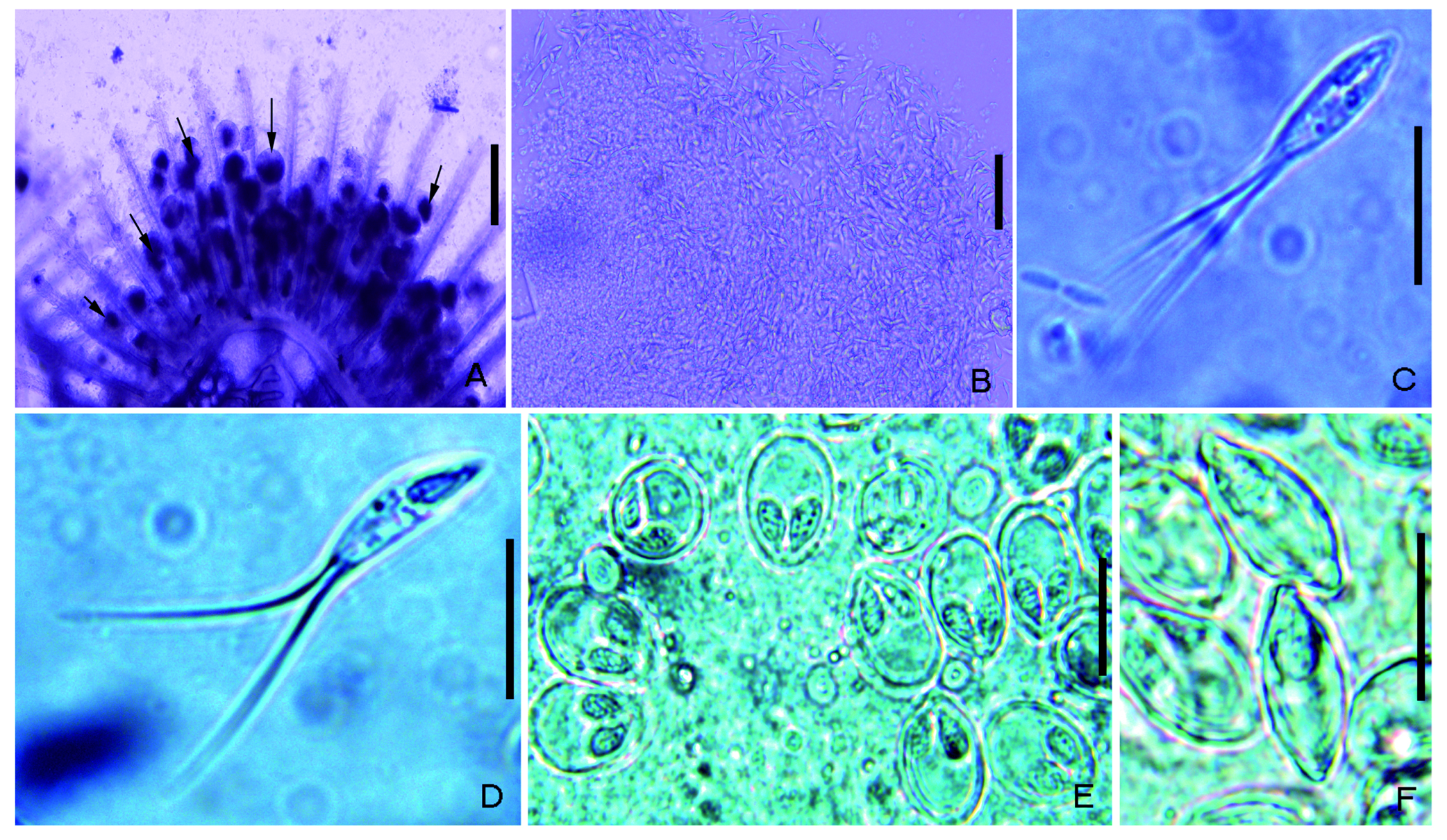
Type host: Chanda nama (= Ambassis nama) Hamilton, 1822; chanda (local name). Site of infection: Gill filaments. Locality: Ganga River at Bairaj, Bijnor (29 ° 23 ’ N, 79 ° 11 ’ E) in the state of Uttar Pradesh (U. P.), India. Prevalence of infection: A total of 40 specimens of Chanda nama shows prevalence of infection 34 / 40 (of the 5 – 6 cm size in length with a prevalence of 85 %; Intensity of infection: High). Material deposited: Digital images (Photos) of spores were deposited in the parasitological collection of the Museum, Department of Zoology, Chaudhary Charan Singh University, Meerut (U. P.), India collection no. (Coll. No. HSS / ZOO / MYX / 01 / 19). The 18 S rDNA sequence was deposited in GenBank under accession numbers MN 218392 and MN 218393. Description: H. namae cyst present in the lamellae of gill filaments roundish in shape, interlamellar measuring 70 – 150 µm, small and large due to synchronicity development and contained myxospores that clearly showed high infection (Fig. 1 A, B). Spore body elongated with two caudal appendages (Fig. 1 C, D; Fig. 2 A, B). In frontal view, anterior end of spores appears blunt while the caudal end somewhat rounded and gradually continued into long, bifurcated caudal appendages (Fig. 2 A, B). Total spores length, 27.82 – 33.17 (30.6 ± 1.71) (N = 30); spore body length, 12.34 – 15.6 (14.15 ± 0.96) (N = 30); caudal appendages length, 15.12 – 17.92 (16.64 ± 1.03) (N = 30); spore width, 4.94 – 5.98 (5.41 ± 0.32) (N = 30) and spore thickness, 3.9 – 4.34 (4.07 ± 0.15) (N = 20). Spore wall smooth, composed of two uniformly thin valves, sutural line prominent and thick. Polar capsule two in number, elongated, pear shaped, pointed at anterior end and unequal in size. Larger capsule 3.18 – 4.16 (3.66 ± 0.34) (N = 30) long and 1.0 – 1.14 (1.08 ± 0.05) (N = 15) wide. Smaller capsule 2.84 – 3.73 (3.27 ± 0.31) (N = 30) long and 0.98 – 1.1 (1.04 ± 0.04) (N = 15) wide. Number of polar filament coils seen 8 – 9 in large and 6 – 7 in small one. H. namae was identified on basis of the above characteristics. All the morphometrical measurements with closely related species listed in the supplementary table 1. Remarks: H. namae was compared with other Henneguya spp. described parasitizing freshwater fish. Originally H. namae was described by Haldar et al. 1983 from gills of C. nama. Approximately> 20 species described thus far in Indian fish, the spores of H. namae infecting the gills of C. nama revealed the greatest similarity to the spores of H. ophiocephali Chakravarty 1939, H. notopterae Lalitha Kumari 1965, H. qadrii Qadri 1965, H. singhi Lalita Kumari 1969 and H. thermalis Seenappa et al. 1981. Moreover, the strict morphological comparisons showed that the shape, size of spore body and the length of caudal appendage of above respective species can be easily differentiated H. namae from others (Supplementary table 1). In the Supplementary table 1, we add Henneguya species reported from India that was similar to H. namae and shows species with unequal polar capsules i. e., H. ophiocephali, H. notopterae, H. qadrii, H. singhi and H. thermalis. H. namae could be distinguished from H. ophiocephali in the size and shape of the spore body as it is more rounded anteriorly in H. ophiocephali. H. notopterae have more pointed at the anterior end in spore shape in comparison to H. namae and additionally with a long duct in polar capsule. Spores of H. qadrii are smaller in size as compared to H. namae but size of polar capsules of H. qadrii is larger as compared to H. namae. Moreover, the spore body of H. namae is more elongated as compare to H. singhi as well as distinguished with each other in polar capsule shape. However, H. thermalis have a more rounded shape of spores while H. namae have little roundish and more pointed, both differ in the shape and size of polar capsules too. With regard to H. chaudhuryi (Bajpai and Haldar 1982) and Henneguya sp. RA- 2015 (KR 704889) Bala (2015), the differences are the presence of equal polar capsules while H. namae comprise unequal polar capsules. There are no molecular data available for the species H. ophiocephali, H. notopterae, H. qadrii, H. singhi and H. thermalis. Therefore, on the basis of above mentioned characteristics of H. namae, it can be readily distinguished from other species (see in supplementary table 1). Molecular analysis: 18 S rDNA of two different pools of isolates of H. namae were sequenced (1305 and 1315 bp). No intraspecific divergence was found among the newly generated sequences from isolates of H. namae and shown to be closely related with other Henneguya species described from Perciformes and Cypriniformes hosts.
Garg, Anupma, Chaudhary, Anshu, Gupta, Abhishek, Kumar, Abhinav, Sharma, Bindu, Singh, Hridaya Shanker (2020): Molecular Characterization of Two Myxosporean Species, Henneguya namae Haldar et al. 1983 and Myxobolus sophorae Jayasri, 1982 (Myxosporea: Myxobolidae). Acta Protozoologica 59 (1): 39-53, DOI: 10.4467/16890027AP.20.003.12159, URL: http://dx.doi.org/10.4467/16890027ap.20.003.12159
Export occurrence data
Darwin Core Archive (ZIP)
CLASSIFICATION
Taxonomic Classification Tree

MULTIMEDIA
Media Files(2)
Fig. 1. Photographs of myxobolids: A – Cysts of H. namae of different sizes between gill filaments of the host fish show by arrows, B – Spores released from ruptured cysts of H. namae, C – H. namae frontal view, D – H. namae sutural view, E – M. sophorae frontal view, F – M. sophorae sutural view. Scale bars (A) 300 µm, (B) 50 µm, (C–F) 10 µm.
Imageimage/png© Garg, Anupma;Chaudhary, Anshu;Gupta, Abhishek;Kumar, Abhinav;Sharma, Bindu;Singh, Hridaya ShankerGarg, Anupma;Chaudhary, Anshu;Gupta, Abhishek;Kumar, Abhinav;Sharma, Bindu;Singh, Hridaya Shanker
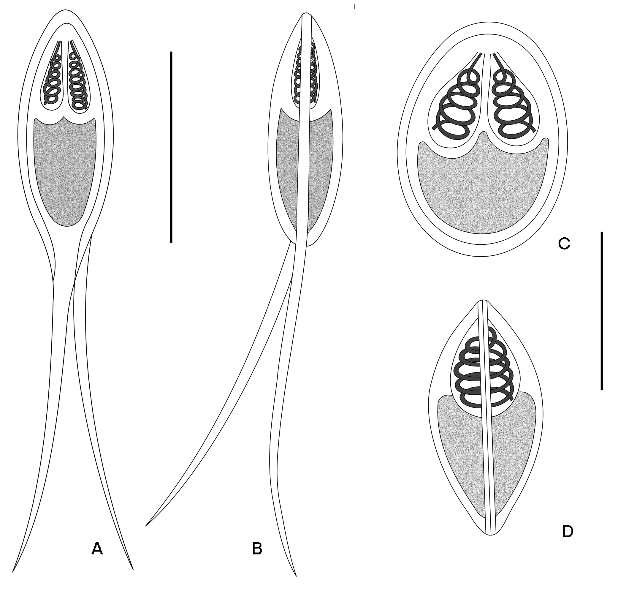
Fig. 2. A schematic drawing of Henneguya namae and Myxobolus sophorae myxospores found infect Chanda nama and Puntius sophore. In frontal view:A – H. namae, C – M. sophorae. In sutural view: B – H. namae, D – M. sophorae. Scale bars (A–D) 10 µm.
Imageimage/png© Garg, Anupma;Chaudhary, Anshu;Gupta, Abhishek;Kumar, Abhinav;Sharma, Bindu;Singh, Hridaya ShankerGarg, Anupma;Chaudhary, Anshu;Gupta, Abhishek;Kumar, Abhinav;Sharma, Bindu;Singh, Hridaya Shanker
IMAGES