AnimaliaNot EvaluatedacceptedspeciesAccepted
Henneguya voronini

Henneguya voronini

Borkhanuddin, Cech, Molnár & Shaharom-Harrison, 2020

GBIF:233768089

0year

ABOUT

Descriptions(1)

Type host: barramundi, Lates calcarifer (Bloch 1790). Site of infection: Base of the gill filament. Type locality: Setiu Wetlands, Terengganu, Malaysia. Prevalence of infection: 2.8 % (1 / 35). Type material: Digital images of syntype spores and histological sections were deposited in the parasitological collection of the Zoological Department, Hungarian Natural History Museum, Budapest, collection no. HNHM- 71893. The 18 S rDNA sequence was deposited in GenBank under accession number MH 743110. Etymology: The species is named in honour of V. A. Voronin, an eminent Russian fish parasitologist. Description of spores: Myxospores symmetric, with two equal caudal appendages and equal-sized polar capsules (Fig. 2). Spore wall thin (0.3 – 0.4 μm), smooth and composed of 2 equal valves. Apical end of spore body blunt, the caudal end tapers and extends into the caudal appendages. Total length 35 – 39 μm, length of spore 9.9 ± 0.3 (9.5 – 10.3) μm and width 5.9 ± 0.3 (5.8 – 6.0) μm; thickness could not be measured as no spores were observed in sutural plane. Polar capsules pear shaped, blunt at the posterior end and tapered anteriorly, length 3.7 ± 0.2 (3.5 – 4.0) μm and width 2.1 ± 0.1 (2.0 – 2.2) μm. Polar tubules coiled in 6 turns perpendicular to the long axis of the polar capsules. Sporoplasm binucleate, with a small iodinophilous vacuole. Caudal appendages straight, tapering, length 27.2 ± 1.4 (25.0 – 29.0) μm, about 4 times as long as the spore body. Plasmodium ellipsoidal 250 – 300 μm × 130 – 150 μm. Remarks: Myxospores of H. voronini n. sp. could not be distinguished morphometrically using most measurements with the two other species observed in the host. Both H. voronini n. sp. and H. setiuensis n. sp. were found in the gills but the specific tissue locality differed: the large ellipsoidal plasmodia (up to 300 μm) of H. voronini n. sp. were localized to the cartilaginous base of the gill filaments, while the smaller spherical plasmodia of H. setiuensis n. sp. (up to 75 μm) developed between lamellae of the gill filaments. Considering Henneguya spp. from other Lates species, spores of H. voronini n. sp. could be differentiated in at least two dimensions (Table 2). Molecular analysis: 1696 bp 18 S rDNA were sequenced, including the primers. A BLAST search indicated that highest sequence similarities were to other Henneguya species in GenBank, but all <90 %. Pairwise analysis showed H. voronini n. sp. was molecularly very similar to H. calcarifer n. sp. (97.7 %; 1658 / 1969 bp; p - distance 0.013), described from the same fish (below). Histology: Ellipsoidal plasmodia were located in the cartilaginous gill arch between gill filaments (Fig. 4 c). We suspect that development began in the multilayered connective tissue covering the gill arch and then a large part of the plasmodium moved into the gill filaments and was covered by a multilayered epithelium as it matured. Microscopy: SEM revealed that the valve cell surfaces were smooth, which is a morphological feature typical of genus Henneguya.
Borkhanuddin, Muhammad Hafiz, Cech, Gábor, Molnár, Kálmán, Shaharom-Harrison, Faizah (2020): Henneguya (Cnidaria: Myxosporea: Myxobolidae) infections of cultured barramundi, Lates calcarifer (Perciformes: Latidae) in an estuarine wetlands system of Malaysia: description of Henneguya setiuensis n. sp., Henneguya voronini n. sp. and Henneguya calcarifer n. sp. Parasitology Research (85) 119 (1): 85-96, DOI: 10.1007/s00436-019-06541-1, URL: http://dx.doi.org/10.1007/s00436-019-06541-1

Export occurrence data

Darwin Core Archive (ZIP)

CLASSIFICATION

Taxonomic Classification Tree

MULTIMEDIA

Media Files(2)

Fig. 2 Henneguya voronini n. sp. (a–b) Line drawings of mature myxospores in frontal view showing polar capsules with coiled polar tubules. (c) Fresh, unstained myxospores in frontal view showing the two pyriform polar capsules. (d) Scanning electron microscope image of the spores showing simple, smooth valve cell surfaces, each contiguous with a caudal process; features typical of the genus

Imageimage/png© Borkhanuddin, Muhammad Hafiz;Cech, Gábor;Molnár, Kálmán;Shaharom-Harrison, FaizahBorkhanuddin, Muhammad Hafiz;Cech, Gábor;Molnár, Kálmán;Shaharom-Harrison, Faizah

Fig. 4 Histological sections of gill filaments from Lates calcarifer infected by plasmodia of H. setiuensis n. sp. and H. voronini n. sp. (a) Plasmodia of H. setiuensis n. sp. (arrows) producing compression and damage to the lamellae (*). (b) Development of plasmodia of H. voronini n. sp. (arrows) in the sub-epithelial layer at the base of the filament. Note that the plasmodia also impinge into the gill arch (g)

Imageimage/png© Borkhanuddin, Muhammad Hafiz;Cech, Gábor;Molnár, Kálmán;Shaharom-Harrison, FaizahBorkhanuddin, Muhammad Hafiz;Cech, Gábor;Molnár, Kálmán;Shaharom-Harrison, Faizah

IMAGES

Gallery(2)

See Gallery

Occurrences with images

Source Information

Henneguya (Cnidaria: Myxosporea: Myxobolidae) infections of cultured barramundi, Lates calcarifer (Perciformes: Latidae) in an estuarine wetlands system of Malaysia: description of Henneguya setiuensis n. sp., Henneguya voronini n. sp. and Henneguya calcarifer n. sp.

checklist

This dataset contains the digitized treatments in Plazi based on the original journal article Borkhanuddin, Muhammad Hafiz, Cech, Gábor, Molnár, Kálmán, Shaharom-Harrison, Faizah (2020): Henneguya (Cnidaria: Myxosporea: Myxobolidae) infections of cultured barramundi, Lates calcarifer (Perciformes: Latidae) in an estuarine wetlands system of Malaysia: description of Henneguya setiuensis n. sp., Henneguya voronini n. sp. and Henneguya calcarifer n. sp. Parasitology Research (85) 119 (1): 85-96, DOI: 10.1007/s00436-019-06541-1, URL: http://dx.doi.org/10.1007/s00436-019-06541-1

Abstract

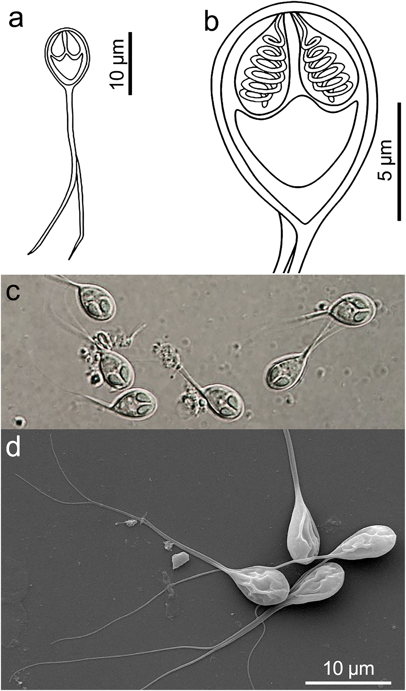
Examination of 35 barramundi (Lates calcarifer) from aquaculture cages in Setiu Wetland, Malaysia, revealed a single fish infected with three Henneguya spp. (Cnidaria: Myxosporea). Characterization of the infections using tissue tropism, myxospore morphology and morphometry and 18S rDNA sequencing supported description of three new species: Henneguya setiuensis n. sp., Henneguya voronini n. sp. and H. calcarifer n. sp. Myxospores of all three species had typical Henneguya morphology, with two polar capsules in the plane of the suture, an oval spore body, smooth valve cell surfaces, and two caudal appendages. Spores were morphometrically similar, and many dimensions overlapped, but H. voronini n. sp. had shorter caudal appendages compared with H. calcarifer n. sp. and H. setiuensis n. sp. Gross tissue tropism distinguished the muscle parasite H. calcarifer n. sp. from gill parasites H. setiuensis n. sp. and H. voronini n. sp.; and these latter two species were further separable by fine-scale location of developing plasmodia, which were intra-lamellar for H. setiuensis n. sp. and basal to the filaments for H. voronini n. sp. small subunit ribosomal DNA sequences distinguished all three species: the two gill species H. setiuensis n. sp. and H voronini n. sp. were only 88% similar (over 1708 bp), whereas the muscle species H. calcarifer n. sp. was most similar to H. voronini n. sp. (98% over 1696 bp). None of the three novel species was more than 90% similar to any known myxosporean sequence in GenBank. Low infection prevalence of these myxosporeans and lack of obvious tissue pathology from developing plasmodia suggested none of these parasites are currently a problem for barramundi culture in Setiu Wetland; however additional surveys of fish, particularly at different times of the year, would be informative for better risk assessment.

Borkhanuddin M H, Cech G, Molnár K, Shaharom-Harrison F, felipe (2019). Henneguya (Cnidaria: Myxosporea: Myxobolidae) infections of cultured barramundi, Lates calcarifer (Perciformes: Latidae) in an estuarine wetlands system of Malaysia: description of Henneguya setiuensis n. sp., Henneguya voronini n. sp. and Henneguya calcarifer n. sp.. Plazi.org taxonomic treatments database. Checklist dataset https://doi.org/10.15468/j469zn accessed via GBIF.org on 2026-06-17.

CC0Published 11/25/2019View dataset
GBIF Usage Key
233768089
Dataset Key
0a7f870a-286b-46e9-8019-591781f9e2cd
Origin
source
Taxon ID
03881A177749E76E2C0CFF5E862F5685.taxon
Last Crawled
6/9/2026
Last Interpreted
6/9/2026