AnimaliaNot EvaluatedacceptedspeciesAccepted
Henneguya calcarifer
Borkhanuddin, Cech, Molnár & Shaharom-Harrison, 2020
GBIF:233768090
0year
ABOUT
Descriptions(1)
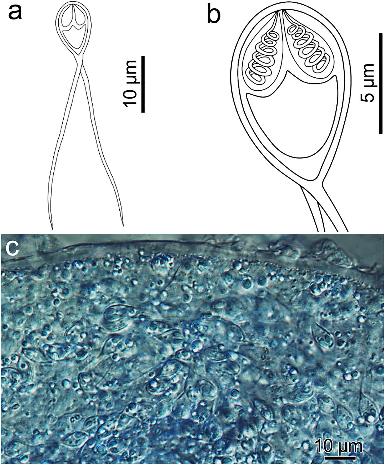
Type host: barramundi, Lates calcarifer (Bloch 1790). Site of infection: Skeletal muscle. Type locality: Setiu Wetlands, Terengganu, Malaysia. Prevalence of infection: 2.8 % (1 / 35). Type material: Digital images of syntype spores were deposited in the parasitological collection of the Zoological Department, Hungarian Natural History Museum, Budapest, collection no. HNHM- 71894. The 18 S rDNA sequence was deposited in GenBank under accession number MH 743109 Etymology: The species is named after the host. Description of spores: Fig. 3. Myxospores symmetric, with two equal caudal appendages, and equal-sized polar capsules. Spore wall 0.3 – 0.4 μm, smooth and composed of two equal valves. Apical end of spore body blunt, the caudal end tapers and extends into the caudal appendages, total length 34 – 45 μm. Spore length 9.4 ± 0.6 (8.3 – 10.0) μm, width 5.2 ± 0.3 (4.8 – 5.5) μm and thickness 3.8 ± 0.1 (3.7 – 4.0) μm. Two polar capsules pear shaped, blunt at the posterior end and taper anteriorly, length 3.4 ± 0.2 (3.1 – 3.7) μm and width 1.4 ± 0.2 (1.1 – 1.7) μm. Polar tubules coiled in 6 turns perpendicular to the long axis of the capsule. Sporoplasm binucleate with a small iodinophilous vacuole. Caudal appendages straight, tapering, length 30.9 ± 3.0 (28.0 – 35.0) μm, ~ 4 times longer than the spore body. Plasmodia spherical 300 × 400 μm. Remarks: H. calcarifer n. sp. resembles morphologically and morphometrically both H. setiuensis n. sp. and H. voronini n. sp. (Table 2) but has different tissue site of development (muscles not gill). This is the first muscle-infecting Henneguya described from Lates calcarifer and is relatively distinct from the only other species from a Lates congener: H. ghaffari from muscle and gills of L. niloticus in Egypt (Table 2). Molecular analysis: 1696 bp 18 S rDNA were sequenced. A BLAST search indicated that the most similar species were other Henneguya species, but all <89 %. Pairwise analysis showed H. calcarifer n. sp. was molecularly very similar (97.7 % over 1696 bp) with H. voronini n. sp., described from the same fish (above). Histology: Low intensity of parasite plasmodia in the host skeletal muscle meant no plasmodia were visible in histological sections.
Borkhanuddin, Muhammad Hafiz, Cech, Gábor, Molnár, Kálmán, Shaharom-Harrison, Faizah (2020): Henneguya (Cnidaria: Myxosporea: Myxobolidae) infections of cultured barramundi, Lates calcarifer (Perciformes: Latidae) in an estuarine wetlands system of Malaysia: description of Henneguya setiuensis n. sp., Henneguya voronini n. sp. and Henneguya calcarifer n. sp. Parasitology Research (85) 119 (1): 85-96, DOI: 10.1007/s00436-019-06541-1, URL: http://dx.doi.org/10.1007/s00436-019-06541-1
Export occurrence data
Darwin Core Archive (ZIP)
CLASSIFICATION
Taxonomic Classification Tree

MULTIMEDIA
Media Files(1)
Fig. 3 Henneguya calcarifer n. sp. (a–b) Line drawings of mature myxospores in frontal view showing polar capsules with coiled polar tubules. (c) Unstained, compressed plasmodium, densely packed with mature myxospores
Imageimage/png© Borkhanuddin, Muhammad Hafiz;Cech, Gábor;Molnár, Kálmán;Shaharom-Harrison, FaizahBorkhanuddin, Muhammad Hafiz;Cech, Gábor;Molnár, Kálmán;Shaharom-Harrison, Faizah
IMAGES