AnimaliaNot EvaluatedacceptedspeciesAccepted
Brinkhurstia americanus

Brinkhurstia americanus

(Brinkhurst, 1964)

GBIF:245596615

0year

0

Synonyms

ABOUT

Descriptions(2)

Length 24 – 30 mm, width 0.56 – 0.75 mm, segments 100 – 130. Prostomium short or large and rounded. Ventral setal couples of XIII replaced by a single penial seta on each side; in the ovarian segments aa: ab: bc: cd: dd = 4.7: 1: 5: 1.1: 6.2; dd = 0 – 25 u; length of a lateral seta from XI, base to node = 87 µm. Clitellum annular, slightly less developed ventrally; segments 1 / 2 XII – XV. Lateral lines present. Male pores ventrolateral in XIII. Female pores anterior in XIV. Spermathecal pore single, middorsal, anteriorly in IX. Septum 3 / 4 excessively delicate and doubtfully complete; 4 / 5 and 14 / 15 hardly appreciably, 6 / 7 – 8 / 9 fairly strongly thickened (two tiers of muscle fibres) the remainder intermediate; 10 / 11 has one tier of muscle fibres and is not backwardly deflected. Septal glands in V – VIII. Alimentary canal expanding greatly in XVI but no definite transition from oesophagus to intestine. Gizzard, oesophageal diverticula and typhlosole absent. Perienteric blood sinus in IX posteriad, not appreciable in front of this. Testes discrete elongated tongues of tissue dependent from the anterior wall of X. Gonadial tissue absent from XI. Seminal vesicles absent or present in XI. Vas deferens running on body wall in XI and XII. Atria very thin much coiled tubes extending from mid – XIII into XIV; discharging in the close proximity of the corresponding vas deferens through the posterior wall of a globular non-muscular ectal chamber; a simple, straight, unornamented penial seta in a follicle approximately twice the length of the chamber, penetrates the anterior wall of the chamber. Each prostate has a cuboidal lining epithelium surrounded in turn by a very thin muscular sheath and an external layer of prostatic cells only one to a few cells thick, though considerable masses of prostatic cells are formed between adjacent coils. Width muscular tube of atrium (external) 28 µm; width ectal chamber 37 µm; length penial seta 115 µm; greatest width 18 µm. Ovaries dependent from the anterior wall of XIII and extending posteriad into ovisacs formed by the backward evagination of septum 13 / 14 through as many as 8 segments: diameter of the largest oocyte – nuclei 33 µm. First nephridia (from peritoneal masses) in IX or X. Spermatheca in IX a simple elongated pouch, 87 µm wide, with no distinct ampulla, lined internally by a short columnar epithelium invested in a single layer of circular muscle and an outer glandular? peritoneal sheath.
Jamieson, Barrie G. M., Fragoso, Carlos (2024): A monograph of the Oligochaete family Alluroididae. Zootaxa 5529 (3): 401-435, DOI: 10.11646/zootaxa.5529.3.1, URL: http://dx.doi.org/10.11646/zootaxa.5529.3.1
Distribution. Brazil; Saint Lucia (Caribbean); Argentina; Guyana.
Jamieson, Barrie G. M., Fragoso, Carlos (2024): A monograph of the Oligochaete family Alluroididae. Zootaxa 5529 (3): 401-435, DOI: 10.11646/zootaxa.5529.3.1, URL: http://dx.doi.org/10.11646/zootaxa.5529.3.1

Export occurrence data

Darwin Core Archive (ZIP)

CLASSIFICATION

Taxonomic Classification Tree

MULTIMEDIA

Media Files(9)

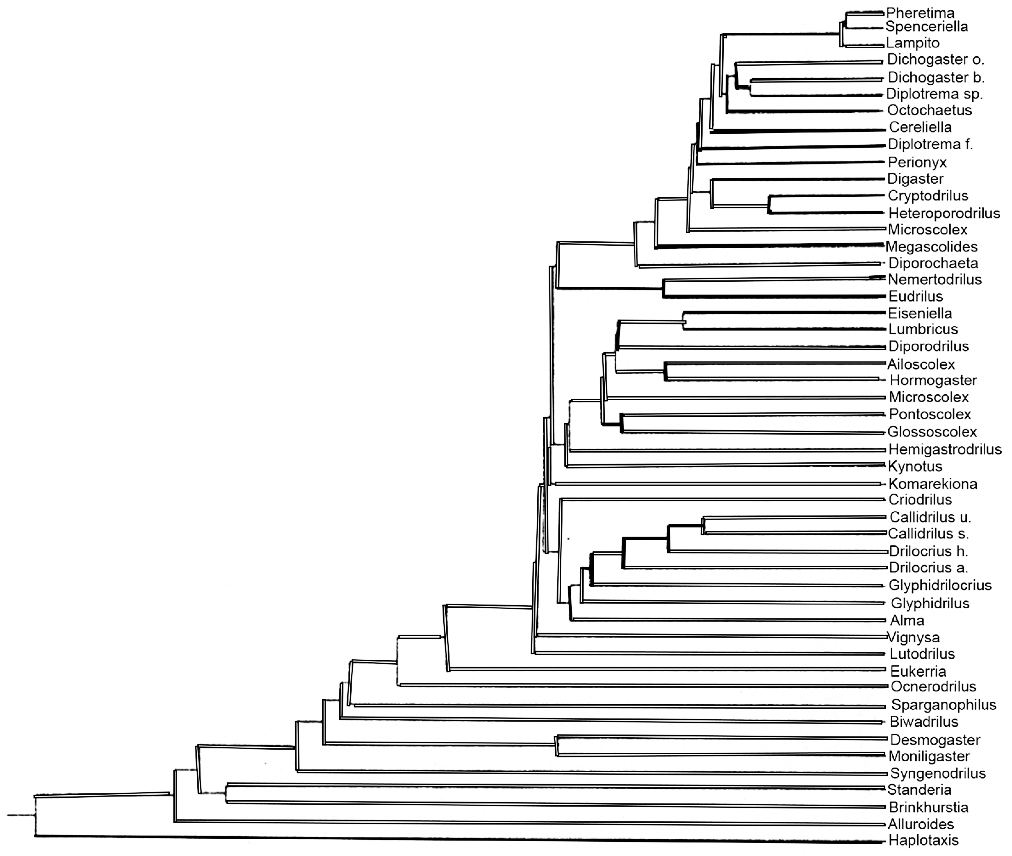
FIGURE 1A. Hennigram (morphological phylogram) for all families of the opisthoporous oligochaetes constructed by nearestneighbour clustering. For details see Jamieson (1978). Redrawn.

Imageimage/png© Jamieson, Barrie G. M.;Fragoso, CarlosJamieson, Barrie G. M.;Fragoso, Carlos

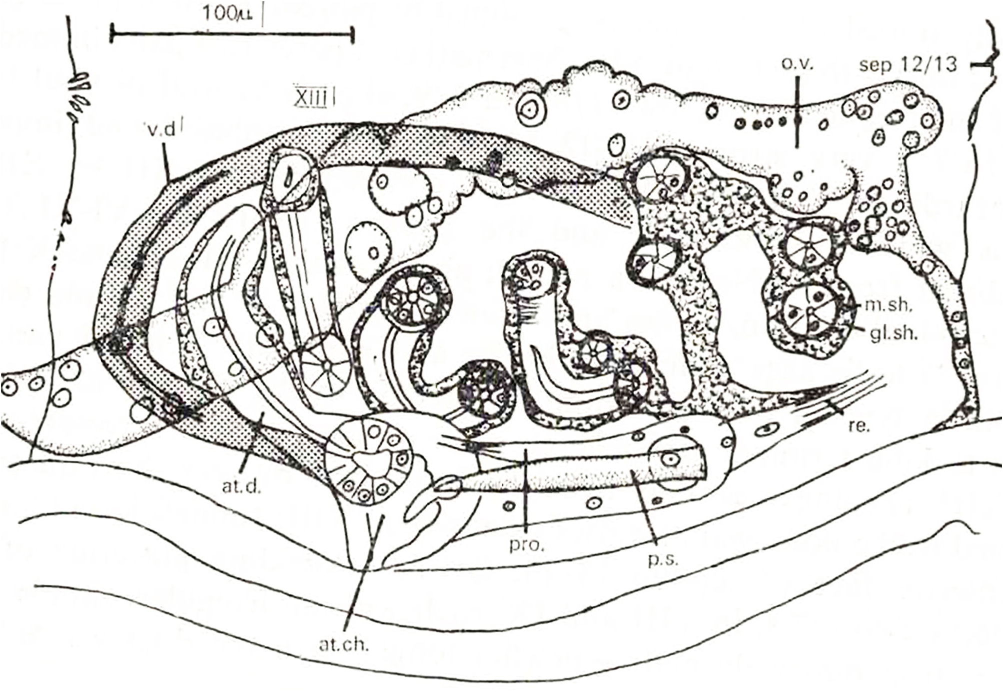
FIGURE 3. Brinkhurstia americanus (Brinkhurst 1964). Longitudinal section through the male terminalia. at.ch, terminal chamber of atrium; at.d, atrial duct; gl.sh, glandular sheath; m.sh, muscular sheath; o.v, ovary; pro, protractor muscle; p.s, penial seta; re, retractor muscle; sep, septum; v.d, vas deferens. From Jamieson (1968).

Imageimage/png© Jamieson, Barrie G. M.;Fragoso, CarlosJamieson, Barrie G. M.;Fragoso, Carlos

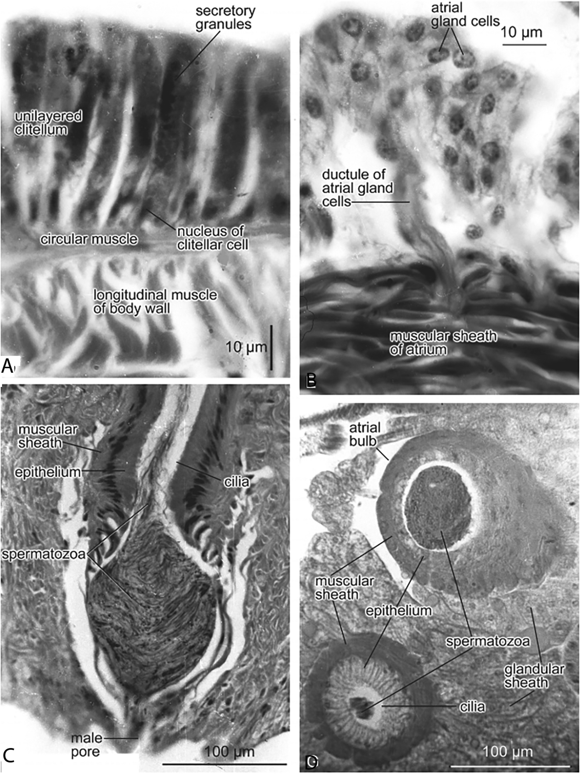
FIGURE 5. Alluroides brinkhursti brinkhursti Jamieson (1968). A. Transverse section (TS) of clitellum, showing single cell layer; the cells with conspicuous secretory granules and each with a basal nucleus. B. TS through the wall of the atrium, showing a group of atrial gland cells with ductule penetrating the muscular sheath of the atrium. C. Longitudinal section through the male pore, showing the ectal end of the atrium, which forms a penis with muscular sheath, ciliated epithelium and rope of spermatozoa in the lumen, forming in the ectal chamber a sperm mass. D. Alluroides pordagei. Oblique section through the atrial bulb, containing a large sperm mass, and the associated atrium. From Jamieson (2006).

Imageimage/png© Jamieson, Barrie G. M.;Fragoso, CarlosJamieson, Barrie G. M.;Fragoso, Carlos

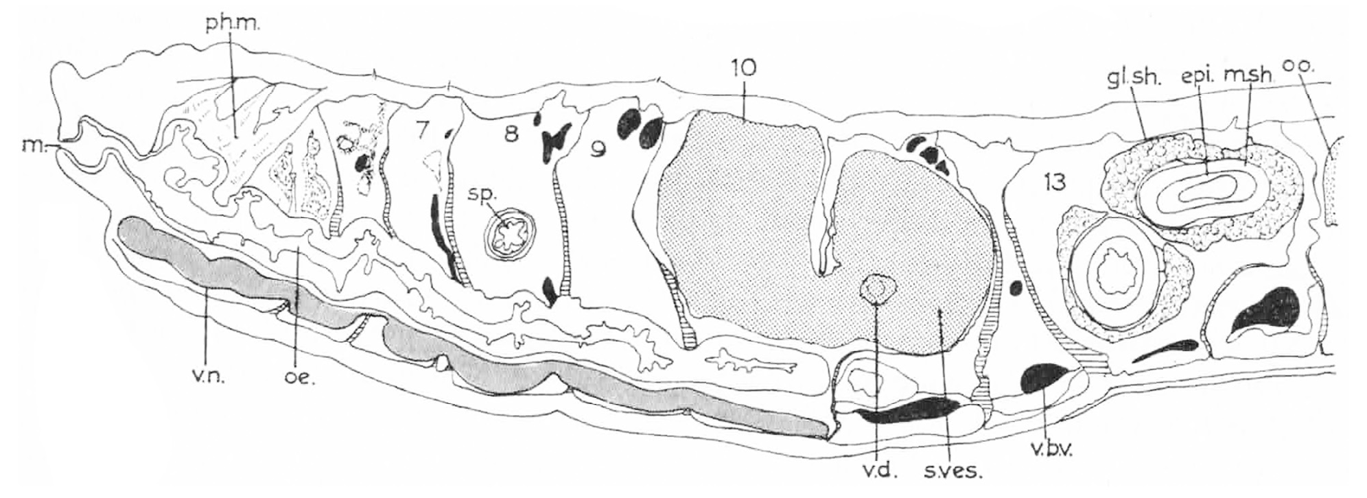
FIGURE 6. Alluroides brinkhursti abyssinicus Jamieson (1968). Longitudinal section of holotype. Abbreviations: epi, epithelium of atrium; gl sh, glandular sheath of atrium; m, mouth; msh, muscular sheath of atrium; oe, oesophagus; oo, oocyte: ph m, pharyngeal musculature; s.ves, seminal vesicle; sp, spermatheca; vbv, ventral blood vessel; vd, vas deferens; v n, ventral nerve cord. Slightly modified from Jamieson (1968).

Imageimage/png© Jamieson, Barrie G. M.;Fragoso, CarlosJamieson, Barrie G. M.;Fragoso, Carlos

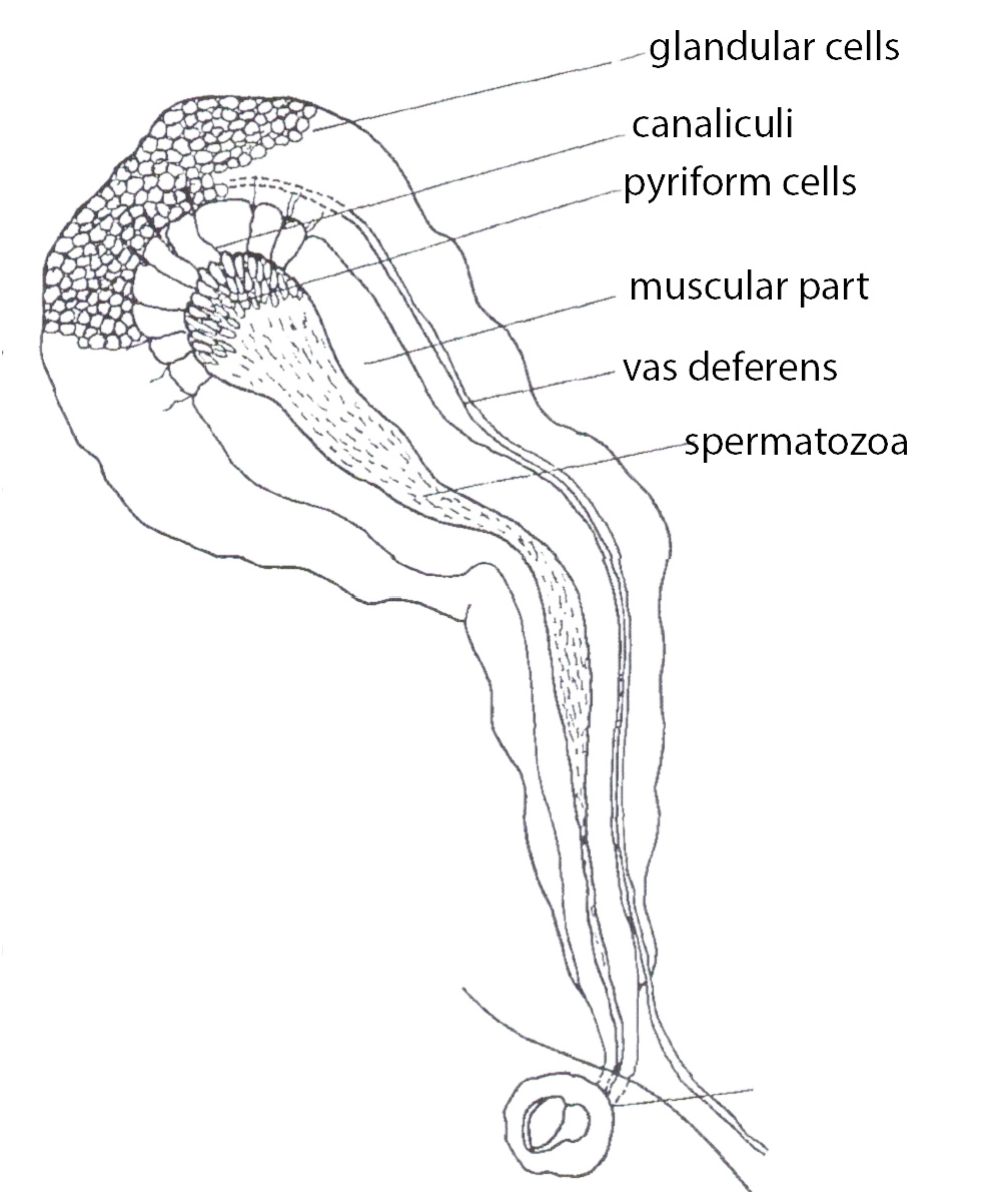
FIGURE 7. Alluroides lauzannei Ljungström, 1971. Detail of an atrium. Note the vas deferens entering the apex of the atrium via canaliculi. Relabelled from Lauzanne (1968).

Imageimage/png© Jamieson, Barrie G. M.;Fragoso, CarlosJamieson, Barrie G. M.;Fragoso, Carlos

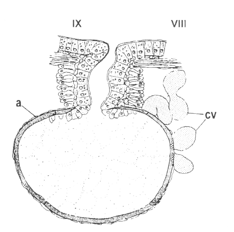
FIGURE 10. Brinkhurstia americanus. (Brinkhurst 1964). Spermatheca opening mid–dorsally in the anterior of IX. Abbreviations: a, ampulla; cv, commissural vessel. From Omodeo & Coates (2001), reproduced with permission.

Imageimage/png© Jamieson, Barrie G. M.;Fragoso, CarlosJamieson, Barrie G. M.;Fragoso, Carlos

IMAGES

Gallery(9)

See Gallery

Occurrences with images

Source Information

A monograph of the Oligochaete family Alluroididae

checklist

This dataset contains the digitized treatments in Plazi based on the original journal article Jamieson, Barrie G. M., Fragoso, Carlos (2024): A monograph of the Oligochaete family Alluroididae. Zootaxa 5529 (3): 401-435, DOI: 10.11646/zootaxa.5529.3.1, URL: http://dx.doi.org/10.11646/zootaxa.5529.3.1

Abstract

The Alluroididae is a small oligochaete family with 8 genera and 13 known species occurring in freshwaters and in riverine soils in Africa, the Caribbean and Central and South America. It is here argued that their anatomy shows a transition from that of microdriles to that of megadriles (Metagynophora). A detailed description of comparative anatomy is given, followed by the systematics and some aspects of the ecology of all known genera, species and subspecies.

Jamieson B G M, Fragoso C, plazi (2024). A monograph of the Oligochaete family Alluroididae. Plazi.org taxonomic treatments database. Checklist dataset https://doi.org/10.15468/uxwm4f accessed via GBIF.org on 2026-06-14.

CC0Published 10/29/2024View dataset
GBIF Usage Key
245596615
Dataset Key
63a39145-1bdf-45c9-9843-6d0cf997c020
Origin
source
Backbone Key
2308694
Taxon ID
6725879F7942FFE6CED9F921FC35F9DA.taxon
Last Crawled
6/9/2026
Last Interpreted
6/9/2026