AnimaliaNot EvaluatedacceptedspeciesAccepted
Alluroides pordagei

Alluroides pordagei

Beddard, 1894

GBIF:245596637

0year

0

Synonyms

ABOUT

Descriptions(3)

Length 25 – 48 mm, width 1.0 mm, segments 202. Prolobous. Form approximately cylindrical throughout, anterior segments at least biannulate with the transverse furrow shortly postsetal. Lateral line in bc, nearer to b than to c. Anus terminal. Ventral setal couples of XIII absent at maturity; all setae sigmoid, with approximately central, well – developed node; none modified as genital setae. Outer setal couples dorsal, dd: u = ca 0.2. Clitellum annular, XII – XVI. Male pores a pair precisely lateral, or slightly ventrolateral, nearer to b than to c lines in the setal arc of XIII, each surrounded by a circular tumescence almost filling the segment longitudinally. Female pores in 13 / 14 directly in line with the male pores, inconspicuous. Spermathecal pore paired or single, dorsal and anterior in VIII, in the type paired near the dorsal midline. Septa 4 / 5 (the first clearly developed), 9 / 10 exceptionally strongly thickened, 10 / 11 thin and attenuated, 11 / 12 and (though less so) 12 / 13 moderately thickened, the succeeding septa thin. Septal glands posterior in V – IX? Nephridia commencing in XII – XVI. Pharynx anterior to septum 4 / 5. Intestine commencing in XIII, internal epithelium with tall cilia; oesophagus similarly ciliated in XII in front of the oesophageal valve; ciliation not certainly present further anteriorly. Brown – granuled chloragogen cells commencing (always?) at 1 / 2 IX. Dorsal blood vessel large and adherent to the gut in XIII posteriorly; slender and freer anteriorly to this. Hearts (posterior commissural) very tortuous extending posteriorly into XI (XII?). Septum 10 / 11 bulging posteriorly far into XI to form a wide – mouthed (unpaired?) seminal vesicle. Muscular tubes of the atria forming slender somewhat coiled tubes extending from XIII into XIV or as far as XVI and widening immediately before opening at the male pores. With the exception of the terminal ectal chamber each tubular atrium surrounded by a sheath of unicellular prostate cells, which approximately doubled the width of the apparatus, the cells communicating with the atrial lumen by minute ductules. Vasa deferentia running in the glandular prostatic duct and presumably entering the atrium entally. Ovaries and funnels in XIII. Oviduct penetrating the body wall immediately behind septum 13 / 14. Female gonadial cells, at maturity, in XIII – XXI, probably in ovisacs. Spermatheca single or paired, dorsally in VIII.
Jamieson, Barrie G. M., Fragoso, Carlos (2024): A monograph of the Oligochaete family Alluroididae. Zootaxa 5529 (3): 401-435, DOI: 10.11646/zootaxa.5529.3.1, URL: http://dx.doi.org/10.11646/zootaxa.5529.3.1
Remarks. Because of the uncertain status of Michaelsen’s material this account (from Jamieson 1968) was derived solely from Beddard’s type – series, and his description, and incorporated results of examinations by Brinkhurst (1964) and Jamieson (Athi River material). The extreme septal thickening, and its distribution, now emerge as diagnostic features of A. pordagei (condition still unknown, however, in A. tanganyikae s. Beddard and s. Michaelsen). Intraspecific variation from a paired to a single spermatheca (in VIII) is confirmed and reinforces Brinkhurst’s conclusion (1964) that A. tanganyikae s. Michaelsen (spermatheca single in VIII), but not s. Beddard (spermatheca single in IX), should be included in A. pordagei. The decision by Michaelsen (1936) to recognize three subspecies, pordagei, tanganyikae and congicus, has not been followed here though it still deserves consideration. A. tanganyikae has been retained as a distinct species but recognition of congicus, from Leopoldsville, as a subspecies appears reasonable. Michaelsen distinguished it by the spermatheca with a thin – walled, sac – like ampulla and sharply demarcated muscular duct. He added that congicus was 90 – 100 mm long, with ca 210 segments, the largest form, whereas the typical subspecies was 22 mm long, the smallest form, while tanganyikae had a length of 45 mm, with ca 150 segments, intermediate between the two other forms. Some of these figures no longer hold.
Jamieson, Barrie G. M., Fragoso, Carlos (2024): A monograph of the Oligochaete family Alluroididae. Zootaxa 5529 (3): 401-435, DOI: 10.11646/zootaxa.5529.3.1, URL: http://dx.doi.org/10.11646/zootaxa.5529.3.1
Distribution. Kenya: swamp 4 miles inland from Mombasa (Type locality, Beddard 1894); lower forest region of Mt. Kenya at 2400 m (Michaelsen 1914 a, b; Michaelsen 1915, as A. tanganyikae); Kahawa stream, Athi River tributary, near Nairobi, collector B. G. M. Jamieson, 11 Dec. 1967. Republic of the Congo: Mulongo, Niunzu; Albertville (Michaelsen 1935); Leopoldville (Michaelsen 1936). Rhodesia: swampy earth, Zambezi River, near Victoria Falls (as A. tanganyikae, Michaelsen 1913).
Jamieson, Barrie G. M., Fragoso, Carlos (2024): A monograph of the Oligochaete family Alluroididae. Zootaxa 5529 (3): 401-435, DOI: 10.11646/zootaxa.5529.3.1, URL: http://dx.doi.org/10.11646/zootaxa.5529.3.1

Export occurrence data

Darwin Core Archive (ZIP)

CLASSIFICATION

Taxonomic Classification Tree

MULTIMEDIA

Media Files(7)

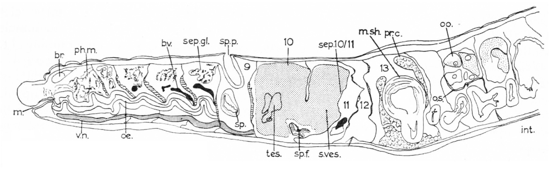
FIGURE 4. Alluroides brinkhursti brinkhursti Jamieson (1968). Longitudinal section of the holotype. Abbreviations: br, brain; bv, blood vessel; m, mouth; msh, muscular sheath of atrium; oe, oesophagus; oo, oocyte: os, ovisac; ph m, pharyngeal musculature; pr c, prostatic cells; sep, septum; sep gl, septal gland; sp p, spermathecal pore; s. ves, seminal vesicle; sp, spermatheca; sp f, sperm funnel; tes, testis; vd, vas deferens; v n, ventral nerve cord. Adapted from Jamieson (1968).

Imageimage/png© Jamieson, Barrie G. M.;Fragoso, CarlosJamieson, Barrie G. M.;Fragoso, Carlos

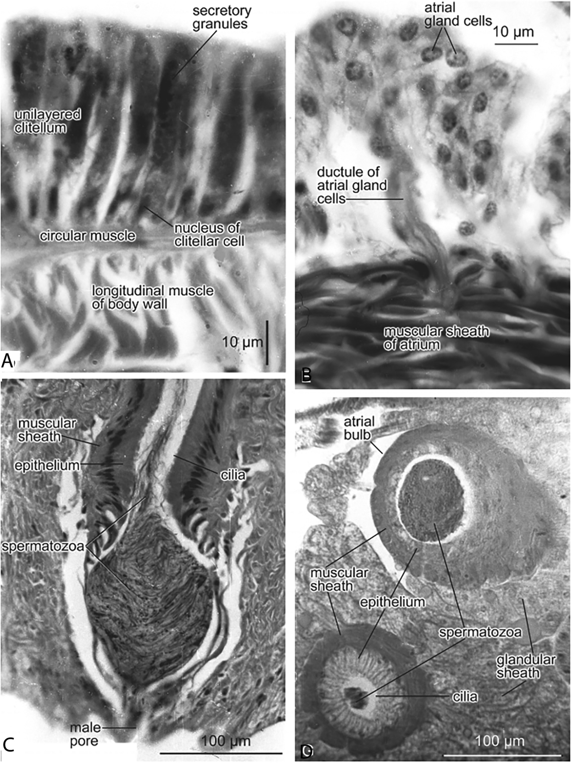
FIGURE 5. Alluroides brinkhursti brinkhursti Jamieson (1968). A. Transverse section (TS) of clitellum, showing single cell layer; the cells with conspicuous secretory granules and each with a basal nucleus. B. TS through the wall of the atrium, showing a group of atrial gland cells with ductule penetrating the muscular sheath of the atrium. C. Longitudinal section through the male pore, showing the ectal end of the atrium, which forms a penis with muscular sheath, ciliated epithelium and rope of spermatozoa in the lumen, forming in the ectal chamber a sperm mass. D. Alluroides pordagei. Oblique section through the atrial bulb, containing a large sperm mass, and the associated atrium. From Jamieson (2006).

Imageimage/png© Jamieson, Barrie G. M.;Fragoso, CarlosJamieson, Barrie G. M.;Fragoso, Carlos

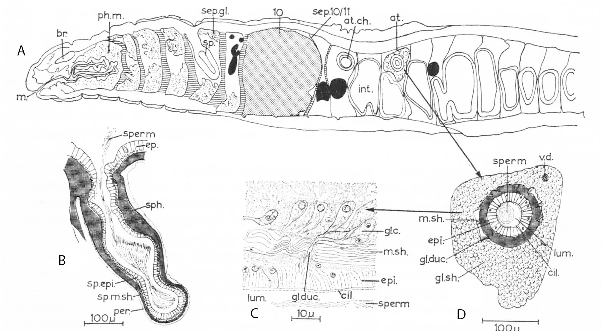
FIGURE 8. Alluroides pordagei Beddard 1894. A. Longitudinal section of the anterior region of the body of Beddard’s paratype. B. Spermatheca. C. Longitudinal section of wall of atrium. D. Transverse section of atrium.Abbreviations: at, atrium; at ch, atrial chamber; br, brain; cil, cilia; ep, epidermis; epi, epithelium; gl duc, glandular ductule(s); gl sh, glandular sheath; int, intestine; lum, lumen; m, mouth; ph m, pharyngeal musculature; per, peritoneum; sep.gl, septal gland; sp, spermatheca; sperm, spermatozoa; sph, sphincter; sp epi, spermathecal epithelium; sp m sh, muscular sheath of spermatheca; v.d, vas deferens.. Slightly modified from Jamieson (1968).

Imageimage/png© Jamieson, Barrie G. M.;Fragoso, CarlosJamieson, Barrie G. M.;Fragoso, Carlos

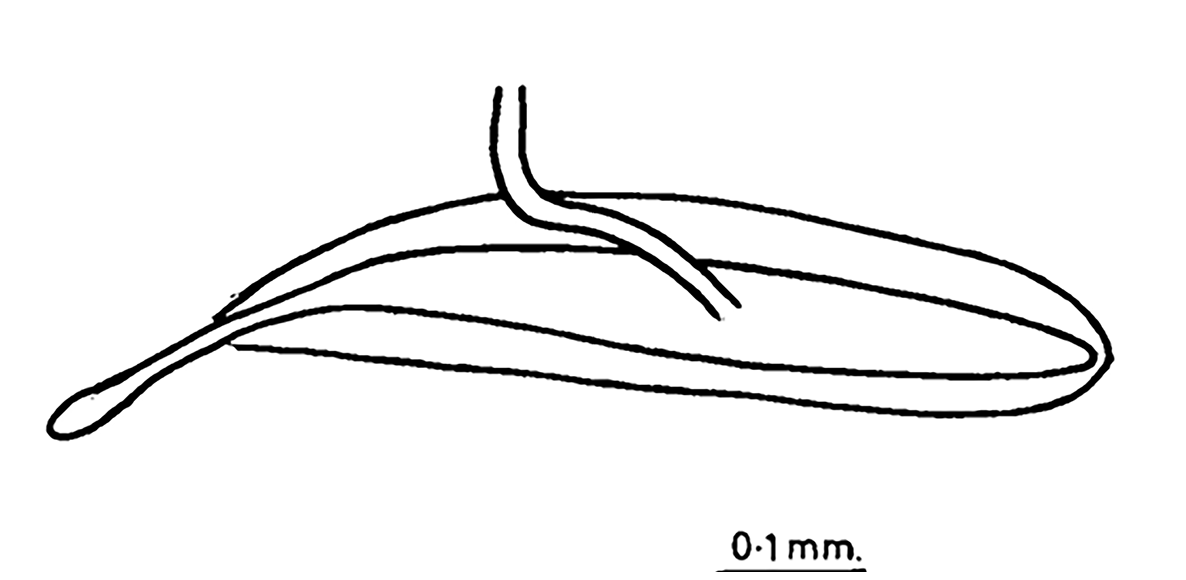
FIGURE 9. Alluroides ruwenzoriensis Brinkhurst 1964. Atrium and vas deferens entering at midlength. From Brinkhurst (1964).

Imageimage/png© Jamieson, Barrie G. M.;Fragoso, CarlosJamieson, Barrie G. M.;Fragoso, Carlos

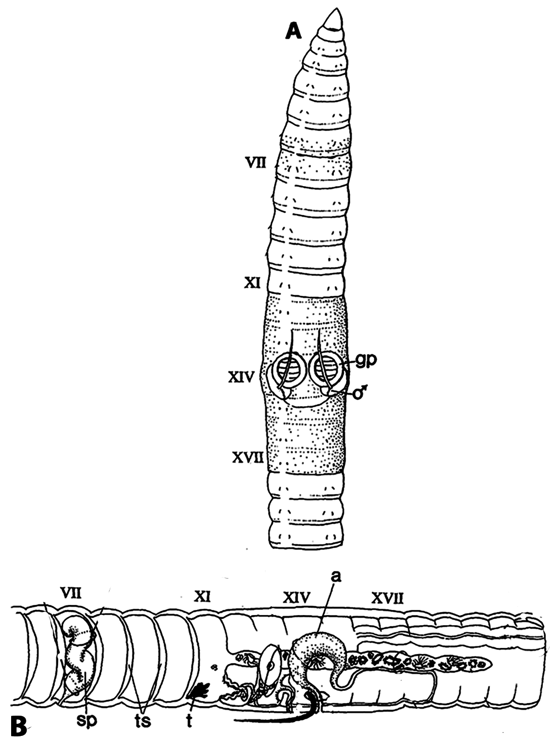
FIGURE 13. Kathrynella guyanae Omodeo 1996.A. Ventral view of anterior segments.Note the pale lateral line.B. arrangement of genital organs. Abbreviations: a, atrium; gp, genital papilla; ♂, male pore; sp, spematheca; t, testis; ts, thickened septum. The penial setae are seen projecting from the male pore. Modified from Omodeo (1996).

Imageimage/png© Jamieson, Barrie G. M.;Fragoso, CarlosJamieson, Barrie G. M.;Fragoso, Carlos

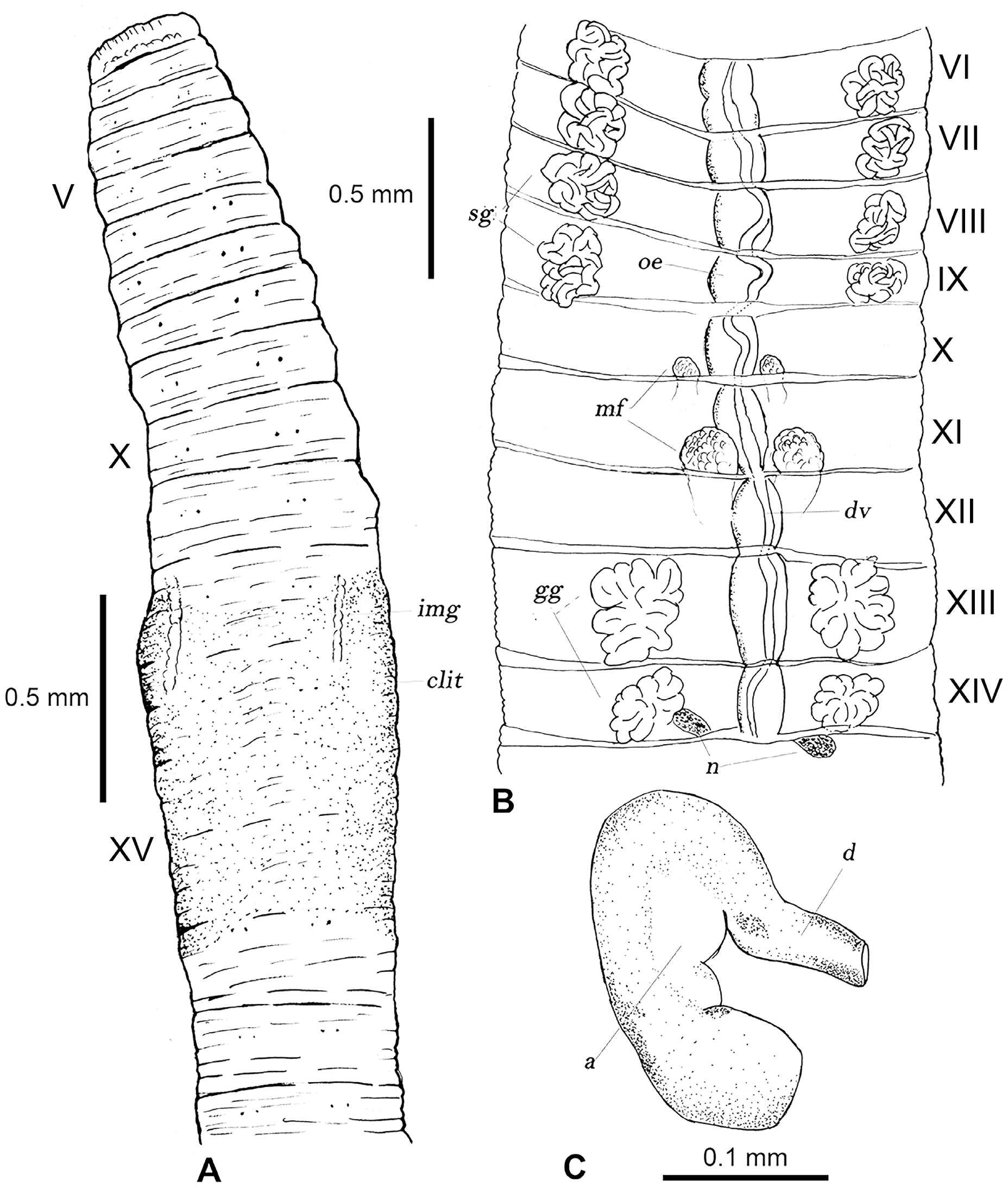
FIGURE 14. Lacandodrilus paludosus Fragoso & Rojas 2023. A. External view of the anterior ventral region of a clitellate adult. B. Internal view of the anterior region of an adult. C. Spermatheca. Abbreviations: a, ampulla: clit, clitellum; d, duct; dv, dorsal vessel; gg, genital glands (prostates?); img, internal male gonoducts; mf, male funnels; n, nephridia; oe, oesophagus; sg, septal glands. Modified from Fragoso & Rojas (2023).

Imageimage/png© Jamieson, Barrie G. M.;Fragoso, CarlosJamieson, Barrie G. M.;Fragoso, Carlos

IMAGES

Gallery(7)

See Gallery

Occurrences with images

Source Information

A monograph of the Oligochaete family Alluroididae

checklist

This dataset contains the digitized treatments in Plazi based on the original journal article Jamieson, Barrie G. M., Fragoso, Carlos (2024): A monograph of the Oligochaete family Alluroididae. Zootaxa 5529 (3): 401-435, DOI: 10.11646/zootaxa.5529.3.1, URL: http://dx.doi.org/10.11646/zootaxa.5529.3.1

Abstract

The Alluroididae is a small oligochaete family with 8 genera and 13 known species occurring in freshwaters and in riverine soils in Africa, the Caribbean and Central and South America. It is here argued that their anatomy shows a transition from that of microdriles to that of megadriles (Metagynophora). A detailed description of comparative anatomy is given, followed by the systematics and some aspects of the ecology of all known genera, species and subspecies.

Jamieson B G M, Fragoso C, plazi (2024). A monograph of the Oligochaete family Alluroididae. Plazi.org taxonomic treatments database. Checklist dataset https://doi.org/10.15468/uxwm4f accessed via GBIF.org on 2026-06-14.

CC0Published 10/29/2024View dataset
GBIF Usage Key
245596637
Dataset Key
63a39145-1bdf-45c9-9843-6d0cf997c020
Origin
source
Backbone Key
2308692
Taxon ID
6725879F7946FFE1CED9FA71FD08FD8D.taxon
Last Crawled
6/9/2026
Last Interpreted
6/9/2026