AnimaliaNot EvaluatedacceptedspeciesAccepted
Kathrynella guyanae

Kathrynella guyanae

Omodeo, 1996

GBIF:245596645

0year

ABOUT

Descriptions(3)

Length 85 – 89 mm; diameter 600 µm at X, 520 µm at mid – body, segments 282, 259. No cutaneous pigmentation, integument transparent. Prostomium zygolobous, conical; peristomium short. Setae paired with lumbricin arrangement. Pygidium small, anus a vertical cleft. Clitellum annular, extending from the posterior part of XI to XVII; the ventral side of XIV with two large, circular papillae with swollen borders and a very thick, whitish, central part crossed by 4 – 5 transverse furrows; behind and beside these papillae two crescent – shaped fields visible, each bearing a single, large, grooved, penial seta. In all specimens examined, penial setae were broken but their length estimated as longer than 300 µm. No other ventral setae visible in XIV. Dorsal setal couples of midclitellar segments slanting relative to the long body axis, setae c located more posteriorly than setae d. Male pore as a slit on hinder portion of each crescent – shaped field in XIV; paired female pores on anterior border of XIV, near midventral line. No dorsal pores. Nephridiopores inconspicuous. Spermathecal pores in furrow 6 / 7 on setal line a. Setal ratio aa: ab: bc: cd: dd = 5.6: 1.1: 5.6: 1: 10 at segment X; 2.5: 1: 1.5: 1: 12 in posterior segments (where all setae are displaced ventrad). The length of setae increases from II, where it is 37 µm, to VII where setae a and b measure 92 µm and setae c and d 43 µm. Further posteriorly, the setal length decreases gradually (penial setae of XIV excepted), to become constant (ca 50 µm) behind the clitellum. Only caudal setae show distal ornamentation. Body wall thin; cuticle approximately 0.5 µm thick; layer of circular muscles very thin. Septa 5 / 6 – 10 / 11 thickened, funnel – shaped. No pharyngeal pad distinguishable. Neither a gizzard, nor any apparent distinction between oesophagus and intestine present. Ciliated epithelium of intestine ending in clitellar region. Peri – intestinal blood sinus occurring from XII backwards. Holonephridia, apparently avesiculate, beginning in XII or XIII; initially poorly developed and acquiring normal size only behind clitellum. The circulatory apparatus exceptionally well developed. Dorsal and ventral vessels run the whole length of the worm and have very large diameters, only slightly less than that of the intestine. Seven pairs of long commissural vessels in V – XI, wound in balls whose size increases backwards; each ball covered with chloragocytes. A pair of large vessels originates in VII from the peri – intestinal blood sinus, and from XII to XVI run along the sperm sacs. Many lesser vessels seen everywhere, but no capillaries. Testes anterior in XI, facing the large seminiferous funnels, covered with sperm and bulging into segment XII. Sperm ducts coiled in XII in their proximal course, then running laterally within the body wall till XVII where they emerge again in the coelomic cavity, become thicker, and run forwards till XIV, where they form the atria. Atria possess thick muscular walls and appear as shining, bent, spindles which open in the middle of XIV, behind the penial setae; atria (which correspond to the ‘ euprostates’ of Eudrilidae) with a thin external coating of non – glandular cells. Two long sperm sacs begin from septum 11 / 12 and run till XX, parallel to the egg sacs and to the posterior course of vasa deferentia. Ovaries located in the ventral forepart of XIII; two long cylindrical egg sacs, apparently beginning in XVII, extending as far back as XXXV; possessing thick glandular walls and containing up to a dozen large ripe eggs (ca 530 µm wide) between XXV – XXXV. Two large, convoluted spermathecae in VII; consisting of a short, partly ciliated duct and elongate ampulla; the ectal section of the latter containing the bulk of spermatozoa, whereas the ental section is filled with a hyaline secretion. The spermathecal length exceeds the body diameter and possibly the length of penial setae. It can be inferred that during copulation the sucker – shaped genital papillae in XIV help to fasten the two partners together, while at the same time the penial grooved setae are introduced into the spermathecae for sperm transfer.
Jamieson, Barrie G. M., Fragoso, Carlos (2024): A monograph of the Oligochaete family Alluroididae. Zootaxa 5529 (3): 401-435, DOI: 10.11646/zootaxa.5529.3.1, URL: http://dx.doi.org/10.11646/zootaxa.5529.3.1
Remarks. In his elegant paper, Omodeo (1996) considered that Kathrynella guyanae showed traits, discussed here in a previous section, which were reminiscent of various ‘ microdrile’ taxa. Omodeo saw two main differences (autapomorphies) of Kathrynella from the Alluroididae previously described i. e. the location of male pores in XIV and the metandric condition of the male apparatus which contrasted with the proandric condition of the Alluroidinae and the holoandric, supposedly plesiomorphic (cf. Jamieson 1980), condition of the Syngenodrilinae. He noted a resemblance of the ‘ atria’ of Kathrynella to the typical ‘ euprostates’ of the Eudrilidae, an African family of unknown kinship.
Jamieson, Barrie G. M., Fragoso, Carlos (2024): A monograph of the Oligochaete family Alluroididae. Zootaxa 5529 (3): 401-435, DOI: 10.11646/zootaxa.5529.3.1, URL: http://dx.doi.org/10.11646/zootaxa.5529.3.1
Distribution. Known only from the type locality in Guyana.
Jamieson, Barrie G. M., Fragoso, Carlos (2024): A monograph of the Oligochaete family Alluroididae. Zootaxa 5529 (3): 401-435, DOI: 10.11646/zootaxa.5529.3.1, URL: http://dx.doi.org/10.11646/zootaxa.5529.3.1

Export occurrence data

Darwin Core Archive (ZIP)

CLASSIFICATION

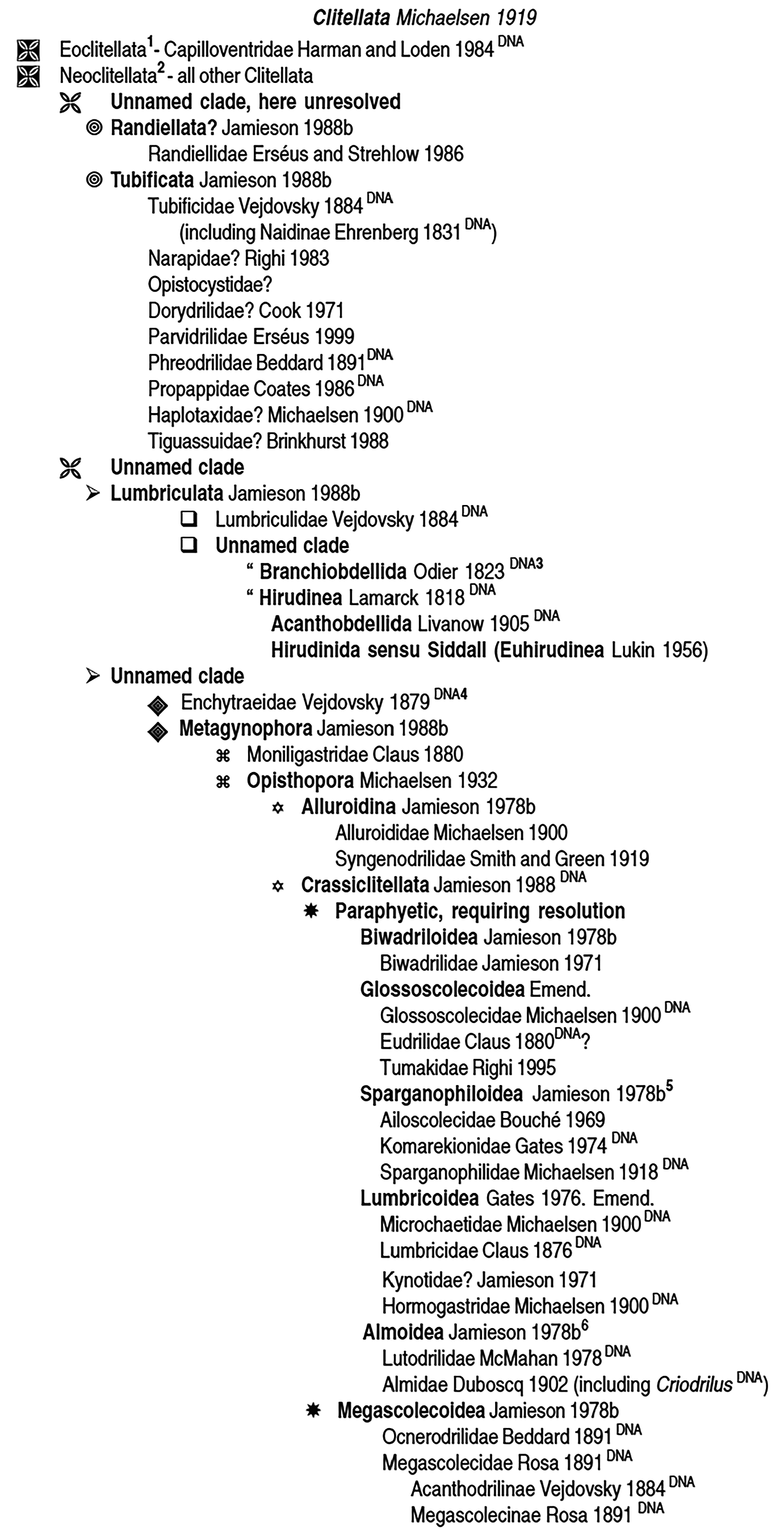
Taxonomic Classification Tree

MULTIMEDIA

Media Files(6)

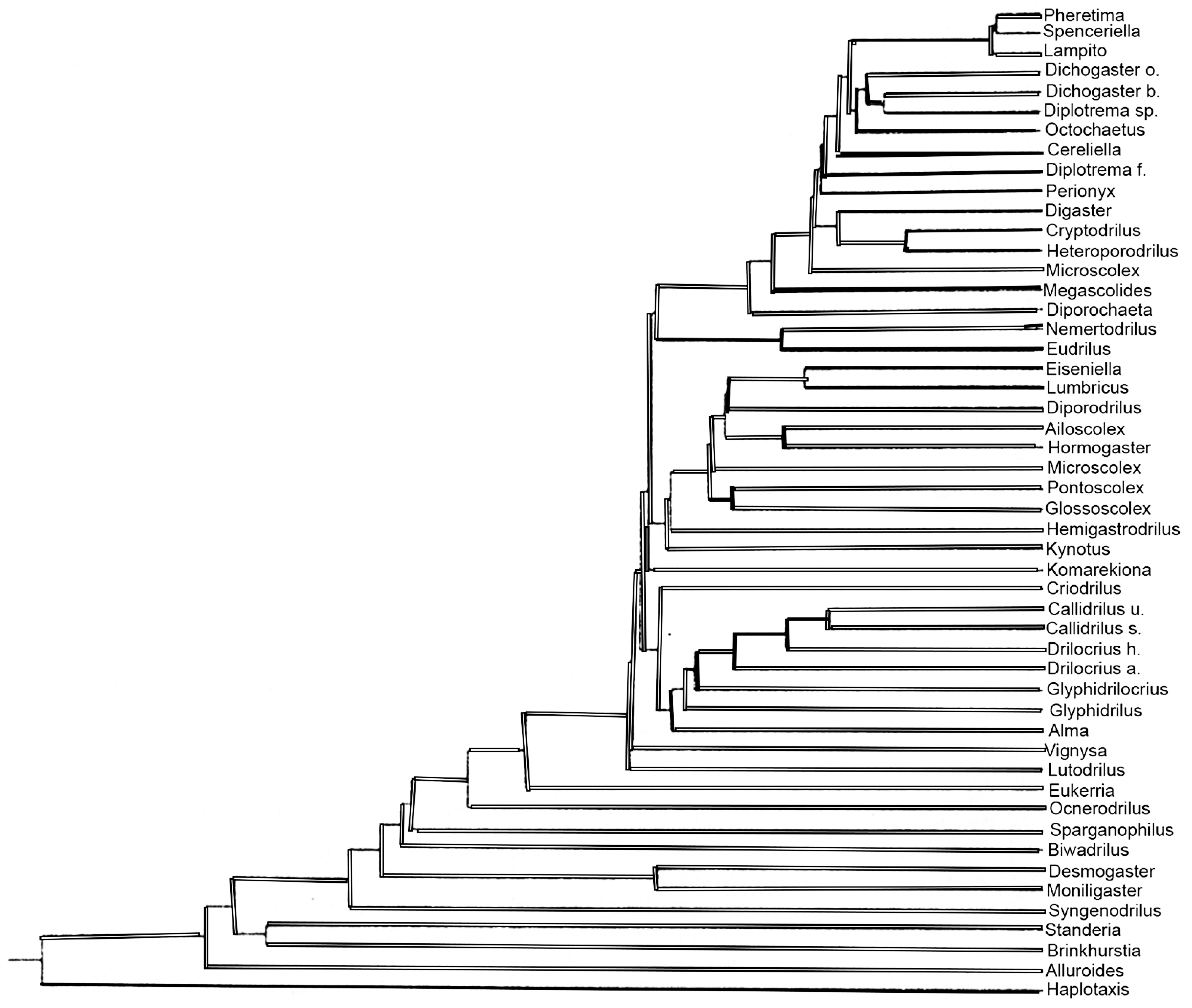
FIGURE 1A. Hennigram (morphological phylogram) for all families of the opisthoporous oligochaetes constructed by nearestneighbour clustering. For details see Jamieson (1978). Redrawn.

Imageimage/png© Jamieson, Barrie G. M.;Fragoso, CarlosJamieson, Barrie G. M.;Fragoso, Carlos

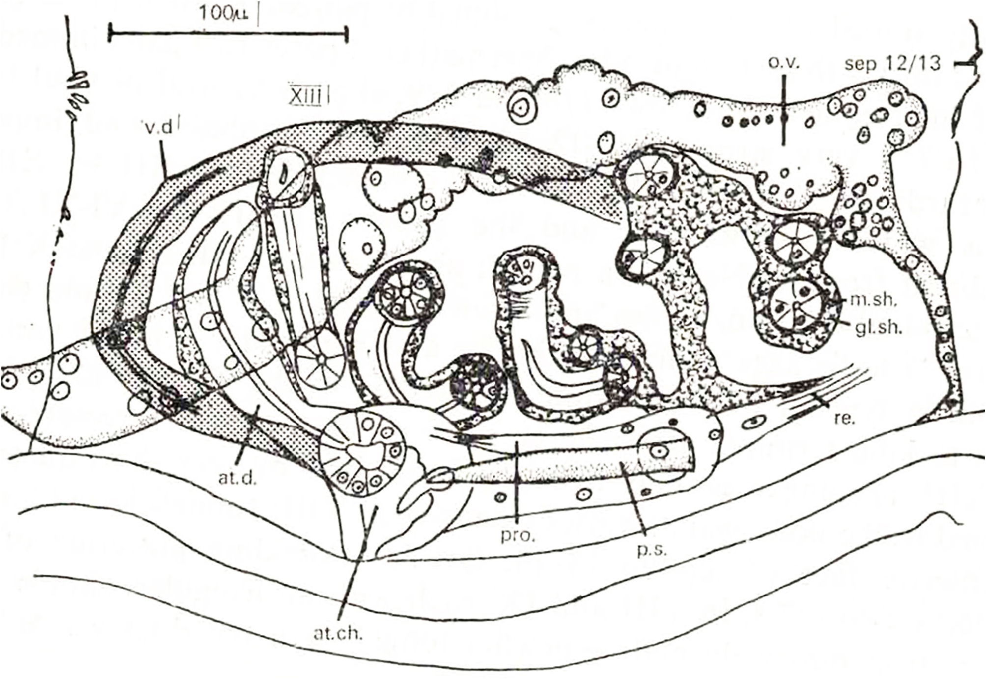
FIGURE 3. Brinkhurstia americanus (Brinkhurst 1964). Longitudinal section through the male terminalia. at.ch, terminal chamber of atrium; at.d, atrial duct; gl.sh, glandular sheath; m.sh, muscular sheath; o.v, ovary; pro, protractor muscle; p.s, penial seta; re, retractor muscle; sep, septum; v.d, vas deferens. From Jamieson (1968).

Imageimage/png© Jamieson, Barrie G. M.;Fragoso, CarlosJamieson, Barrie G. M.;Fragoso, Carlos

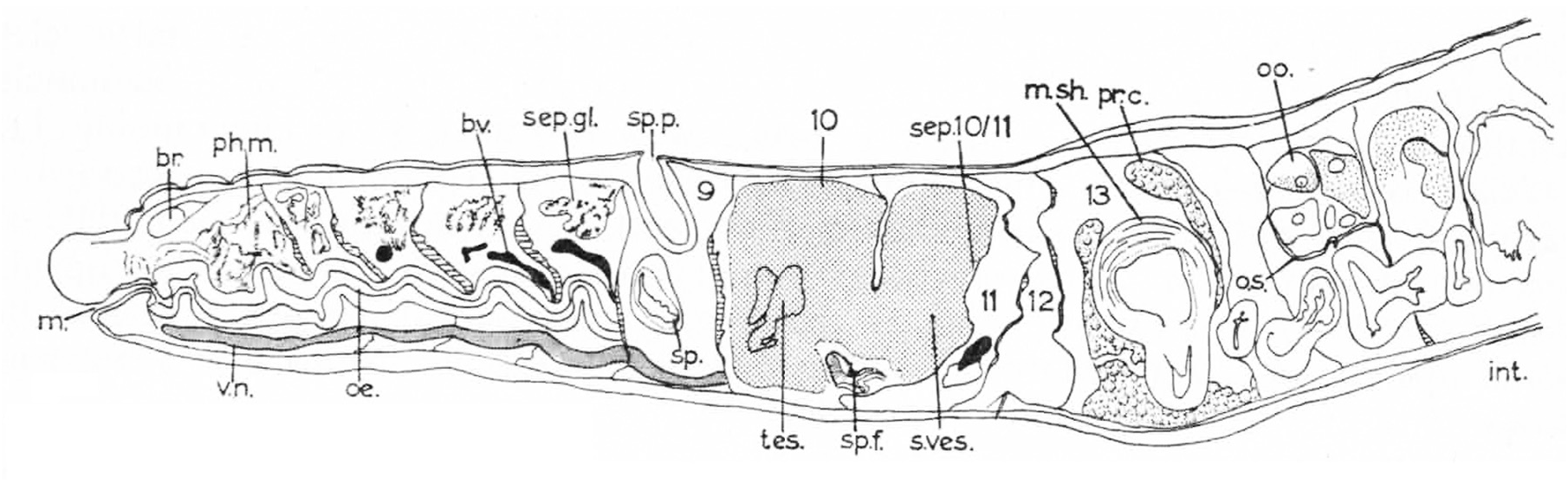
FIGURE 4. Alluroides brinkhursti brinkhursti Jamieson (1968). Longitudinal section of the holotype. Abbreviations: br, brain; bv, blood vessel; m, mouth; msh, muscular sheath of atrium; oe, oesophagus; oo, oocyte: os, ovisac; ph m, pharyngeal musculature; pr c, prostatic cells; sep, septum; sep gl, septal gland; sp p, spermathecal pore; s. ves, seminal vesicle; sp, spermatheca; sp f, sperm funnel; tes, testis; vd, vas deferens; v n, ventral nerve cord. Adapted from Jamieson (1968).

Imageimage/png© Jamieson, Barrie G. M.;Fragoso, CarlosJamieson, Barrie G. M.;Fragoso, Carlos

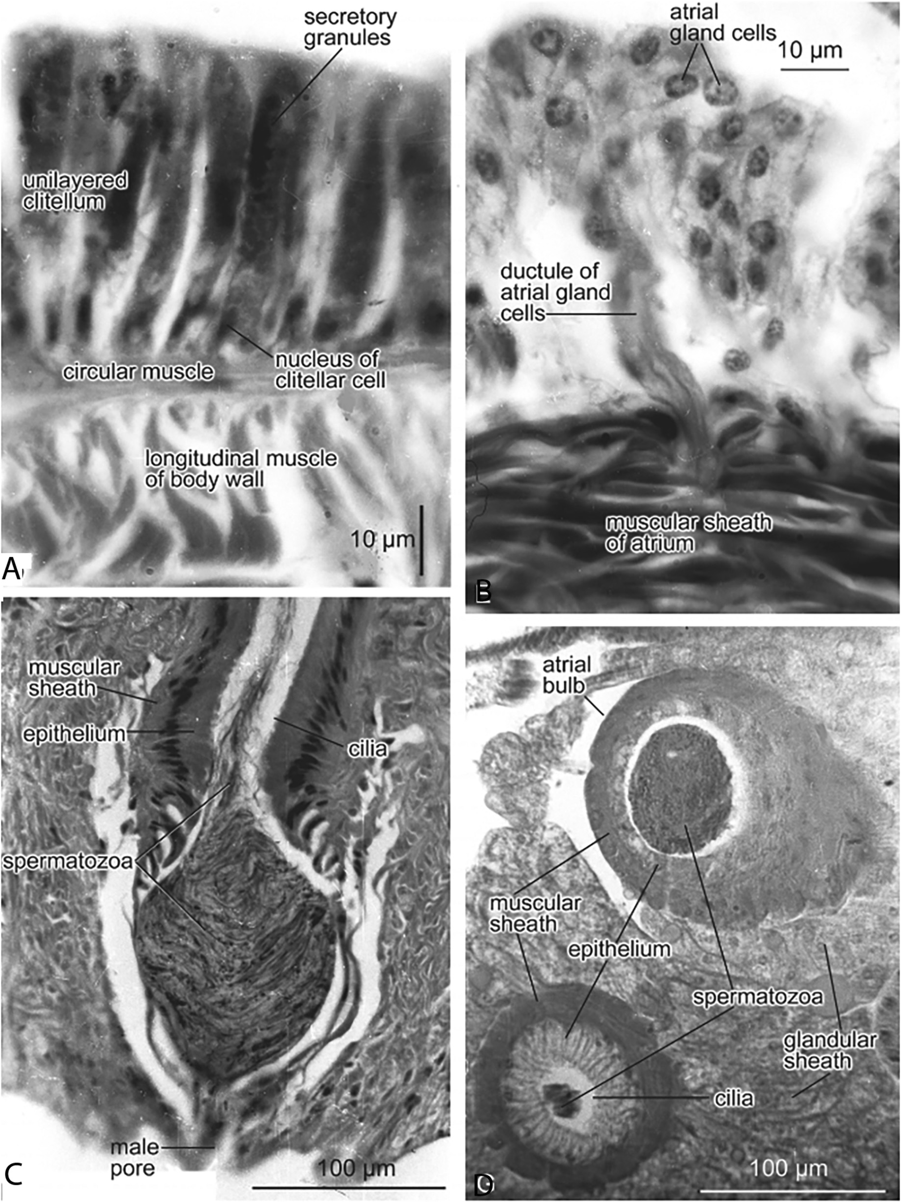
FIGURE 5. Alluroides brinkhursti brinkhursti Jamieson (1968). A. Transverse section (TS) of clitellum, showing single cell layer; the cells with conspicuous secretory granules and each with a basal nucleus. B. TS through the wall of the atrium, showing a group of atrial gland cells with ductule penetrating the muscular sheath of the atrium. C. Longitudinal section through the male pore, showing the ectal end of the atrium, which forms a penis with muscular sheath, ciliated epithelium and rope of spermatozoa in the lumen, forming in the ectal chamber a sperm mass. D. Alluroides pordagei. Oblique section through the atrial bulb, containing a large sperm mass, and the associated atrium. From Jamieson (2006).

Imageimage/png© Jamieson, Barrie G. M.;Fragoso, CarlosJamieson, Barrie G. M.;Fragoso, Carlos

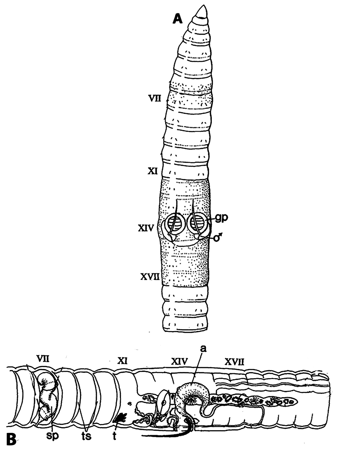
FIGURE 13. Kathrynella guyanae Omodeo 1996.A. Ventral view of anterior segments.Note the pale lateral line.B. arrangement of genital organs. Abbreviations: a, atrium; gp, genital papilla; ♂, male pore; sp, spematheca; t, testis; ts, thickened septum. The penial setae are seen projecting from the male pore. Modified from Omodeo (1996).

Imageimage/png© Jamieson, Barrie G. M.;Fragoso, CarlosJamieson, Barrie G. M.;Fragoso, Carlos

FIGURE 2. A phylogenetic classification of the Clitellata. (From Jamieson 2006).

Imageimage/png© Jamieson, Barrie G. M.;Fragoso, CarlosJamieson, Barrie G. M.;Fragoso, Carlos

IMAGES

Gallery(6)

See Gallery

Occurrences with images

Source Information

A monograph of the Oligochaete family Alluroididae

checklist

This dataset contains the digitized treatments in Plazi based on the original journal article Jamieson, Barrie G. M., Fragoso, Carlos (2024): A monograph of the Oligochaete family Alluroididae. Zootaxa 5529 (3): 401-435, DOI: 10.11646/zootaxa.5529.3.1, URL: http://dx.doi.org/10.11646/zootaxa.5529.3.1

Abstract

The Alluroididae is a small oligochaete family with 8 genera and 13 known species occurring in freshwaters and in riverine soils in Africa, the Caribbean and Central and South America. It is here argued that their anatomy shows a transition from that of microdriles to that of megadriles (Metagynophora). A detailed description of comparative anatomy is given, followed by the systematics and some aspects of the ecology of all known genera, species and subspecies.

Jamieson B G M, Fragoso C, plazi (2024). A monograph of the Oligochaete family Alluroididae. Plazi.org taxonomic treatments database. Checklist dataset https://doi.org/10.15468/uxwm4f accessed via GBIF.org on 2026-06-14.

CC0Published 10/29/2024View dataset
GBIF Usage Key
245596645
Dataset Key
63a39145-1bdf-45c9-9843-6d0cf997c020
Origin
source
Backbone Key
9419865
Taxon ID
6725879F7941FFEACED9F884FCEAFC79.taxon
Last Crawled
6/9/2026
Last Interpreted
6/9/2026